Biology 9700/31 — May/June 2012
Cambridge AS Level · Advanced Practical Skills · worked solutions for every part, with the mark scheme
Topics Use of the Light Microscope · Presentation of Data and Observations · Analysis, Conclusions and Evaluation · Manipulation, Measurement and Observation
When onion cells are placed into a sodium chloride solution with a higher concentration than the cells, water leaves the vacuoles and the cells become plasmolysed.
You are provided with three samples of onion tissue, each soaked in a different concentration of sodium chloride solution, labelled S1, S2 and S3.
The three concentrations of sodium chloride solution are , and .
The onion tissue in S1 is in sodium chloride solution.
You are required to:
- observe and record the cells in S1 and the effect of adding water, W, to these cells
- observe and record the onion cells to identify the concentration of the sodium chloride solutions, S2 and S3.
You are advised to read steps 1 to 12 before proceeding.
Proceed as follows:
- Label one dry and clean microscope slide S1 and put the slide on a paper towel.
- Put a few drops of the sodium chloride solution from S1 onto the slide.
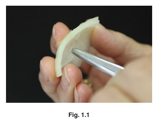
- Remove a piece of the onion tissue from solution S1 and, using forceps or fingers, peel off the inner concave epidermis as shown in Fig. 1.1.
- Cut one piece of the epidermis to fit under the coverslip. Replace the remaining epidermis into the solution in S1.
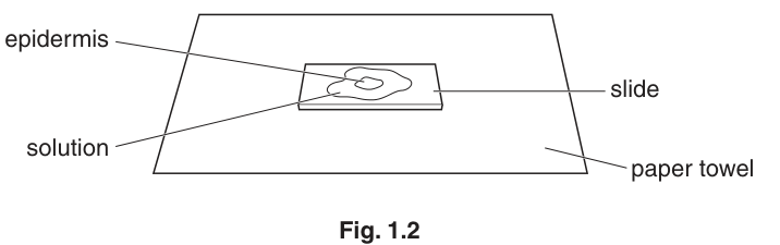
- Place the epidermis into the sodium chloride solution on the slide as shown in Fig. 1.2. If the epidermis is folded, you may need to add more drops of sodium chloride solution so that it floats and uncurls. It is important to stop the epidermis from drying out.
- Cover the epidermis on the slide with a coverslip and use a paper towel to remove any excess liquid that is outside the coverslip.
- View the slide using the microscope. You may need to reduce the amount of light entering the microscope to observe the cells clearly.
Make a large drawing of two adjacent (touching) cells which show the effect of soaking the cells in the sodium chloride solution, S1.
Label a cell membrane.
Describe how you will obtain quantitative results to identify the concentration of the sodium chloride solutions, S2 and S3, compared with S1.
- Record the result for S1.
- Take the slide off the microscope and remove the coverslip.
- Remove as much of the sodium chloride solution as you can and put a few drops of W onto the epidermis. Replace the coverslip and leave the slide until step 12.
- Repeat steps 1 to 8 with S2 and S3 (note: you do not need to repeat (a)(i)).
Prepare the space below to record your quantitative results as you have described in (a)(ii).
Identify the concentrations of sodium chloride solution in S2 and S3.
Complete the table.
| solution | concentration of sodium chloride solution / |
|---|---|
| S1 | 1.00 |
| S2 | |
| S3 |
Describe how you identified the concentrations of sodium chloride.
- Put the S1 slide onto the microscope and select two adjacent cells to show the difference in the cells after adding the water compared with those drawn in (a)(i).
Make a large labelled drawing of these two adjacent (touching) cells.
Label a cell wall.
Use your knowledge and understanding of plasmolysis to explain how and why the cells changed when they were left in water.
Suggest how you would modify this investigation to find the water potential of the onion cells.
The rest of this paper
1 more questions- Q2Use of the Light Microscope · Analysis, Conclusions and Evaluation · Presentation of Data and Observations18M