Biology 9700/23 — May/June 2012
Cambridge AS Level · AS Level Structured Questions · worked solutions for every part, with the mark scheme
Topics Biological Molecules · Infectious Diseases · Transport in Mammals · Cell Structure · Transport in Plants · Gas Exchange · +4 more
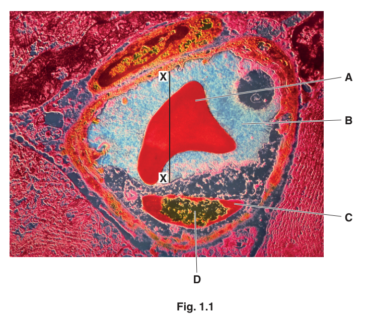
Fig. 1.1 is an electron micrograph of a cross section through a blood vessel.
Name the type of blood vessel shown in Fig. 1.1 and describe one visible feature which is characteristic of this type of vessel.
type of vessel = ______
characteristic feature = ______
Answer
type of vessel = capillary
characteristic feature = wall is one cell thick (only endothelium present) / the lumen is only just wider than a red blood cell
capillary; wall is one cell thick (only endothelium present)
Background Concept
Mammalian blood vessels come in three main types: arteries, veins and capillaries. They differ in the structure and thickness of their walls, which is closely tied to their function.
- Arteries carry blood away from the heart at high pressure, so they have thick walls with prominent layers of smooth muscle and elastic fibres.
- Veins carry blood back to the heart at low pressure, so their walls are thinner and contain less muscle and elastic tissue; they often have a visibly larger, more irregular lumen.
- Capillaries are the site of exchange between blood and surrounding tissues. They consist of a single layer of endothelial cells (a one-cell-thick endothelium) sitting on a thin basement membrane, with no smooth muscle or elastic layers. Their lumen is so narrow — typically about 5–10 µm in diameter — that red blood cells (about 7 µm across) often have to deform to pass through in single file.
Understanding the Question
The electron micrograph in Fig. 1.1 shows a cross-section of a blood vessel. We are asked to (i) name the type of vessel and (ii) describe ONE visible feature that is characteristic of it. The mark scheme accepts any one clear, observable feature that distinguishes this vessel from an artery or vein.
Approach
Look at the image and identify the most diagnostic features:
- The wall is very thin — only a single layer of cells is visible between the lumen and the surrounding tissue.
- The lumen is just large enough to hold the red blood cell(s); in capillaries the red cells are squeezed against the wall.
- There is no visible layer of smooth muscle or elastic tissue around the endothelium.
The simplest single observation is that the wall is only one cell thick — this is the textbook defining feature of a capillary.
Step-by-Step Reasoning
- The vessel is not an artery: there is no thick muscular wall, no clearly visible internal or external elastic lamina, and the lumen is irregular rather than circular and held open by a thick wall.
- The vessel is not a vein: veins have a much larger, more irregular lumen, a thinner relative wall, but still show a distinct endothelium plus some smooth muscle; here only endothelium is visible.
- The wall is one cell thick (only endothelium), and the lumen is so narrow that a red blood cell fills most of it — both are diagnostic of a capillary.
- Either of those two features earns the second mark; the cleanest single answer is that the wall is one cell thick.
Key Takeaways
- A capillary is defined structurally by having a wall of a single layer of endothelial cells on a basement membrane — there is no smooth muscle, no elastic tissue, and no outer connective-tissue sheath.
- The very narrow lumen (just wider than a red blood cell) is a useful secondary diagnostic, and it is the structural basis of efficient exchange by diffusion.
Common Mistakes
- Writing "artery" or "vein": these would be wrong because the wall here is far too thin and there is no visible muscle layer.
- Saying "the wall is thin" without more detail: this is true but does not specify what the wall consists of. Better: "wall is one cell thick" or "only endothelium present".
- Confusing the small dark feature D (nucleolus) with the nucleus of the endothelial cell — the mark scheme accepts both, but the more precise answer is nucleolus.
Things to Be Careful About
- The question asks for a visible feature. Do not describe invisible features (e.g. "site of exchange") — that is a function, not an observable structural characteristic.
- Stick to one clear feature; the mark is for the single best, observable point.
Name:
structure A
Answer
red blood cell / erythrocyte
red blood cell (erythrocyte)
Background Concept
Red blood cells (erythrocytes) are the most numerous cells in mammalian blood. In mammals they are biconcave discs about 7 µm in diameter, lack a nucleus when mature, and are filled with haemoglobin, which gives them their characteristic red colour. In electron micrographs they appear as smooth, anucleate, often slightly deformed shapes — frequently squeezed into the shape of the vessel lumen because they are flexible.
Understanding the Question
Structure A is the large, red, irregularly shaped body lying in the centre of the vessel lumen. It is not surrounded by any membrane other than its own plasma membrane, and it has no visible nucleus.
Approach
Apply the diagnostic features of a red blood cell in a micrograph: biconcave/irregular shape, no nucleus, occupies the lumen of a small vessel.
Step-by-Step Reasoning
- The shape (irregular, deformed) is consistent with a flexible cell being squeezed inside a narrow vessel — typical of a red blood cell passing through a capillary.
- No nucleus is visible inside A, which is the hallmark of a mature mammalian red blood cell.
- The label points to a single cell within the lumen of a vessel whose identity we have already confirmed as a capillary. This is the only cell type that fits.
Key Takeaways
- A mature mammalian red blood cell is anucleate (lacks a nucleus) — a key feature visible in micrographs.
- Red blood cells are flexible and deform to pass through capillaries only slightly wider than themselves.
Common Mistakes
- Calling it a "white blood cell" or "leucocyte". White blood cells are larger, have a clearly visible nucleus (often lobed), and are far less numerous than red blood cells.
- Calling it simply a "cell". The question wants the specific identity.
Things to Be Careful About
- The acceptable answers are "red blood cell", "erythrocyte" or "red blood corpuscle". Avoid informal descriptions like "red blob".
the main component of substance B.
Answer
water
water
Background Concept
Blood is composed of cellular elements (red cells, white cells, platelets) suspended in a liquid matrix called plasma. Plasma is approximately 90% water, with the remaining ~10% made up of dissolved plasma proteins (e.g. albumins, globulins, fibrinogen), inorganic ions (Na⁺, Cl⁻, HCO₃⁻, K⁺, Ca²⁺), nutrients (glucose, amino acids, lipids), hormones, urea and dissolved gases. Water is therefore overwhelmingly the main component by mass and by volume.
Understanding the Question
Substance B is the pale, light-blue material surrounding the red blood cell in the lumen — i.e. the liquid portion of blood. The question asks for its main component, not for the name of the substance itself.
Approach
Recognise B as plasma, then state the single chemical that makes up the largest fraction of plasma.
Step-by-Step Reasoning
- The micrograph shows B occupying all the space in the lumen not taken up by the red blood cell — characteristic of plasma.
- The single largest constituent of plasma is water (~90%).
- The mark scheme accepts "water" as the answer and also "plasma" as an alternative wording.
Key Takeaways
- Plasma is the liquid matrix of blood in which cells are suspended.
- Plasma is ~90% water; the remaining ~10% is dissolved proteins, ions, nutrients, hormones, urea and gases.
Common Mistakes
- Writing "plasma" as the only answer: this is accepted (the mark scheme allows it), but the more chemically precise answer the question is looking for is "water".
- Writing "blood" — too vague, since blood is the whole mixture of cells plus plasma.
Things to Be Careful About
- The question says "main component", not "name". The component, in chemical terms, is water. Both "water" and "plasma" earn the mark, but "water" is the more accurate response to "main component".
Cell C in Fig. 1.1 is an endothelial cell.
Name structure D.
Answer
nucleolus (nucleus also accepted)
nucleolus (nucleus accepted)
Background Concept
The nucleus of a eukaryotic cell contains one or more dense, darker-staining bodies called nucleoli (singular: nucleolus). The nucleolus is the site of ribosomal RNA (rRNA) synthesis and the assembly of ribosomal subunits. Inside a thin section of an endothelial cell viewed by electron microscopy, the nucleolus typically appears as a small, dark, rounded body sitting within the larger, paler nucleus.
Understanding the Question
Structure D is labelled on the micrograph at a small, dark, rounded feature inside the endothelial cell (C). The question asks us to name it.
Approach
Compare the appearance and position of D to known sub-nuclear structures: it is a discrete dark spot located inside the nucleus of an endothelial cell. That is exactly where a nucleolus is found.
Step-by-Step Reasoning
- C is the endothelial cell that forms part of the capillary wall.
- D is positioned within the cell's nucleus and is a small, dense, roughly circular body — visually distinct from the surrounding nucleoplasm.
- Such a structure inside a nucleus is, by definition, a nucleolus.
- The mark scheme prefers "nucleolus" but accepts "nucleus" as an alternative.
Key Takeaways
- The nucleolus is a dense body inside the nucleus; the nucleus is the larger organelle containing it.
- The nucleolus is the site of rRNA synthesis and ribosome subunit assembly.
Common Mistakes
- Saying "the nucleus" alone: this is accepted, but the more precise answer is "nucleolus" because the label clearly points to a sub-structure within the nucleus, not to the nucleus as a whole.
- Confusing the nucleolus with other dark cytoplasmic inclusions (e.g. a secretory granule or a mitochondrion) — those would not be located inside the nucleus.
Things to Be Careful About
- Look at where the label line ends. Here D ends inside the dark spot, not at the outer nuclear envelope, so the structure being asked about is the spot, i.e. the nucleolus.
The magnification of Fig. 1.1 is .
Calculate the diameter of the lumen along the line X–X.
Show your working and give your answer in micrometres () to the nearest whole number.
answer = ______
Working
Image length along X–X on the printed micrograph = 39 mm (accept ±1 mm, i.e. 38–40 mm).
Convert mm to µm: 39 mm = 39 × 1000 µm = 39 000 µm.
Answer
7 µm
7 µm
Background Concept
Electron micrographs are highly magnified images of very small specimens. The relationship between the size of an object as it appears in a printed image (the image size), its real size (the actual size), and the magnification is:
To find actual size, rearrange to:
Because magnifications are often very large, and because cells and organelles are usually measured in micrometres (1 µm = 1/1000 mm = 10⁻⁶ m), unit conversion is an essential part of every calculation. The most common error in these questions is forgetting to convert millimetres (in which the image is measured with a ruler) into micrometres (the unit the answer must be in).
Understanding the Question
We are told the magnification of Fig. 1.1 is ×6000 and are asked to calculate the diameter of the lumen along the line X–X, showing working and giving the answer in µm to the nearest whole number. To do this we need to:
- Measure the length of line X–X on the printed micrograph with a ruler (in mm).
- Convert that image size from mm to µm.
- Divide by the magnification to obtain the actual size.
Approach
Use the rearranged magnification formula
actual size = image size / magnification.
Measure the line on the page, convert the image size to the same units you want for the answer, then divide. Keep a careful eye on units — image size in mm must be converted to µm before the answer is given in µm.
Step-by-Step Reasoning
- The mark scheme accepts an image measurement of 39 mm (±1 mm) along X–X, so a measured value anywhere in the range 38–40 mm is treated as correct.
- Convert 39 mm to µm: 39 mm × 1000 = 39 000 µm. (If you measured 38 mm, use 38 000 µm; if 40 mm, use 40 000 µm — both still earn the working mark.)
- Apply the formula:
- The question asks for the answer to the nearest whole number, so 6.5 µm rounds to 7 µm.
- For 38 mm: 38 000/6000 = 6.33 µm → 6 µm.
- For 40 mm: 40 000/6000 = 6.67 µm → 7 µm.
Because the mark scheme allows ±1 mm in the measurement, a final answer of either 6 µm or 7 µm is acceptable, with the official answer given as 7 µm.
Key Takeaways
- Always state the formula and show the substitution so the working mark is safe even if the final answer is slightly off.
- Convert units before dividing, not after: a clean sequence is mm → µm → divide by magnification.
- The answer must be quoted to the precision the question requests (here, the nearest whole µm).
Common Mistakes
- Dividing 39 by 6000 without converting mm to µm first. This gives 0.0065 mm, which is the right answer in mm but wrong because the question requires the answer in µm. Always check the unit requested.
- Forgetting to show working. The mark scheme allows the working mark independently of the final numerical mark, so showing the substitution protects a measurement error.
- Rounding incorrectly: 6.5 µm rounds to 7 µm (round half up), not 6 µm.
- Quoting 6.5 µm when the question explicitly asks for the answer "to the nearest whole number".
Things to Be Careful About
- Use a ruler and measure to the nearest mm; the mark scheme tolerates ±1 mm in the measurement.
- Magnification is given as ×6000 — a pure number, with no units, so the image size and actual size must end up in the same units.
- Re-read the unit requested in the question. Here it is micrometres (µm), not millimetres.
The rest of this paper
5 more questions- Q2Transport in Plants · Gas Exchange11M
- Q3Biological Molecules · Cell Membranes and Transport · Infectious Diseases10M
- Q4Nucleic Acids and Protein Synthesis9M
- Q5Immunity · Biological Molecules · Infectious Diseases11M
- Q6(outdated) Ecology12M