Biology 9700/23 — May/June 2010
Cambridge AS Level · AS Level Structured Questions · worked solutions for every part, with the mark scheme
Topics Cell Structure · Transport in Mammals · Biological Molecules · Infectious Diseases · Immunity · (outdated) Ecology · +2 more
Fig. 1.1 shows part of an animal cell viewed with an electron microscope.
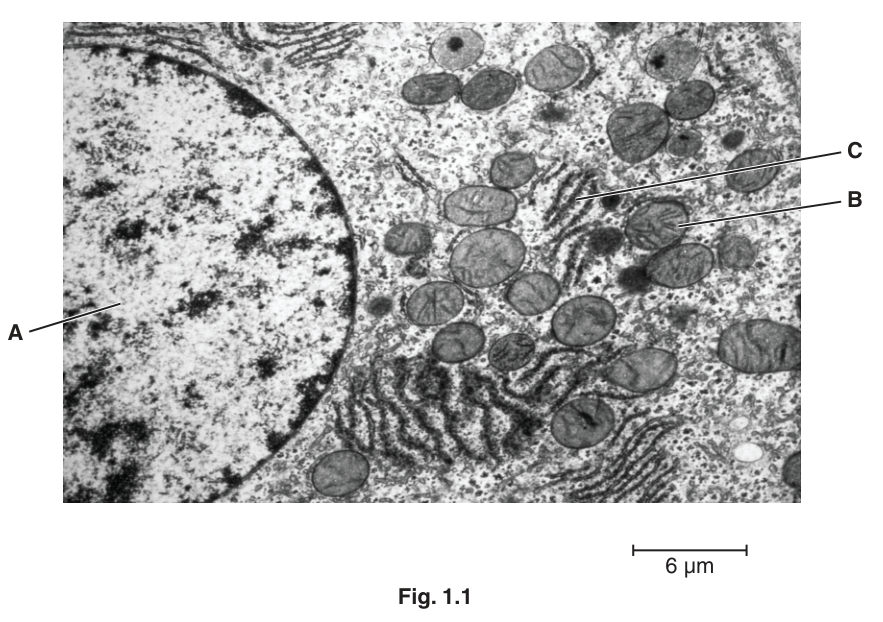
Name the structures A to C.
A ______
B ______
C ______
Answer
- A: nucleus (or euchromatin)
- B: mitochondrion
- C: rough endoplasmic reticulum (RER)
A: nucleus; B: mitochondrion; C: rough endoplasmic reticulum (RER)
Background Concept
A transmission electron micrograph (TEM) lets us see the internal ultrastructure of organelles at very high resolution because it uses a beam of electrons (whose wavelength is far shorter than visible light) instead of light. Animal cells contain several membrane-bound organelles, each with a distinctive appearance in TEM:
- The nucleus is the largest organelle, bounded by a double-membrane nuclear envelope perforated with nuclear pores. Inside, the genetic material is dispersed as chromatin — euchromatin (pale and dispersed, transcriptionally active) and heterochromatin (dark and condensed, largely inactive). A small, dense, rounded body within the nucleus is the nucleolus, the site of ribosome subunit assembly.
- Mitochondria are oval-shaped organelles with a smooth outer membrane and a highly folded inner membrane forming cristae. In section, the cristae give them a characteristic striated appearance.
- The rough endoplasmic reticulum (RER) is a continuous network of flattened membrane sacs (cisternae) whose cytoplasmic surface is studded with ribosomes, giving the membrane a dotted or rough texture. RER is the site of synthesis of proteins destined for secretion, for insertion into membranes, or for delivery to other organelles. The smooth ER (SER) lacks ribosomes and looks smooth-walled.
Understanding the Question
Fig. 1.1 is a TEM image of an animal cell. The candidate is shown an image (with a scale bar) and asked to name the three structures indicated by the labels A, B and C. This is a structure-recognition question; the mark scheme accepts the principal alternative names for each structure but explicitly rejects a few close-miss answers.
Approach
For each label, look at the distinctive structural features in the image (size, shape, internal pattern, surface texture) and match them to your knowledge of organelle ultrastructure:
- A is the largest organelle in the image, with a clear envelope and dark internal material — this is the nucleus.
- B is an oval body with internal parallel membranes — this is a mitochondrion.
- C is a stack of flattened sacs whose surfaces are dotted — this is the RER.
Step-by-Step Reasoning
- A — nucleus. The label points into a large rounded organelle on the left of the image, bounded by a clear envelope. The dark granular material inside is chromatin. The mark scheme accepts "nucleus" (the whole organelle) or "euchromatin" (the dispersed chromatin it points into) but explicitly rejects "nucleolus" — a nucleolus would be a discrete, denser, rounded body inside the nucleus, not the diffuse chromatin region shown.
- B — mitochondrion. The label points to an oval structure with internal parallel lines. These lines are the cristae formed by the inner mitochondrial membrane. No other organelle in an animal cell has this combination of an outer envelope and an extensively folded inner membrane.
- C — rough endoplasmic reticulum (RER). The label points to flattened membrane sacs whose surfaces appear covered in small dark dots — the ribosomes. The dot-studded surface is the defining feature that distinguishes RER from SER. The mark scheme accepts "rough endoplasmic reticulum" or "(R)ER" but explicitly rejects "smooth ER".
Key Takeaways
- Recognise the nucleus by its size, its envelope, and the chromatin pattern inside.
- Recognise mitochondria by their double membrane and cristae.
- Recognise RER by the ribosomes (small dots) on its cytoplasmic surface — this is the key feature distinguishing RER from SER.
- Always read the mark scheme's reject list: "nucleolus" is wrong for A, and "smooth ER" is wrong for C.
Common Mistakes
- Writing "nucleolus" for A — this is rejected by the mark scheme. The image does not show a discrete dense body within the nucleus.
- Writing "smooth ER" or just "ER" for C — the visible ribosomes make it specifically RER, and the mark scheme rejects "smooth/S".
- Confusing mitochondria with other round structures (e.g., lysosomes, vesicles) — only mitochondria have cristae.
- Naming individual ribosomes as the structure at C — the candidate should name the whole RER network, not a single ribosome.
Things to Be Careful About
- Note the mark scheme's R (reject) markings and avoid those answers.
- The mark scheme accepts two answers for A ("nucleus" or "euchromatin"); in a written answer either would earn the mark, but "nucleus" is the safer, more general answer.
- Magnification of the image is not relevant here — the structures must be identified by their morphology, not by their size.
State the function of structure C.
Answer
Synthesis of proteins / polypeptides.
Protein (polypeptide) synthesis
Background Concept
The rough endoplasmic reticulum (RER) is a continuous network of flattened membrane sacs whose cytoplasmic surface is studded with ribosomes. Each ribosome consists of a large and a small subunit of ribosomal RNA and protein, and it is the ribosome that catalyses peptide bond formation between amino acids during translation. When ribosomes are bound to the RER, the proteins they make are threaded directly into the lumen of the RER through protein translocators in the membrane. Inside the lumen, the proteins are folded, modified (e.g. by addition of carbohydrates to form glycoproteins), and packaged into transport vesicles that bud off and travel to the Golgi apparatus for further processing.
Proteins synthesised on the RER are typically those destined for secretion, insertion into membranes, or delivery to lysosomes — that is, proteins that must enter the endomembrane system. Proteins that function in the cytosol (e.g. glycolytic enzymes, haemoglobin in red cell precursors) are made on free ribosomes in the cytosol, not on the RER.
Understanding the Question
This part is the simplest of the question — it asks for the function of structure C, identified in part (a) as the rough endoplasmic reticulum. The mark is for a single, correctly-stated function.
Approach
Recall that the rough ER is the site of protein synthesis (by its bound ribosomes) and also a site of protein modification and transport. State the function in one precise phrase.
Step-by-Step Reasoning
The defining feature of the RER is the presence of ribosomes on its surface. Ribosomes are the organelles of protein synthesis, so the function of the RER is protein (or polypeptide) synthesis. The mark scheme also accepts "protein transport/modification" as an alternative because the RER lumen is where synthesised proteins are folded and modified before being sent to the Golgi. Either formulation is correct.
Key Takeaways
- The RER is the site of synthesis of proteins that are secreted, membrane-bound, or sent to other organelles.
- The defining structural feature (ribosomes) determines the function (protein synthesis).
- This is a classic example of structure relating to function at the subcellular level.
Common Mistakes
- Writing "transport of proteins" alone, without reference to synthesis — the mark scheme accepts modification/transport as alternatives, but "transport only" misses the primary function.
- Confusing RER with smooth ER, which is involved in lipid synthesis, detoxification and Ca²⁺ storage — the two are functionally distinct.
- Writing "makes proteins" in a vague way — the mark scheme's precise terms are "protein" or "polypeptide" and "synthesis".
Things to Be Careful About
- Use the precise biological term "synthesis" rather than informal terms like "making".
- A common error is to confuse the functions of RER and Golgi apparatus — the RER is the primary site of synthesis; the Golgi further modifies, sorts and packages proteins from the RER.
Explain why structure C cannot be seen using a light microscope.
Answer
- The (maximum) resolution of a light microscope is about , whereas the resolution of an electron microscope is about — the light microscope has much lower resolution.
- Ribosomes are only about – in diameter (and the ER membrane is only – thick), which is smaller than the resolution of a light microscope, so structure C cannot be resolved by a light microscope.
Light microscope resolution (~200 nm) is too low to resolve the structure; ribosomes are 20-30 nm / ER membrane is 7-10 nm, both smaller than 200 nm.
Background Concept
Resolution (or resolving power) is the minimum distance between two points at which they can still be distinguished as separate. It is a property of the microscope itself, set by the wavelength of the radiation used and the numerical aperture of the lenses:
For a light microscope using visible light (–), the best achievable resolution is about . For an electron microscope, the electrons have a much shorter effective wavelength (set by the accelerating voltage), and the best resolution is about or better — roughly a thousand-fold improvement.
Magnification is different: it is the number of times the image is enlarged, and it can be increased almost without limit. However, increasing magnification does not reveal new detail if the resolution is too low — you simply get a bigger, blurrier image. This is sometimes called "empty magnification".
The sizes of cellular structures relevant to this question are:
- Ribosomes: about – in diameter
- ER membrane thickness: about –
Both are well below the resolution limit of a light microscope, so neither can be resolved using light microscopy.
Understanding the Question
This part asks the candidate to explain why structure C (the RER, with its bound ribosomes) cannot be seen using a light microscope. The mark scheme explicitly says to ignore references to magnification — the key concept is resolution, not magnification. Two marks are available.
Approach
The two marking points are:
- State that the resolution of a light microscope is too low (and ideally compare with that of an electron microscope, with values).
- State that the structure (ribosomes and/or ER membrane) is too small to be resolved by a light microscope (with sizes).
Step-by-Step Reasoning
- Marking point 1 — resolution comparison. The light microscope's resolution is limited by the wavelength of visible light to about . The electron microscope, because it uses electrons with a much shorter wavelength, has a resolution of about . Therefore, the light microscope's resolution is several hundred times poorer.
- Marking point 2 — size of the structure. Ribosomes have a diameter of only –, and the ER membrane is – thick. Both are far smaller than , so a light microscope cannot resolve them. In other words, the wavelength of visible light is longer than the size of the structure, so the structure cannot be seen.
The candidate could equally phrase marking point 2 as: "the wavelength of light is longer than the size of the ribosomes / the RER membrane" or "ribosomes are smaller than in diameter".
Key Takeaways
- Resolution is the key concept — not magnification. A common misconception is that light microscopes "can't magnify enough"; the real limit is resolution.
- Resolution of a light microscope is about ; of an electron microscope about .
- Ribosomes (–) and ER membrane (–) are well below the resolution limit of a light microscope.
Common Mistakes
- Writing that the magnification of the light microscope is too low — this is rejected by the mark scheme (the mark scheme says "ignore refs to magnification").
- Omitting numerical sizes (the mark scheme credits quoting sizes or quoting the comparison of resolutions).
- Confusing ribosomes (which are the small dots seen on the RER in the TEM) with the RER membrane itself — both points can earn credit, and either is acceptable as the size reference.
- Writing "the light microscope is not powerful enough" — vague, does not name resolution.
Things to Be Careful About
- Use the precise biological term resolution (or resolving power); avoid the vague word "power".
- The mark scheme will ignore any mention of magnification, so do not waste words on it.
- Quote at least one numerical comparison: either resolutions ( vs ) or sizes of the structures (– / –), or both.
Suggest one disadvantage of the electron microscope compared to the light microscope for the study of cells.
Answer
Only dead / non-living specimens can be viewed (because the sample is mounted in a vacuum inside the electron microscope).
[Other acceptable answers: more expensive to buy / maintain; produces only black-and-white / monochrome images; samples are more difficult / take longer to prepare; requires technical training to operate.]
Only dead / non-living specimens can be viewed.
Background Concept
The electron microscope offers much higher resolution than the light microscope, but it does so at a cost. The electron beam can travel only in a vacuum (otherwise the electrons would collide with air molecules and be scattered), so the specimen chamber is held under high vacuum. This forces several constraints on how specimens must be prepared and observed.
Specimens must be:
- Dead (because they are fixed, dehydrated and held in a vacuum — they cannot survive these conditions).
- Extremely thin (typically – sections for TEM) because the electrons must pass through them.
- Stained with heavy metals (e.g. uranyl acetate, lead citrate) to scatter electrons and produce contrast — these are toxic.
Other practical limitations of electron microscopy include:
- The instrument is expensive to buy, house and maintain.
- It produces monochrome / black-and-white images (contrast comes from heavy-metal staining, not from colour).
- Operating it requires technical training and stable electrical supplies.
- Specimen preparation is lengthy and can introduce artefacts (features caused by preparation, not present in the living cell).
Understanding the Question
The question asks for one disadvantage of the electron microscope compared with the light microscope. The mark scheme lists many possible answers, of which any one is sufficient for the single mark.
Approach
Recall the practical constraints of electron microscopy listed in the Background Concept and pick the most concrete, specific one to write down. A good answer names the constraint and (briefly) why it is a disadvantage.
Step-by-Step Reasoning
The cleanest, most specific answer is: "only dead specimens can be viewed", because the vacuum and chemical fixation kill the cells before they can be imaged. This is a major limitation in cell biology, where researchers often want to observe living processes.
Other valid choices that would each earn the single mark include:
- (More) expensive to buy, maintain or run
- Requires (more) technical training to operate
- Specimens are more difficult / take longer to prepare
- Produces only black-and-white (monochrome) images
- Cannot be used on coloured specimens or to study coloured processes
- Sensitive to external magnetic fields / vibration
- Not portable
The mark scheme is generous: any single, specific, relevant disadvantage is credited. Vague answers such as "not as good" or "hard to use" without elaboration are not credited.
Key Takeaways
- The electron microscope's main advantage is its very high resolution, but it comes with the constraint that specimens must be dead, fixed, dehydrated and stained.
- Other practical disadvantages are cost, complexity, monochrome imaging, and lengthy preparation.
- Choose one specific, well-stated disadvantage rather than several vague ones.
Common Mistakes
- Writing vague answers such as "hard to use" or "complicated" without specifying why (e.g. "requires technical training").
- Stating a disadvantage of the light microscope instead (e.g. "lower resolution") — that is the advantage of the electron microscope.
- Writing "lower magnification" — this is incorrect; electron microscopes have higher magnification than light microscopes.
- Listing multiple disadvantages in one short answer; the mark is for one well-expressed point.
Things to Be Careful About
- Keep the answer focused on the electron microscope's limitations, not its advantages.
- "Human error" or "less accurate" are too vague to earn credit.
- A specific disadvantage is required — name the practical consequence (e.g. "expensive to buy") rather than just saying "it's not good".
Calculate the magnification of the image in Fig. 1.1.
Show your working and give your answer to the nearest whole number.
Answer = ______
Working
Measure the scale bar on the printed Fig. 1.1 with a ruler: (i.e. ).
Answer
× 3333
Background Concept
Magnification is the number of times an image is larger than the object:
In a printed image, the scale bar tells you the actual size that a particular length on the page represents. By measuring the scale bar on the printed image with a ruler, you can find the image size, and then divide by the actual size (read from the scale bar's label) to obtain the magnification of the image.
Unit conversion is essential in these calculations. The standard units for cellular measurements are:
So a measurement in millimetres on the printed page must be converted to micrometres (or nanometres) before it can be divided by the actual size in the same units.
Understanding the Question
Fig. 1.1 includes a scale bar labelled . The candidate is asked to calculate the magnification of the image, showing working, and to give the answer to the nearest whole number. Two marks are available: one for the working (and correct unit conversion), and one for the final numerical answer.
Approach
- Use a ruler to measure the length of the scale bar on the printed page (in mm).
- Convert this measurement from mm to µm.
- Substitute into the magnification formula.
- Round to the nearest whole number.
Step-by-Step Reasoning
- Measure the scale bar. With a ruler, the scale bar on a typical A4 printing of this paper measures approximately . The mark scheme allows a tolerance of on this reading, so any value in the range – is acceptable.
- Convert to µm.
- Apply the magnification formula.
- Round to the nearest whole number.
The mark scheme accepts the following alternatives based on the measurement tolerance:
| Scale-bar reading | Calculation | Magnification |
|---|---|---|
Key Takeaways
- The magnification of a printed image is found by measuring a known feature (e.g. a scale bar) and dividing by its labelled actual size.
- Unit conversion is essential: always convert mm to µm (or µm to mm) so that image size and actual size are in the same units before dividing.
- The mark scheme rewards working (a formula or a clear substitution) as well as the final numerical answer.
- Round to the nearest whole number, as the question requests.
Common Mistakes
- Forgetting to convert mm to µm — dividing by gives a magnification of only , which is wildly wrong. The mark scheme awards partial credit (one mark) for a correctly measured scale bar divided by but without correct unit conversion.
- Not showing working — the question explicitly says "Show your working"; a bare numerical answer with no formula or substitution forfeits one of the two marks.
- Rounding errors — the mark scheme wants the answer to the nearest whole number, not to one or more decimal places. An answer such as "" would earn only one mark (the formula mark) but not the final-answer mark.
- Measuring the wrong length — for example, measuring the whole image rather than the scale bar.
Things to Be Careful About
- Always quote the formula for magnification, even if it seems obvious.
- Make sure the units cancel cleanly: the µm in the numerator and denominator cancel, leaving a dimensionless magnification.
- The mark scheme allows a small measurement tolerance, so an answer of or (within the reading range) is fully acceptable.
- Include the multiplication sign () in the final answer to make clear it is a magnification.
The rest of this paper
5 more questions- Q2Transport in Mammals11M
- Q3Biological Molecules6M
- Q4Infectious Diseases · Immunity12M
- Q5(outdated) Ecology · Enzymes11M
- Q6Gas Exchange11M