Biology 9700/22 — May/June 2010
Cambridge AS Level · worked solutions for every part, with the mark scheme
Topics Cell Structure · Transport in Mammals · Infectious Diseases · Immunity · Gas Exchange · (outdated) Ecology
Fig. 1.1 is a diagram of an electron micrograph of a plant cell.
Fig. 1.2 is a diagram of an electron micrograph of an animal cell.
Both diagrams are incomplete.
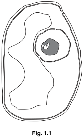
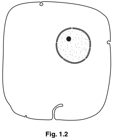
Explain how Fig. 1.1 can be identified as a plant cell.
Answer
Fig. 1.1 can be identified as a plant cell because:
- it has a (cellulose) cell wall, external to the plasma membrane;
- it has a large / central vacuole, surrounded by a tonoplast (vacuolar membrane);
- plasmodesmata pass through the cell wall, connecting the cytoplasm to neighbouring cells.
Any two of the above for 2 marks.
Cell wall and large central vacuole (with tonoplast); plasmodesmata also acceptable.
Background Concept
Plant and animal cells share many features (plasma membrane, cytoplasm, nucleus, mitochondria, ribosomes, RER, Golgi, etc.) but plant cells have several extra structures that the cell uses for support, water storage and intercellular transport. The features that mark a cell out as plant are:
- Cell wall made of cellulose, lying outside the plasma membrane, giving a fixed, often polygonal, outline.
- Large / central vacuole bounded by a selectively permeable membrane, the tonoplast; the vacuole stores cell sap, maintains turgor and can occupy most of the cell volume.
- Plasmodesmata – narrow cytoplasmic channels through cell walls that link adjacent plant cells into a continuous symplast.
- Chloroplasts – the site of photosynthesis (in green, photosynthetic cells).
Animal cells have none of these. The shape of the outline, and the presence of a single large fluid-filled compartment, are the most useful giveaways on an electron micrograph.
Understanding the Question
You are shown a simplified line drawing based on an electron micrograph of a plant cell (Fig. 1.1) and asked to explain how it can be recognised as plant. The command word "explain" here is light-touch: it is enough to state the plant-cell features visible in the figure and to make clear that these features are absent from animal cells. The figure in the question shows the cell wall, the plasma membrane, a large vacuole and a nucleus; the question wording ("incomplete") hints that plasmodesmata and the tonoplast are also acceptable answers because they are typical plant features you would expect to see.
Approach
Look at the diagram and pick out anything that an animal cell could not have. The diagram clearly shows a thick outer wall (animal cells do not have walls) and a large fluid-filled space (animal cells have small, temporary vesicles, not a central vacuole). You can name the wall's material (cellulose) for precision. Mention the tonoplast and plasmodesmata if you want to show knowledge beyond what is literally drawn.
Step-by-Step Reasoning
- Cell wall: the rigid outer boundary outside the plasma membrane is diagnostic of a plant (and fungal) cell. Cellulose is the correct plant-wall material; do not say chitin, peptidoglycan or lignin – these score no mark.
- Large / central vacuole: the figure shows a very large, single internal compartment that pushes the cytoplasm to the edge. Animal cells do not have a large permanent vacuole; the term "permanent" is ignored by the mark scheme, so "vacuole" alone is fine.
- Tonoplast: the membrane that bounds the central vacuole. It is implicit in the figure even if not labelled.
- Plasmodesmata: pores through the cell wall. They are not actually drawn, but they are a recognised plant-cell feature, and the mark scheme allows them.
Any two of these four points earn the full 2 marks.
Key Takeaways
- A cell wall of cellulose, a large central vacuole with a tonoplast, and plasmodesmata are the defining plant-cell features you can list when asked to identify a plant cell from a micrograph.
- Animal cells share most other organelles with plant cells, so it is the extras that distinguish them.
Common Mistakes
- Writing "it has chloroplasts" – chloroplasts are not visible in Fig. 1.1, and a non-photosynthetic plant cell (e.g. a root cell) has no chloroplasts, so a chloroplast is not a defining feature of a plant cell.
- Writing "it has a nucleus" – both plant and animal cells have a nucleus, so this is not diagnostic.
- Saying the wall is made of "chitin" or "peptidoglycan" – these are fungal and bacterial wall materials respectively, and the mark scheme rejects them.
Things to Be Careful About
- The question is not asking you to list every organelle; it is asking for the identifying features, i.e. those that distinguish plant from animal cells.
- Use the precise term tonoplast (or "vacuolar membrane") rather than vague wording such as "membrane around the vacuole" if you can.
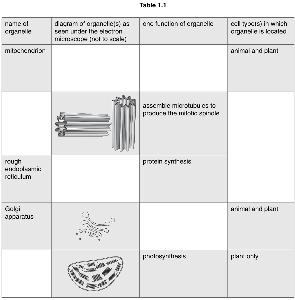
Some organelles are missing from Figs 1.1 and 1.2. Information about these organelles is shown in the shaded boxes in Table 1.1.
Complete the empty boxes in Table 1.1 by adding the correct information below each column heading.
Answer
| name of organelle | diagram of organelle(s) as seen under the electron microscope (not to scale) | one function of organelle | cell type(s) in which organelle is located |
|---|---|---|---|
| mitochondrion | oval shape with two membranes close together and the inner membrane infolded into two or more cristae | aerobic respiration / ATP (production) | animal and plant |
| centrioles | (centrioles shown as two cylinders of microtubules at right angles to each other) | assemble microtubules to produce the mitotic spindle | animal |
| rough endoplasmic reticulum | (continuous network of) two membranes with ribosomes on the external surface | protein synthesis | animal and plant |
| Golgi apparatus | (stack of flattened cisternae with vesicles budding from the edges) | processing / modification / packaging of proteins (or other molecules) | animal and plant |
| chloroplast | (chloroplast with double membrane and internal grana of thylakoids) | photosynthesis | plant only |
Completed table as shown above.
Background Concept
A eukaryotic cell is divided into membrane-bound compartments (organelles), each with a specific function. The table asks you to match five of these organelles with a recognisable electron-microscope appearance, a function and the cell type(s) in which they occur. The five organelles are mitochondrion, centrioles, rough endoplasmic reticulum (RER), Golgi apparatus and chloroplast – four of them are in both plant and animal cells, while centrioles are confined to animal cells and chloroplasts to plant cells.
Key features to know:
- Mitochondrion – oval, double membrane, the inner one thrown into folds called cristae; the matrix inside houses enzymes of the Krebs cycle, the intermembrane space is where oxidative phosphorylation builds the proton gradient that drives ATP synthesis. They are the site of aerobic respiration / ATP production.
- Centrioles – a pair of short, hollow cylinders made of microtubules, set at right angles to each other; they organise the mitotic spindle at cell division. Found in animal cells (lower plants such as mosses and ferns also have them; flowering plants do not).
- Rough endoplasmic reticulum (RER) – a continuous system of flattened membrane sacs (cisternae) studded with ribosomes on the cytoplasmic (outer) surface; ribosomes translate proteins destined for secretion, membranes or lysosomes, and the RER also begins their folding and initial modification. Present in both cell types.
- Golgi apparatus – a stack of flattened cisternae with small transport vesicles budding from the edges; it modifies (e.g. glycosylates) and packages proteins/lipids received from the RER and dispatches them in vesicles to the membrane, lysosomes or secretion.
- Chloroplast – a double-membrane organelle containing internal stacks of thylakoid membranes (grana) joined by stromal lamellae, suspended in the stroma; the site of photosynthesis. Only in green/photosynthetic plant cells.
Understanding the Question
You are given a partially completed table. Some cells are filled in and some are blank; you have to fill the blanks. Each completed row earns marks:
- Row 1 (mitochondrion): diagram description and function.
- Row 2: name of the organelle and the cell type in which it occurs.
- Row 3 (RER): diagram description and cell type.
- Row 4 (Golgi): function.
- Row 5 (chloroplast): name of the organelle.
The shaded boxes show what is already given; do not rewrite the given information, just complete the empty boxes.
Approach
For each row, identify which two of the four columns are empty, then:
- If the name is blank (rows 2 and 5) – read the diagram and function, and write the organelle name. The diagram of two cylinders at right angles plus the spindle function is unmistakably centrioles; the green organelle with grana doing photosynthesis is a chloroplast.
- If the diagram is blank (rows 1 and 3) – describe the appearance using the mark-scheme's required features. The mitochondrion description must include all three of: oval/circular shape, two membranes close together, inner membrane folded into two or more cristae. The RER description needs both of: a membrane system (two membranes / cisternae) and ribosomes on the outside; ribosomes drawn excessively large are rejected.
- If the function is blank (rows 1 and 4) – give the precise function. For the mitochondrion say aerobic respiration or ATP production (do not say "produces energy"). For the Golgi say modification / processing / packaging of proteins (or molecules); do not say "protein synthesis" – that is the RER's job and the mark scheme explicitly rejects it here.
- If the cell type is blank (rows 2 and 3) – state the cell type. Centrioles are in animal cells; RER is in animal and plant cells (i.e. both).
Step-by-Step Reasoning
Row 1 – mitochondrion
- Diagram: needs all three features for one mark – oval/circular shape, two membranes close together, inner membrane infolded as cristae.
- Function: aerobic respiration or ATP (production / synthesis). "Produces energy" is rejected (energy is not a substance that can be synthesised). Acceptable alternatives include oxidative phosphorylation, β-oxidation of fats, and the urea / ornithine cycle.
- Cell type: given as animal and plant.
Row 2 – centrioles
- Name: centrioles (or centriole, or centrosome – mark scheme accepts these).
- Function: given – assemble microtubules to produce the mitotic spindle.
- Cell type: animal (centrioles are characteristic of animal cells; flowering-plant cells lack them).
Row 3 – rough endoplasmic reticulum
- Diagram: needs both – a (continuous) network of two membranes (or cisternae) and ribosomes on the external (cytoplasmic) surface.
- Function: given – protein synthesis.
- Cell type: animal and plant / both – RER is universal in eukaryotes (it is even found in yeast).
Row 4 – Golgi apparatus
- Name and diagram: given.
- Function: processing / modification / packaging of proteins (or other molecules). Examples the mark scheme allows include glycosylation, production of secretory/Golgi vesicles, and production of lysosomes. "Protein synthesis" is rejected because that is the RER's role.
- Cell type: given – animal and plant.
Row 5 – chloroplast
- Name: chloroplast.
- Function: given – photosynthesis.
- Cell type: given – plant only (specifically green / photosynthetic plant cells; non-photosynthetic plant cells such as root cells do not contain chloroplasts, so the cell-type answer is a useful clue).
Key Takeaways
- Five core eukaryotic organelles to recognise and recall: mitochondrion, centrioles, RER, Golgi, chloroplast.
- For each, you should be able to: name it, sketch the EM appearance with the diagnostic structural features, state a precise function, and say whether it is in animal cells, plant cells or both.
- Distinguish protein synthesis (RER / ribosomes) from protein processing and packaging (Golgi).
- Distinguish ATP production (mitochondrion – aerobic respiration) from energy (a process, not a molecule; "produces energy" is rejected).
Common Mistakes
- Writing "produces energy" for the mitochondrion – the mark scheme rejects this; ATP is the molecule, energy is the process.
- Writing "protein synthesis" for the Golgi apparatus – the mark scheme rejects this; protein synthesis is the RER's job.
- Drawing the RER's ribosomes as huge dots so that they look almost as big as the cisternae; the mark scheme rejects "excessively large" ribosomes.
- Describing the mitochondrion with only one or two of the three required features (e.g. only mentioning cristae) – all three are needed for the mark.
- Putting centrioles in plant cells, or chloroplasts in animal cells.
- Spelling "chloroplast" as "chloreplast" etc. – examiners will accept it, but the spelling should ideally be correct.
Things to Be Careful About
- Where the function is already given, do not restate it; the mark is for completing the empty box, not for repeating the full row.
- Use the precise terms: tonoplast, Golgi apparatus (not "Golgi body"), rough endoplasmic reticulum (spelt out or RER).
- "Animal and plant" is the safest phrasing; "both" or "eukaryotic" is also accepted.
The rest of this paper
5 more questions- Q2Transport in Mammals9M
- Q3Infectious Diseases · Immunity13M
- Q4—13M
- Q5Gas Exchange8M
- Q6(outdated) Ecology7M