Biology 9700/12 — May/June 2010
Cambridge AS Level · Multiple Choice (AS Level) · answer key with instant marking and worked solutions
Topics Biological Molecules · Cell Structure · Nucleic Acids and Protein Synthesis · Gas Exchange · Cell Membranes and Transport · Transport in Plants · +6 more
Tap an option under each question to check it — your score builds as you go.
The diagram shows a section through a type of epithelium.
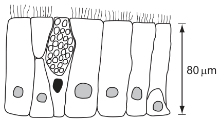
Where is this type of epithelium found in the respiratory system?
Options
| trachea | bronchus | all bronchioles | alveolus | |
|---|---|---|---|---|
| A | ✓ | ✓ | ✓ | ✗ |
| B | ✓ | ✓ | ✗ | ✗ |
| C | ✗ | ✗ | ✓ | ✓ |
| D | ✗ | ✗ | ✗ | ✓ |
key
✓ = present
✗ = absent
Working
The diagram shows tall cells (≈80 µm high) with cilia on the apical surface and an interspersed goblet cell containing mucus granules — this is ciliated columnar (pseudostratified) epithelium with goblet cells.
In the respiratory system:
- Trachea — lined by ciliated columnar epithelium with many goblet cells ✓
- Bronchus — same lining as the trachea ✓
- All bronchioles — only the larger bronchioles carry ciliated epithelium; smaller bronchioles have ciliated cuboidal cells and lose their goblet cells ✗
- Alveolus — lined by simple squamous epithelium (Type I pneumocytes), not ciliated columnar ✗
Answer
B
B
Background Concept
The conducting division of the human respiratory system is lined by a sequence of epithelia that become progressively simpler as the airways branch and decrease in diameter:
- Trachea and bronchi are lined by ciliated pseudostratified columnar epithelium with numerous goblet cells. The cilia beat in a coordinated manner to move the mucus layer (produced by goblet cells and submucosal glands) upward towards the pharynx — the mucociliary escalator.
- Bronchioles lose cartilage and submucosal glands. Larger bronchioles are lined by ciliated columnar or ciliated cuboidal epithelium with fewer goblet cells. Smaller (terminal and respiratory) bronchioles have ciliated cuboidal epithelium and may lack goblet cells entirely.
- Alveoli are the gas-exchange surface, lined by an extremely thin simple squamous epithelium (mainly Type I pneumocytes, with Type II pneumocytes that secrete surfactant). This thinness minimises the diffusion distance for O₂ and CO₂.
Understanding the Question
The diagram (Fig. 1.1) shows tall, column-shaped cells with cilia on the apical (luminal) surface and a single goblet cell packed with mucus granules, with an overall epithelial height of 80 µm. The question asks where this ciliated columnar epithelium with goblet cells is found in the respiratory system, by ticking its presence or absence in the trachea, bronchus, all bronchioles and alveolus.
The command word is implicit (identify the correct option), and the mark is awarded only for the option that correctly maps the distribution.
Approach
- Identify the tissue from the diagram: ciliated columnar epithelium with goblet cells.
- Recall (or apply) the distribution of this tissue along the respiratory tree.
- Eliminate options where the tick pattern contradicts the known distribution.
Step-by-Step Reasoning
- Trachea — the trachea is lined by ciliated pseudostratified columnar epithelium with goblet cells, so this must be ticked ✓.
- Bronchus — branches of the trachea, structurally similar; also ciliated columnar with goblet cells ✓.
- All bronchioles — this is the critical distractor. Only the larger bronchioles carry ciliated columnar (or cuboidal) epithelium; the smaller bronchioles are lined by ciliated cuboidal cells and lose goblet cells. Because the option specifies all bronchioles, it cannot be ticked ✗.
- Alveolus — gas exchange occurs across a simple squamous epithelium; no cilia, no goblet cells ✗.
Combining these: trachea ✓, bronchus ✓, all bronchioles ✗, alveolus ✗. Only option B matches this pattern.
Key Takeaways
- Ciliated columnar epithelium with goblet cells lines the trachea and bronchi (and the very largest bronchioles).
- The word "all" in option B's row for bronchioles is a deliberate trap — smaller bronchioles have ciliated cuboidal epithelium and lack goblet cells.
- Alveoli are lined by simple squamous epithelium to keep the diffusion distance short.
Common Mistakes
- Ticking "all bronchioles" because the larger ones do carry ciliated columnar epithelium — the question requires the epithelium to be present in every bronchiole.
- Confusing alveolar Type I pneumocytes with columnar cells — alveoli are squamous, not columnar.
- Selecting the option that has a single tick in the right place without checking the rest of the row.
Things to Be Careful About
- Read the row/column heading carefully: "all bronchioles" means every bronchiole, not just the larger ones.
- Distinguish the conducting zone (trachea, bronchi, bronchioles — ciliated) from the respiratory zone (alveoli — squamous, no cilia).
- The 80 µm height given in the figure is consistent with a tall columnar epithelium, ruling out the much thinner squamous lining of alveoli.
The rest of this paper
39 more questions- Q2Gas Exchange1M
- Q3Biological Molecules1M
- Q4Cell Membranes and Transport1M
- Q5Cell Membranes and Transport1M
- Q6Transport in Plants1M
- Q7Transport in Plants1M
- Q8Transport in Plants1M
- Q9(outdated) Ecology1M
- Q10(outdated) Ecology1M
- Q11(outdated) Ecology1M
- Q12Cell Structure1M
- Q13Cell Structure1M
- Q14Cell Structure1M
- Q15Cell Structure1M
- Q16Cell Structure1M
- Q17Cell Structure1M
- Q18Cell Structure · Cell Membranes and Transport1M
- Q19The Mitotic Cell Cycle1M
- Q20Nucleic Acids and Protein Synthesis1M
- Q21The Mitotic Cell Cycle1M
- Q22Nucleic Acids and Protein Synthesis1M
- Q23Nucleic Acids and Protein Synthesis1M
- Q24Enzymes1M
- Q25Enzymes1M
- Q26Biological Molecules1M
- Q27Biological Molecules1M
- Q28Biological Molecules1M
- Q29Biological Molecules1M
- Q30Biological Molecules1M
- Q31Biological Molecules1M
- Q32Nucleic Acids and Protein Synthesis1M
- Q33Infectious Diseases1M
- Q34Immunity1M
- Q35Immunity1M
- Q36Infectious Diseases1M
- Q37Transport in Mammals1M
- Q38Transport in Mammals1M
- Q39Transport in Mammals1M
- Q40Gas Exchange1M