Biology 9700/43 — May/June 2025
Cambridge A-Level · A Level Structured Questions · worked solutions for every part, with the mark scheme
Topics Selection and Evolution · Genetic Technology · Classification, Biodiversity and Conservation · Energy and Respiration · Inheritance · Homeostasis · +2 more
Fig. 1.1 is a diagram of part of a liver cell.
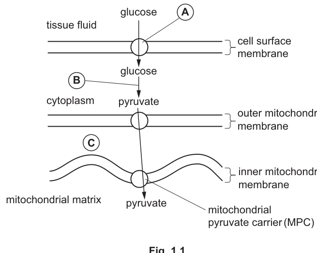
With reference to Fig. 1.1, name:
the type of membrane transport protein represented by A ______
process B ______
area C ______
Answer
A: GLUT (carrier protein)
B: glycolysis
C: intermembrane space
A: GLUT (carrier protein); B: glycolysis; C: intermembrane space
Background Concept
Liver cells take up glucose from the blood (here shown as tissue fluid) through specific transport proteins in the cell surface membrane. The most important of these are the GLUT family of carrier (facilitated-diffusion) proteins, which allow glucose to move down its concentration gradient without using ATP. Once inside the cytoplasm, glucose is broken down by glycolysis into two molecules of pyruvate, with a small net gain of ATP and reduced NAD.
Pyruvate is then transported into the mitochondrion. A mitochondrion has a smooth outer membrane and a highly folded inner membrane. Between them is a narrow compartment — the intermembrane space — in which protons () accumulate during electron transport. The inner membrane encloses the matrix, the site of the link reaction and Krebs cycle.
Understanding the Question
The question gives a labelled diagram of part of a liver cell and asks for three named structures or processes:
- A is the transport protein allowing glucose to enter the cell from the tissue fluid.
- B is the cytoplasmic process that converts glucose to pyruvate.
- C is the compartment between the two mitochondrial membranes.
These are all direct identifications from the diagram, with the mark scheme accepting specific CIE terms.
Approach
Match each letter to the feature it labels, then supply the single technical term the mark scheme accepts. A common trap is writing "channel" for A — the mark scheme explicitly ignores "channel" because glucose is too polar to pass through an open aqueous channel and must bind to a carrier protein (GLUT).
Step-by-Step Reasoning
- A is a carrier protein in the cell surface membrane that transports glucose into the cytoplasm. The CIE-accepted name is GLUT (or simply "carrier protein").
- B is the metabolic pathway in the cytoplasm that splits glucose into two pyruvate molecules. This is glycolysis.
- C is the space between the outer and inner mitochondrial membranes. This is the intermembrane space (the alternative spelling "intermembranal space" is also accepted, but "inner membrane" is rejected because the inner membrane is a structure, not a space).
Key Takeaways
- Glucose enters cells via GLUT carrier proteins (facilitated diffusion).
- Glycolysis occurs in the cytoplasm and produces pyruvate.
- A mitochondrion has two membranes; the compartment between them is the intermembrane space, the site of the proton gradient used in oxidative phosphorylation.
Common Mistakes
- Writing "channel" for A — channels only allow small/charged species to pass; glucose needs a carrier.
- Writing "inner membrane" for C — C asks for a space, not a membrane.
- Writing "oxidative decarboxylation" or "respiration" for B — too vague; the precise name is glycolysis.
Things to Be Careful About
Use the exact terminology the mark scheme accepts. "GLUT" on its own is sufficient; "GLUT carrier" or "GLUT protein" is also fine, but avoid the wrong name such as "sodium–glucose cotransporter" (SGLT, which is found in intestinal/ kidney cells, not the liver cell surface membrane).
The mitochondrial pyruvate carrier (MPC), shown in Fig. 1.1, allows the passage of pyruvate into the mitochondrial matrix. When pyruvate enters the mitochondrial matrix, it takes part in the link reaction.
Describe the link reaction.
Answer
- Pyruvate undergoes decarboxylation — carbon dioxide () is removed/released.
- Pyruvate is oxidised by dehydrogenation, and reduced NAD is produced (NAD is the hydrogen acceptor).
- An acetyl (acetate) group is formed from the remaining 2-carbon fragment.
- The acetyl group combines with coenzyme A to form acetyl CoA.
Pyruvate is decarboxylated (CO2 released) and dehydrogenated (reduced NAD formed) to produce an acetyl group, which combines with coenzyme A to form acetyl CoA.
Background Concept
The link reaction is the bridge between glycolysis (in the cytoplasm) and the Krebs cycle (in the mitochondrial matrix). For every glucose molecule, two pyruvate molecules are produced, and each one is processed once by the link reaction. The whole process is sometimes called "oxidative decarboxylation" because oxidation and loss of carbon dioxide happen together. The reaction is catalysed by a multi-enzyme complex (pyruvate dehydrogenase complex) attached to the inner mitochondrial membrane.
Understanding the Question
Part (b) asks for a description of the link reaction. With four marks, the mark scheme wants four distinct points from a list of five: decarboxylation, dehydrogenation, formation of an acetyl group, formation of reduced NAD, and combination with coenzyme A to give acetyl CoA. "Describe" means giving the key events in order, not just naming one feature.
Approach
Recall the two chemical changes (decarboxylation and dehydrogenation) and the products they generate ( and reduced NAD). Then state what is left of the pyruvate — an acetyl group — and what it combines with — coenzyme A — to give the final product, acetyl CoA, which then enters the Krebs cycle.
Step-by-Step Reasoning
- Pyruvate (3 carbons) loses one carbon as . This is decarboxylation; the enzyme class is decarboxylase.
- The remaining 2-carbon fragment (acetate/acetyl) is oxidised by the removal of hydrogen. This is dehydrogenation (dehydrogenase). The hydrogen is accepted by the coenzyme NAD, producing reduced NAD (NADH + ).
- The oxidised 2-carbon fragment is the acetyl (acetate) group.
- The acetyl group then combines with coenzyme A (CoA) to form acetyl CoA, the substrate that enters the Krebs cycle.
Key Takeaways
- The link reaction is oxidative decarboxylation of pyruvate.
- Products: , reduced NAD, and acetyl CoA.
- Acetyl CoA carries the acetyl group into the Krebs cycle.
- The reaction occurs once per pyruvate, so twice per glucose.
Common Mistakes
- Saying "oxygen is added" instead of "oxidation by removal of hydrogen (dehydrogenation)" — at A level, oxidation is defined as loss of electrons or loss of hydrogen (or gain of oxygen), and the relevant one here is hydrogen loss.
- Forgetting to name reduced NAD as a product — the hydrogen must be accepted by NAD.
- Calling the process "Krebs cycle" — the link reaction and the Krebs cycle are two distinct stages.
- Writing "acetyl-CoA enters the link reaction" — acetyl CoA is the product of the link reaction and the substrate of the Krebs cycle, not the other way around.
Things to Be Careful About
Note that the substrate is pyruvate (the ionised form of pyruvic acid) — write "pyruvate", not "pyruvic acid", when describing what enters the mitochondrion. Also remember that the link reaction does not itself produce ATP directly; the ATP is made later during oxidative phosphorylation.
Some tumour cells have a greatly reduced ability to transport pyruvate into the matrix of the mitochondrion.
Suggest how a reduction in pyruvate transport could affect respiration in these tumour cells.
Answer
- Little or no pyruvate enters the mitochondrial matrix, so the link reaction, Krebs cycle and oxidative phosphorylation are greatly reduced or do not occur.
- ATP is produced only / mostly by glycolysis (substrate-linked phosphorylation) in the cytoplasm.
- Less ATP is produced overall (per glucose molecule), because only 2 ATP are made per glucose by glycolysis rather than ~32 by full aerobic respiration.
- Anaerobic respiration occurs in the cytoplasm, with pyruvate being converted to lactate (in human cells) to regenerate NAD for continued glycolysis.
Link reaction/Krebs cycle/oxidative phosphorylation would barely occur; ATP would come almost entirely from glycolysis; total ATP yield per glucose would be much lower; cells would switch to anaerobic respiration producing lactate.
Background Concept
Aerobic respiration in eukaryotes is divided into four stages, each happening in a different location:
- Glycolysis — cytoplasm, produces 2 ATP (net) and 2 reduced NAD per glucose.
- Link reaction — mitochondrial matrix, produces and reduced NAD per pyruvate.
- Krebs cycle — mitochondrial matrix, produces , reduced NAD, reduced FAD, and 1 ATP (as GTP) per acetyl CoA.
- Oxidative phosphorylation — inner mitochondrial membrane, uses the reduced NAD and FAD to generate ~28 ATP per glucose via the electron transport chain and ATP synthase.
If pyruvate cannot enter the matrix (because the MPC is defective), stages 2–4 cannot proceed, and the cell must rely on stage 1 only.
Understanding the Question
Part (c) is a "suggest" question — the candidate must apply what they know to a hypothetical situation. The mark scheme wants three of four possible ideas: a block on stages 2–4, ATP coming from glycolysis only, lower total ATP yield, and a switch to anaerobic respiration (lactate fermentation in animal cells).
Approach
Trace what would happen if pyruvate transport is blocked. The link reaction cannot occur, so no acetyl CoA enters the Krebs cycle, so no reduced NAD/FAD from those stages feeds the electron transport chain, so oxidative phosphorylation grinds to a halt. The only ATP available is the small amount from glycolysis. Reduced NAD from glycolysis must still be reoxidised for glycolysis to continue, and in the absence of mitochondria the cell does this anaerabically, producing lactate from pyruvate.
Step-by-Step Reasoning
- Block on the link reaction, Krebs cycle and oxidative phosphorylation: without pyruvate in the matrix, no is released in the link reaction, no acetyl CoA forms, no reduced NAD/FAD is produced in the mitochondrial matrix, and the proton gradient cannot be maintained. The Krebs cycle and electron transport chain have no substrate, so almost no ATP is made by oxidative phosphorylation.
- ATP from glycolysis only: glycolysis is in the cytoplasm and does not need the mitochondrion, so it continues and makes 2 ATP (net) per glucose by substrate-linked phosphorylation.
- Lower ATP yield: full aerobic respiration yields ~32 ATP per glucose in eukaryotes, but glycolysis alone gives only 2, so the cell's energy supply is severely compromised.
- Switch to anaerobic respiration: the reduced NAD from glycolysis must be reoxidised to keep glycolysis running. Without functioning mitochondria to do this aerobically, the cell uses lactate dehydrogenase to reduce pyruvate to lactate, regenerating NAD.
Key Takeaways
- Pyruvate entry into the mitochondrion is the gateway to the aerobic stages of respiration.
- If that gateway is blocked, the cell survives only on the small amount of ATP from glycolysis, and switches to lactate fermentation.
- This is exactly the Warburg-effect logic in many tumour cells: they rely heavily on glycolysis even when oxygen is available, in part because mitochondrial function is altered.
Common Mistakes
- Saying "respiration stops" — glycolysis continues; only the aerobic stages are blocked.
- Saying the cell will produce ethanol — ethanol fermentation occurs in yeast and some plant cells, not in human liver/tumour cells.
- Saying "less reduced NAD is produced" — the link reaction does produce less reduced NAD, but the Krebs cycle is the major source; the question is about consequences for respiration as a whole, not just the link reaction.
- Forgetting the link to ATP yield — examiners reward connecting the metabolic block to a numerical or comparative drop in ATP.
Things to Be Careful About
The question says "tumour cells", which are human, so the anaerobic product is lactate, not ethanol. Also note that some credit is given simply for saying "less ATP"; stronger answers will add the comparison (2 ATP per glucose from glycolysis vs ~32 from full aerobic respiration).
Natural selection and selective breeding (artificial selection) are processes that result in changes in the gene pool of a population.
Natural selection and selective breeding have implications for humans.
If a person with a bacterial infection does not finish the course of an antibiotic given, it provides the conditions for a population of bacteria to become resistant to this antibiotic.
A mutation in a bacterial gene can give resistance to an antibiotic. Directional selection can occur when the antibiotic is present in the environment.
A bacterium can also gain resistance when it receives genetic material from another bacterium in a process known as horizontal gene transfer.
Outline how directional selection and horizontal gene transfer result in a new population of bacteria that is resistant to an antibiotic.
Answer
- The antibiotic acts as a selection pressure;
- Bacteria with the antibiotic resistance allele have a selective advantage / are selected for;
- (Resistant) bacteria survive and reproduce;
- The resistance allele is passed on / the frequency of the resistance allele increases in the population;
- The resistance gene is often carried on a plasmid;
- Horizontal gene transfer (e.g. by transduction, transformation or conjugation) can transfer the resistance gene between bacteria;
- Horizontal gene transfer causes a rapid increase in the number / proportion of resistant bacteria.
See working
Background Concept
Natural selection is the differential survival and reproduction of individuals due to differences in heritable traits. In bacterial populations, natural selection acts extremely rapidly because bacteria reproduce by binary fission, with some species dividing every 20 minutes under ideal conditions. This means many generations can occur within hours, allowing evolution to be observed in real time.
Directional selection is a specific form of natural selection in which one extreme phenotype is favoured over the others. Over generations, the mean phenotype of the population shifts towards that favoured extreme. In the context of antibiotic resistance, the resistant phenotype is favoured when the antibiotic is present in the environment.
Horizontal gene transfer (HGT) is the movement of genetic material between organisms by routes other than vertical (parent-to-offspring) transmission. The three main mechanisms of HGT in bacteria are:
- Transformation: uptake of free, 'naked' DNA from the environment (often released by dead bacteria)
- Transduction: transfer of DNA from one bacterium to another by a bacteriophage (a virus that infects bacteria)
- Conjugation: direct transfer of DNA between two bacteria that are in physical contact, usually through a sex pilus, with the DNA often being carried on a plasmid
Plasmids are small, circular, double-stranded DNA molecules that exist separately from the main bacterial chromosome. They can replicate independently and are not essential for survival, but they often carry 'useful' genes - including, very commonly, antibiotic resistance genes. Plasmids can be transferred between bacteria of the same or even different species, and a single plasmid may carry several different resistance genes, allowing bacteria to become resistant to multiple antibiotics in one transfer event.
Understanding the Question
This question asks you to outline (give the main points in a logical order) how two distinct processes - directional selection AND horizontal gene transfer - together produce a new population of bacteria that is resistant to an antibiotic. You have 4 marks, so you must give at least 4 distinct, mark-worthy points. The mark scheme lists 7 possible creditable points, giving you flexibility in which 4 you choose to write.
The question stem sets up the context: a person who does not finish a course of antibiotics leaves sub-lethal doses of the drug in their body. Bacteria that have a resistance allele survive while non-resistant ones are killed. Over time, this scenario selects for resistance. The stem also notes two routes to resistance: a random mutation in a bacterial gene, or acquisition of genetic material from another bacterium (horizontal gene transfer).
Approach
Split your answer into the two processes the question names.
For directional selection, work through the chain of logic:
- Identify the selection pressure (the antibiotic)
- Identify which bacteria are favoured (those with the resistance allele)
- State the consequence (they survive and reproduce)
- State the genetic outcome (the resistance allele is passed on / its frequency in the population rises over generations)
For horizontal gene transfer, focus on:
- The vehicle for the gene (plasmids)
- The mechanisms of transfer (transduction, transformation, conjugation)
- The outcome (resistance spreads rapidly through the population, even between unrelated bacteria)
A complete answer mentions both processes and clearly connects them to the antibiotic resistance context.
Step-by-Step Reasoning
Working through each marking point in turn:
Point 1 - Antibiotic as selection pressure: The antibiotic is the environmental factor that determines which bacteria survive. Without the antibiotic, there would be no differential survival between resistant and non-resistant bacteria.
Point 2 - Resistant bacteria have a selective advantage: Bacteria that already have a mutation conferring resistance (or have received a resistance gene via HGT) can grow in the presence of the antibiotic, while non-resistant bacteria are killed. The resistant ones are 'selected for'.
Point 3 - Resistant bacteria survive and reproduce: Because they are not killed by the antibiotic, the resistant bacteria continue to divide, producing a population of resistant cells. This is the differential reproduction that drives selection.
Point 4 - Pass on the resistance allele / allele frequency increases: As resistant bacteria reproduce (vertically), they pass the resistance gene to their offspring. Over many generations, the proportion of bacteria in the population carrying the resistance allele rises. The mark scheme accepts either 'pass on gene/allele' or 'increase in allele frequency' here.
Point 5 - Reference to plasmids: Many resistance genes are located on plasmids rather than the main chromosome. Plasmids replicate independently of the chromosome, so they are maintained in the cell and passed to daughter cells. They are central to the spread of resistance.
Point 6 - Horizontal gene transfer by transduction, transformation or conjugation: These are the three named mechanisms by which one bacterium can pass genetic material (including resistance genes) to another. This is a way for bacteria to acquire resistance without inheriting it from a parent - the resistance can be acquired by an unrelated bacterium in a single event.
Point 7 - Rapid increase in resistant bacteria: Because HGT does not require reproduction, resistance can spread through a population much faster than through natural selection alone. A single conjugation event can confer resistance on a recipient bacterium in one step, and a plasmid carrying multiple resistance genes can make the recipient simultaneously resistant to several antibiotics.
Key Takeaways
- Antibiotic resistance is the classic example of evolution by natural selection observable on human timescales.
- The resistance allele must already exist (through random mutation or HGT) BEFORE the antibiotic is applied - the antibiotic does not 'cause' the mutation, it selects for it.
- Horizontal gene transfer (especially via plasmids) dramatically accelerates the spread of resistance, both within and between bacterial species.
- Not completing an antibiotic course leaves sub-lethal doses that kill sensitive bacteria but allow resistant ones to survive, multiply, and potentially transfer their resistance genes to other bacteria.
- Both vertical gene transfer (reproduction) and horizontal gene transfer contribute to the rise of resistant populations.
Common Mistakes
- Saying the antibiotic 'causes' the resistance mutation. The mutation is random and pre-exists; the antibiotic selects for it.
- Confusing vertical and horizontal gene transfer. Vertical is parent to offspring (reproduction); horizontal is between contemporaries of the same generation.
- Not specifying the role of plasmids in HGT. The mark scheme explicitly credits any reference to plasmids.
- Vague wording such as 'bacteria adapt' or 'bacteria evolve to become resistant' without naming the selection pressure or the pre-existing variation.
- Saying 'survival of the fittest' without explaining what 'fit' means in this context (i.e. carrying the resistance allele).
- Only addressing one of the two processes (the question asks about BOTH directional selection AND horizontal gene transfer).
- Writing only one or two detailed points and assuming they will cover four marks - each marking point is a distinct credit.
Things to Be Careful About
- Use the precise term 'allele' (not just 'gene') in line with the mark scheme wording for several points.
- 'Selection pressure' is more precise than 'selection'; use the full term.
- 'Increase in allele frequency' is the genetic phrasing the mark scheme credits - more precise than 'more resistant bacteria'.
- The question is worth 4 marks, so you need 4 distinct points.
- The mark scheme allows 'ora' (or reverse argument) on some points, so you can argue either direction where it is offered.
Some bacterial diseases can be treated only with one antibiotic, because the bacterial pathogens are resistant to all other antibiotics.
A drug is being developed to help treatment.
- The drug is a small polynucleotide.
- The drug inhibits translation of the messenger RNA (mRNA) produced by transcription of the gene associated with antibiotic resistance.
- The bacteria are then susceptible to more antibiotics.
Suggest and explain how the drug could cause bacteria to become susceptible to more antibiotics.
Answer
- The (polynucleotide) drug binds to the mRNA (by complementary base pairing) / binds to the ribosome, preventing the ribosome attaching or reading the mRNA;
- The protein coded for by the antibiotic resistance gene is not synthesised;
- The antibiotic can now work / the bacterium is no longer resistant (because it lacks the resistance protein).
See working
Background Concept
Translation is the stage of protein synthesis at which the sequence of codons in messenger RNA (mRNA) is decoded to build a polypeptide chain. It takes place on ribosomes - large complexes of ribosomal RNA (rRNA) and protein. During translation:
- The small ribosomal subunit binds to the mRNA, and the ribosome moves along the mRNA reading one codon (three bases) at a time
- A transfer RNA (tRNA) carrying the corresponding amino acid recognises the codon via complementary base pairing between its anticodon and the mRNA codon
- The ribosome catalyses peptide bond formation between successive amino acids, building the polypeptide chain
If translation is inhibited, the protein coded for by the mRNA is not made, and the function of that protein is lost.
In the context of antibiotic resistance, the bacterium often carries a gene that codes for a 'resistance protein' - for example, an enzyme that breaks down the antibiotic (such as β-lactamase, which destroys penicillin), or a membrane pump that actively exports the antibiotic from the cell. If the resistance protein is not made, the bacterium loses its defence against the antibiotic.
A 'polynucleotide' is a polymer of nucleotides - it could be DNA or RNA. A 'small polynucleotide' drug is short enough to be a single-stranded nucleic acid that can bind, via complementary base pairing, to a specific mRNA target. This is the principle of antisense therapy: a short oligonucleotide is designed to be complementary to a target mRNA, base-pairs with it, and physically blocks the ribosome from reading it.
Understanding the Question
You are asked to suggest AND explain how the small polynucleotide drug could make bacteria susceptible to more antibiotics. The stem gives you the key facts:
- The drug is a small polynucleotide (a short nucleic acid)
- It inhibits translation of the mRNA produced from the antibiotic resistance gene
- The outcome is that bacteria become susceptible to more antibiotics (not just the one they were previously resistant to)
You need to connect translation inhibition to the loss of resistance, and to explain why the antibiotic can then work. 2 marks means you need 2 distinct points.
Approach
Think step by step through the mechanism and its consequences:
- Mechanism of translation inhibition: How does a polynucleotide block translation? The most direct way is by binding to the mRNA via complementary base pairing, physically blocking the ribosome. It could also bind to the ribosome itself.
- Consequence for the protein: With translation blocked, the polypeptide coded for by the antibiotic resistance gene cannot be synthesised. The bacterium lacks the functional resistance protein.
- Why the antibiotic now works: Without the resistance protein, the bacterium cannot defend itself. The antibiotic can reach its target inside the bacterium and either kill it (bactericidal) or stop it from growing (bacteriostatic). The bacterium is therefore now susceptible to antibiotics it was previously resistant to.
Step-by-Step Reasoning
The mark scheme awards 2 marks from these three points:
Point 1 - Mechanism of translation inhibition: The drug prevents the mRNA from attaching to the ribosome, OR prevents tRNA from attaching to the mRNA/ribosome, OR binds to the ribosome itself. Since the drug is a polynucleotide, the most likely mechanism is that it base-pairs with the mRNA and physically blocks the ribosome. The mark scheme credits any one of these mechanistic descriptions.
Point 2 - The resistance protein is not made: With translation blocked, the polypeptide coded for by the antibiotic resistance gene cannot be synthesised. The bacterium no longer produces the functional resistance protein (e.g. β-lactamase, or the efflux pump).
Point 3 - The antibiotic can now work: Without the resistance protein, the bacterium's defence against the antibiotic is removed. The antibiotic can now reach its target and kill or inhibit the bacterium. The bacterium is therefore susceptible to the antibiotic.
A complete 2-mark answer would normally include points 1 and 2 (or 2 and 3). For full clarity, all three are listed in the solution above.
Key Takeaways
- This is an example of antisense technology - using a short nucleic acid to block gene expression at the translation stage.
- The drug acts on the mRNA, not the DNA - so the bacterium's DNA is unchanged, and once the drug is removed translation of the resistance gene would resume.
- Inhibiting translation of the resistance gene removes the bacterium's defence, allowing existing antibiotics to work.
- This approach is promising because it does not require the development of new antibiotics - it restores the effectiveness of antibiotics that already exist.
- The bacterium is 'susceptible to more antibiotics' because, without the resistance protein, it cannot defend against any of the antibiotics it was previously resistant to.
Common Mistakes
- Saying the drug 'kills' the bacteria or 'destroys their DNA'. The drug is not itself an antibiotic - it is a translation inhibitor.
- Confusing transcription (making mRNA from DNA) with translation (making protein from mRNA). The stem explicitly states the drug inhibits translation, not transcription.
- Saying the resistance gene is removed or mutated. The drug does not alter the DNA; it only blocks the mRNA temporarily.
- Not specifying which protein is not made. It is specifically the protein coded for by the antibiotic resistance gene - not just 'a protein'.
- Saying the antibiotic 'becomes more powerful' or 'stronger'. The antibiotic is unchanged; it is the bacterium that becomes susceptible.
- Saying the drug 'denatures the ribosome' or 'destroys mRNA'. The drug binds to and blocks the mRNA, it does not destroy it.
Things to Be Careful About
- 'Inhibits translation of the mRNA' specifically means preventing the protein from being made at the ribosome, not preventing mRNA from being made in the first place (which would be transcription inhibition).
- The bacteria do not lose the resistance gene - they just cannot translate its mRNA into protein.
- 'Susceptible to more antibiotics' means the bacteria lose their resistance; it does not mean the drug is itself an antibiotic or that the drug treats multiple infections.
- The mark scheme wording for point 1 is 'drug prevents attachment of mRNA to ribosome OR drug prevents attachment of tRNA to, amino acid / ribosome / mRNA OR drug binds to ribosome' - any of these three mechanistic descriptions is credited.
Answer
- In natural selection, the environment acts as the selection pressure; in selective breeding, humans act as the selection pressure;
- In natural selection, mating is random; in selective breeding, humans select which organisms breed;
- Natural selection takes more generations / is a slower process;
- In natural selection, organisms are not selected for human-defined desirable phenotypes; in selective breeding, humans specifically select for desired features;
- Natural selection does not decrease genetic diversity; selective breeding reduces genetic diversity;
- Natural selection can result in speciation;
- Natural selection has less inbreeding, so greater heterozygosity.
See working
Background Concept
Natural selection and selective breeding (also called artificial selection) are both mechanisms by which the gene pool of a population changes over generations, but they differ in important ways.
Natural selection is the process by which individuals with heritable traits that make them better suited to their environment are more likely to survive and reproduce, passing on those advantageous traits. Key features:
- The environment acts as the selection pressure
- Mating is (broadly) random; all individuals have the opportunity to reproduce
- It is generally a slow process, taking many generations for visible change
- It selects for survival and reproductive success, not human-defined 'desirable' traits
- It maintains or even increases genetic diversity
- Over long time periods, it can lead to speciation (the formation of new species)
Selective breeding (artificial selection) is the process by which humans choose which individuals breed based on traits they consider desirable. Key features:
- Humans act as the selection pressure
- Mating is controlled by humans - only individuals chosen by the breeder reproduce
- It is a relatively fast process, with significant changes possible in just a few generations
- It selects specifically for human-valued traits (e.g. higher milk yield, sweeter fruit, faster growth)
- It often reduces genetic diversity through inbreeding and selection for narrow trait ranges
- It does not typically lead to speciation
Understanding the Question
This question asks you to outline how natural selection differs from selective breeding. The mark scheme offers 8 possible marking points, but you only need to write 3 for the 3 available marks. The mark scheme note 'ora' (or reverse argument) means you can write the difference in either direction - either as 'natural selection does X (selective breeding does Y)' or 'selective breeding does Y (natural selection does X)'.
The most important distinctions to cover are:
- The source of selection pressure (environment vs human)
- The nature of mating (random vs controlled)
- The speed of change (slow vs fast)
- The effect on genetic diversity (maintained vs reduced)
Approach
For a 3-mark answer, choose the 3 distinctions you can articulate most clearly and concisely. Avoid overlapping points. Possible combinations include:
- Selection pressure + mating + speed
- Selection pressure + desirable phenotypes + genetic diversity
- Mating + speed + speciation
The mark scheme accepts any 3 from the 8 listed, so you have flexibility. The most commonly credited combination is: (1) selection pressure, (2) mating, (3) speed.
Step-by-Step Reasoning
Point 1 - Selection pressure:
- Natural selection: the environment (predators, climate, food availability, disease) acts as the selection pressure. Traits that aid survival and reproduction in that environment are favoured.
- Selective breeding: humans act as the selection pressure. Humans decide which traits are desirable and choose which individuals breed.
Point 2 - Mating:
- Natural selection: mating is random (or driven by natural mate choice behaviours). All individuals in the population have the opportunity to mate.
- Selective breeding: humans select which individuals will breed. Most individuals are deliberately prevented from reproducing.
Point 3 - Speed:
- Natural selection: a slower process. Many generations of differential survival and reproduction are needed for visible change.
- Selective breeding: a faster process. Strong selection pressure applied by humans can produce significant changes in just a few generations.
Point 4 - Selection criterion:
- Natural selection: organisms are not selected for human-defined 'desirable' phenotypes. They are selected for traits that aid survival and reproduction.
- Selective breeding: humans specifically select for traits they consider desirable (e.g. yield, appearance, behaviour).
Point 5 - Speciation:
- Natural selection: over very long time periods, can lead to speciation - the formation of new species (e.g. Darwin's finches).
- Selective breeding: generally does not lead to speciation; it just modifies existing populations.
Point 6 - Genetic diversity:
- Natural selection: does not decrease genetic diversity. In fact, heterozygotes and varied genotypes are often maintained because they confer advantages in changing environments (heterozygote advantage, hybrid vigour).
- Selective breeding: decreases genetic diversity because only individuals with the desired narrow trait range are allowed to breed, narrowing the gene pool.
Point 7 - Inbreeding:
- Natural selection: inbreeding is less likely because mating is (broadly) random and individuals from across the gene pool can mate.
- Selective breeding: inbreeding is common because closely related individuals with the desired traits are often chosen to breed together to 'fix' the traits.
Point 8 - Heterozygosity:
- Natural selection: populations tend to have higher heterozygosity (more heterozygous individuals, with two different alleles at many loci).
- Selective breeding: populations tend to have lower heterozygosity (more homozygous at selected loci, because desirable alleles are fixed through inbreeding).
Key Takeaways
- Natural selection is environment-driven and broadly random in mating; selective breeding is human-driven and controlled in mating.
- Selective breeding is faster than natural selection but at the cost of genetic diversity.
- Natural selection can lead to speciation; selective breeding does not typically do so.
- Selective breeding often involves inbreeding, which reduces heterozygosity and can lead to inbreeding depression.
- Both processes change the gene pool of a population, but in very different ways and at very different rates.
Common Mistakes
- Saying 'natural selection is random'. Variation arises randomly (by mutation), but selection is non-random - it consistently favours traits that aid survival.
- Saying selective breeding is 'wrong' or 'unnatural' or 'bad'. The question is about the differences between the two processes, not which is better.
- Not specifying who/what is the selection pressure in each case.
- Vague statements like 'natural selection takes longer' without explaining why (it requires many generations because each generation exerts only a small selection pressure).
- Confusing the effects on genetic diversity (e.g. saying natural selection reduces diversity).
- Not using precise terms like 'heterozygosity', 'inbreeding', and 'allele frequency'.
- Writing only one detailed point and expecting it to count for 3 marks.
Things to Be Careful About
- The mark scheme offers 'ora' (or reverse argument) for most points, so you can write the comparison in either direction. But whichever direction you choose, you must write a complete point.
- You need 3 distinct points for 3 marks. Each marking point is independent.
- The mark scheme accepts any 3 of the 8 listed points, so you do not need to know all 8 - choose the ones you find easiest to articulate.
- 'Selective breeding' and 'artificial selection' are the same process - use whichever term the question uses (the question stem uses both).
Selective breeding is used to produce uniform varieties of maize. The maize plants in a crop ripen at the same time and are the same height. The advantage of this is that harvesting is easy and quick. The disadvantage is that farmers must buy new seeds each year.
Explain why farmers must buy new seeds each year.
Answer
- The F1 plants are heterozygous, so their offspring (the F2) will not be genetically similar to the parents;
- The F2 generation will show variation in the next crop (e.g. different heights and ripening times) / will not be true-breeding;
- Therefore, the uniformity of the F1 crop is lost in the F2, and farmers must buy new F1 seeds to maintain a uniform crop.
See working
Background Concept
Selective breeding involves crossing individuals that display desirable traits, then continuing to select and breed from the offspring that best exhibit those traits. Over many generations, this process 'fixes' the desired traits in the population, making them homozygous (having two identical alleles at the relevant loci).
When two different inbred lines are crossed (for example, inbred line A crossed with inbred line B), the first generation of offspring (the F1 generation) are all genetically similar. They are heterozygous at many loci - they have received one allele from line A and one allele from line B at each gene. Because every F1 individual has received the same combination of alleles, they all look the same. This uniformity is sometimes called 'hybrid vigour' or 'heterosis'.
However, when F1 individuals reproduce with each other (or self-pollinate in the case of plants), the alleles segregate independently during gamete formation (in accordance with Mendel's first law). The F2 offspring inherit a random mix of alleles from each F1 parent, leading to genetic variation. Some F2 plants may be homozygous for one parental allele, some homozygous for the other, and some heterozygous. This produces visible variation in traits such as height and ripening time.
The F1 plants are uniform but do not 'breed true' - their offspring are not all the same as the F1 parent. To maintain uniformity, new F1 seeds must be produced each year by re-crossing the two inbred parental lines.
Understanding the Question
This question asks you to explain why farmers must buy new seeds each year, given that selectively bred maize plants are uniform in height and ripening time. The question stem sets up the context:
- Maize is selectively bred to be uniform (all plants ripen at the same time and are the same height)
- This uniformity makes harvesting easy and quick
- But farmers must buy new seeds each year
The key insight is that the uniformity is a feature of the F1 generation only. If a farmer saves and replants seeds from the F1 crop, the next generation (F2) will not be uniform.
Approach
Think about what happens when the F1 plants reproduce:
- F1 maize plants are heterozygous (they carry one allele from each of the two inbred parental lines)
- They produce gametes with different allele combinations at each locus (by meiosis and independent assortment)
- When F1 plants cross-pollinate, the F2 offspring inherit a random mix of these alleles
- The F2 plants are therefore genetically diverse from each other
- The F2 generation will show variation in the traits that were uniform in the F1 (e.g. different heights and ripening times)
- This loss of uniformity defeats the purpose of the selective breeding programme
- To restore uniformity, the farmer must buy new F1 seeds (produced fresh by crossing the two inbred parental lines)
Step-by-Step Reasoning
Point 1 - The F1 offspring will not be genetically similar:
- The F1 maize plants are heterozygous - they have different alleles from the two parental inbred lines
- When they reproduce, each F1 plant produces gametes with different allele combinations
- The F2 offspring therefore inherit a random mix of alleles from each parent
- The F2 plants are not genetically identical to the F1 parent or to each other
- There is genetic variation in the F2 generation
Point 2 - The F2 plants will show variation in the next crop / will not be true-breeding:
- The genetic variation in the F2 is expressed as visible variation in traits
- Some F2 plants will be taller, others shorter; some will ripen earlier, others later
- The F2 plants are not 'true-breeding' - if they were to reproduce among themselves, the F3 would be even more variable
- The uniformity of the F1 is lost in the F2
- This makes harvesting difficult - plants have different heights and ripen at different times
- The farmer must buy new F1 seeds each year to obtain a uniform crop with the desired traits
Key Takeaways
- F1 hybrid plants are uniform but heterozygous - they do not 'breed true'.
- F2 generation shows genetic variation because of allele segregation and independent assortment during gamete formation in the F1.
- The uniformity benefit of selective breeding is lost if seeds are saved and replanted.
- Commercial F1 hybrid seeds must be bought each year to maintain uniform crops.
- This is a key reason why seed companies can sustain their businesses, and also a cause for concern about reduced genetic diversity in agriculture.
Common Mistakes
- Saying the F1 seeds 'won't grow' - they will grow, but the resulting F2 plants will not be uniform.
- Saying the F1 plants are 'homozygous' - they are heterozygous.
- Not understanding that uniformity is lost in the F2 generation (the key point of the question).
- Saying the F1 plants 'die after one season' - they do not die; they just do not breed true.
- Vague answers like 'the seeds lose quality' or 'the plants get weaker' without explaining the genetic basis (segregation of alleles).
- Confusing F1 and F2 generations.
- Stating only one point when the question requires two distinct points for 2 marks.
Things to Be Careful About
- The maize plants are uniform because they are F1 hybrids, not because of any special 'magic' - the uniformity arises because every F1 plant has received the same allele combination from the two inbred parents.
- The genetic principle is the same as Mendel's first law: alleles segregate during gamete formation and recombine during fertilisation.
- 'True-breeding' means offspring are genetically identical to the parents. F1 hybrids are NOT true-breeding because they are heterozygous.
- The question is worth 2 marks, so you need 2 distinct points - one about lack of genetic similarity in the F2, and one about variation in the next crop.
- The reasoning chain must be: F1 are heterozygous → F2 offspring not genetically similar → F2 shows variation → uniformity is lost → must buy new F1 seeds.
An operon is a section of DNA found in prokaryotes.
Explain why the enzymes coded for by the lac operon are described as inducible enzymes.
Answer
- The enzymes are only produced (expressed) when lactose (or allolactose) is present, i.e. only when required.
- The genes are not expressed (transcribed) continuously; they are only switched on when lactose (or allolactose) binds to the repressor protein, causing the repressor to release from the operator.
The lac operon enzymes are inducible because they are only synthesised when their substrate (lactose/allolactose) is present — i.e. only when the substrate binds to the repressor and the structural genes are switched on.
Background Concept
The lac operon of Escherichia coli is the textbook example of prokaryotic gene regulation, first described by François Jacob and Jacques Monod. It is a cluster of three structural genes (lacZ, lacY, lacA) under the control of a single promoter and operator, plus a separate regulatory gene (lacI) that codes for a repressor protein.
The repressor protein is synthesised continuously (constitutively) and, in the absence of lactose, binds to the operator and physically blocks RNA polymerase from transcribing the structural genes. The operon is therefore switched OFF by default.
Enzymes are classified by how their synthesis is regulated:
- Constitutive enzymes are produced continuously, at a roughly constant rate.
- Inducible enzymes are only synthesised in response to a specific signal, usually the presence of their substrate.
- Repressible enzymes are produced until a specific end-product accumulates, at which point synthesis is switched off.
The lac operon enzymes (β-galactosidase, lactose permease, transacetylase) are the classic example of inducible enzymes.
Understanding the Question
This is a 2-mark "explain" question asking why the lac operon enzymes are described as inducible. The command word "explain" requires both a definition-like statement and the underlying reason — the candidate must show how the system is induced rather than just assert that it is.
Approach
The answer needs two clearly linked points:
- State the defining feature of inducible enzymes: they are only made when the substrate is present / only when required.
- State the mechanism that produces this behaviour: the genes are off by default and only switched on when the substrate (lactose/allolactose) binds to the repressor, releasing it from the operator.
Step-by-Step Reasoning
Point 1 — Substrate-controlled synthesis:
The word "inducible" literally means "able to be induced". The lac operon enzymes fit this because they are only synthesised in response to the presence of lactose (or its isomer allolactose, formed by a small amount of basal β-galactosidase activity). When the bacterium is growing on glucose, there is no need to make lactose-metabolising enzymes, so they are not produced.
Point 2 — The genetic switch mechanism:
- In the absence of lactose, the repressor protein (made by lacI) binds to the operator sequence, physically preventing RNA polymerase from transcribing the structural genes. The operon is OFF.
- When lactose enters the cell, a small fraction is converted to allolactose by the trace amount of β-galactosidase already present. Allolactose binds to the repressor, causing a conformational (shape) change.
- The repressor–allolactose complex can no longer recognise the operator, so it releases.
- RNA polymerase can now bind the promoter and transcribe the structural genes.
- The enzymes, including β-galactosidase, are translated and the operon is ON.
The substrate itself is therefore the inducing molecule: the enzymes are produced only when their substrate is present, which is the definition of inducible. This is a resource-efficient strategy — the bacterium avoids wasting energy and amino acids producing enzymes for a sugar that is not available.
Key Takeaways
- Inducible enzymes are synthesised only when their substrate is present (only when needed).
- The lac operon is inducible because the substrate (lactose/allolactose) is the molecule that removes the repressor from the operator.
- Inducible enzymes contrast with constitutive enzymes (always produced) and repressible enzymes (switched off by their end-product).
Common Mistakes
- Saying "inducible means the enzymes are made all the time" — that is the definition of constitutive, the opposite of inducible.
- Writing only "inducer" without identifying what the inducer is. The mark scheme explicitly ignores "inducer" as a stand-alone answer; candidates must say lactose, allolactose or substrate.
- Confusing the lac operon with the trp operon (which is repressible, with tryptophan as a co-repressor that activates the repressor).
- Saying the repressor is destroyed by lactose — it changes shape but is not destroyed.
Things to Be Careful About
- Use precise terms: lactose or allolactose (not just "inducer"); repressor; operator.
- Cover both points: substrate-controlled synthesis AND the off-by-default / substrate-switched-on mechanism.
- For 2 marks, two well-articulated points are sufficient — do not pad with restatements.
An investigation into the induction and action of the lac operon was carried out using the bacterium, Escherichia coli, grown in a growth medium containing glucose.
When the bacteria had used all the glucose, an excess of lactose was added to the growth medium. The activity of -galactosidase was measured from this time (), as shown in Fig. 3.1.
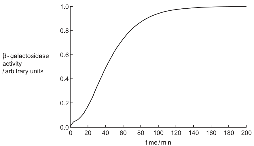
With reference to the lac operon, explain the shape of the curve in Fig. 3.1.
Answer
Lag phase (0–10 min) — time for the induction cascade:
- Lactose enters the bacterium.
- Lactose binds to the repressor, causing a shape change in the repressor.
- The repressor detaches from the operator.
- The promoter is unblocked.
- RNA polymerase binds to the promoter.
- The genes are transcribed.
- -galactosidase is synthesised by translation.
Steep rise (10–160 min):
- -galactosidase breaks down lactose; as more enzyme is made, activity increases.
Plateau (after ~160 min):
- -galactosidase activity reaches a maximum — all the active sites of the enzyme are saturated with substrate, so activity levels off.
The lag represents the time for the induction cascade (lactose entry → repressor binding → shape change → repressor detachment → RNA polymerase binding → transcription → translation); the steep rise reflects the accumulation of β-galactosidase; the plateau represents maximum enzyme activity (all active sites saturated with substrate).
Background Concept
The lac operon of Escherichia coli is a cluster of genes that allows the bacterium to use lactose as an energy and carbon source when the preferred sugar, glucose, is unavailable. The key components are:
- Promoter (P) — the DNA sequence to which RNA polymerase binds to begin transcription.
- Operator (O) — a DNA sequence that overlaps the promoter; the binding site for the repressor protein.
- Structural genes — lacZ (β-galactosidase, hydrolyses lactose to glucose + galactose), lacY (lactose permease, transports lactose into the cell), and lacA (thiogalactoside transacetylase).
- Regulatory gene lacI — located upstream of the operon, codes for the repressor protein. The repressor is produced continuously and binds the operator in the absence of lactose, physically blocking RNA polymerase.
A second layer of regulation, catabolite repression (not tested in detail here), means E. coli preferentially uses glucose; the lac operon is only fully expressed once glucose runs out. The experimental design in the question — growing E. coli on glucose, then adding lactose — exploits this switch.
Understanding the Question
This is a 7-mark "explain" question asking the candidate to interpret Fig. 3.1, a graph of β-galactosidase activity versus time, in terms of the lac operon mechanism. The graph has three clear features:
- A short lag phase (0–10 min) where activity remains at zero.
- A steep sigmoidal rise (10–160 min) where activity climbs to ~1.0 arbitrary units.
- A plateau (after ~160 min) at maximum activity.
The mark scheme allows any 7 of 10 listed points: nine points covering the induction mechanism and one point covering why the curve plateaus. A complete answer links the curve features to the molecular events.
Approach
The strategy is to walk through the induction cascade in order, then explicitly address the plateau:
- List the molecular events in sequence: lactose entry → repressor binding → shape change → repressor detachment → promoter unblocked → RNA polymerase binds → transcription → translation → enzyme accumulation → lactose hydrolysis.
- Map these events to the three phases of the curve: the cascade duration explains the lag; enzyme accumulation explains the rise; enzyme saturation explains the plateau.
Step-by-Step Reasoning
Lag phase (0–10 min) — induction cascade:
At time 0, lactose is added to the medium. The graph shows essentially no β-galactosidase activity for the first 10 minutes. This is because the cell has to run through the full induction cascade before any new enzyme appears:
- Lactose enters the bacterium, primarily through lactose permease (lacY).
- Inside the cell, a small amount of the inducer molecule allolactose is formed (and lactose itself can also act as the inducer for the purposes of the mark scheme).
- Allolactose binds to the repressor protein, which is already bound to the operator.
- This binding causes a conformational (shape) change in the repressor.
- The repressor, now unable to recognise the operator sequence, detaches from the operator.
- The promoter is unblocked, so RNA polymerase can bind to it.
- RNA polymerase transcribes the structural genes lacZ, lacY and lacA into a single mRNA (a polycistronic message).
- The mRNA is translated by ribosomes, and β-galactosidase is synthesised.
The whole cascade takes a few minutes; during this window, the activity of β-galactosidase remains effectively at zero, producing the lag phase.
Steep sigmoidal rise (10–160 min) — enzyme accumulation:
Once β-galactosidase begins to accumulate in the cell, its activity rises steeply. The rise is sigmoidal because:
- More enzyme is being made, so more lactose is being hydrolysed into glucose and galactose.
- The products provide energy and precursors, supporting further protein synthesis.
- As enzyme concentration increases, the rate of lactose hydrolysis increases proportionally.
The graph levels off as the cell reaches a steady state between enzyme synthesis and dilution by growth.
Plateau (after ~160 min) — enzyme saturation:
The activity stops increasing at ~1.0 arbitrary units. This is not because the bacterium has stopped producing the enzyme. It is because β-galactosidase is working at its maximum rate (Vmax) — all of the active sites on the enzyme molecules are saturated with lactose substrate. Once every active site is occupied, adding more substrate (or more enzyme) cannot increase the rate, so the measured activity plateaus.
Key Takeaways
- The lac operon is an inducible system: the substrate (lactose/allolactose) is the inducing signal.
- The lag phase of a gene-induction curve represents the time required for the cascade of events between the inducing signal and the appearance of active enzyme.
- The plateau of an enzyme-activity curve represents enzyme saturation (Vmax), not a switch-off of gene expression.
- A sigmoidal rise is typical of a self-reinforcing system where more enzyme → more product → more resources for more enzyme.
Common Mistakes
- Saying the plateau occurs because the bacterium stops producing β-galactosidase. The plateau is because the enzyme is working at maximum rate (active sites full), not because the gene has been switched off.
- Saying the repressor is destroyed when lactose binds. The repressor changes shape; it is not broken down.
- Confusing the repressor (a protein) with the operator (a DNA sequence).
- Skipping the initial step of lactose entering the cell.
- Saying RNA polymerase binds the operator (it binds the promoter; the operator is the repressor binding site).
- Confusing the lac operon (inducible) with the trp operon (repressible).
- Saying "inducer" alone for what is binding the repressor — the mark scheme requires lactose, allolactose or substrate.
Things to Be Careful About
- Use exact biological terms: repressor, operator, promoter, RNA polymerase, transcription, β-galactosidase.
- State the conformational change of the repressor explicitly — "binds and causes a shape change" is the credit-worthy wording.
- Address the plateau: this is a separate mark and many candidates lose it by only describing the rise.
- The mark scheme allows any 7 of 10 points, so candidates should aim to cover the full mechanism plus the plateau explanation.
- Note that the question stem already specifies the experimental context (glucose used up, then lactose added) — candidates do not need to explain the switch from glucose to lactose catabolite repression in detail, only the lac operon induction.
The Grand Canyon is located in Arizona, USA. It is estimated to have formed over five million years ago as the Colorado River began to create a deep channel (canyon) in the surrounding rocks.
Before the canyon formed, an ancestral species of antelope squirrel lived in the area. An antelope squirrel is a type of rodent and member of the squirrel family, Sciuridae.
It is estimated that around 3.6 million years ago, an ancestral species diverged into the two species that are present today.
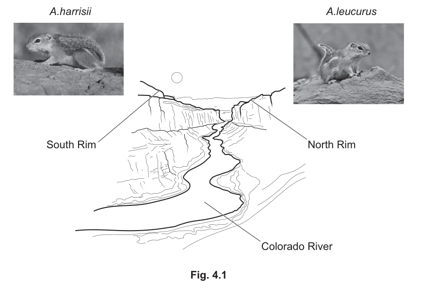
- Harris’s antelope squirrel, Ammospermophilus harrisii, has its habitat range extending from the south rim of the canyon.
- The white-tailed antelope squirrel, Ammospermophilus leucurus, has its habitat range extending from the north rim of the canyon.
Fig. 4.1 shows the location of the Colorado River in the Grand Canyon and the location of these species of antelope squirrel.
Answer
- The formation of the Grand Canyon / Colorado River caused geographical isolation of the ancestral squirrel population, separating it into two populations (one on the south rim, one on the north rim).
- The two isolated populations had no gene flow between them.
- The two populations experienced different selection pressures (different environments on the north and south rims).
- Random / independent mutations occurred in each population.
- This led to different changes in allele frequencies / different gene pools in the two populations, and the populations developed different morphological, physiological and behavioural features.
- Eventually the two populations became so different that they were unable to interbreed to produce fertile offspring, i.e. reproductive isolation had occurred.
- This is an example of allopatric speciation.
Allopatric speciation: geographical isolation of the ancestral population by the Grand Canyon prevented gene flow; different mutations and selection pressures caused allele frequencies to diverge, eventually producing reproductive isolation between A. harrisii and A. leucurus.
Background Concept
Speciation is the process by which one ancestral species gives rise to two or more new species. A species is generally defined as a group of organisms that can interbreed to produce fertile offspring. When two populations can no longer interbreed successfully, they are considered separate species — this is reproductive isolation.
Allopatric speciation occurs when a physical barrier divides a population. With the two sub-populations now separated:
- mutations occur independently in each;
- natural selection acts on each population according to its own local environment;
- the two gene pools drift apart in allele frequency.
Eventually the genetic differences accumulate to the point where, even if the barrier is removed, the two populations can no longer interbreed successfully — speciation is complete.
Understanding the Question
The stem describes an ancestral antelope squirrel population that existed before the Grand Canyon formed. Around 3.6 million years ago the canyon (with the Colorado River at its base) became a barrier. Today, the south-rim population is A. harrisii and the north-rim population is A. leucurus. The command word "suggest and explain" means you must propose a mechanism AND justify each step — not just say "they evolved separately".
Approach
Recognise this as a textbook case of allopatric speciation driven by a geographical barrier. Walk through the standard sequence:
- Identify the barrier and the two isolated populations.
- State that gene flow has been cut off.
- Explain why the populations diverge (different mutations, different selection pressures).
- State the consequence — different gene pools and morphological/physiological/behavioural features.
- Conclude with reproductive isolation, and name the process: allopatric speciation.
Step-by-Step Reasoning
- Barrier: the Colorado River and the steep canyon walls physically separate the two populations. This is geographical isolation (Marking point 1).
- No gene flow: because individuals from the south cannot reach the north (and vice versa), alleles are not exchanged between populations (Marking point 2).
- Independent mutations: each population accumulates its own random mutations, which are not shared with the other (Marking point 4).
- Different selection pressures: the north rim and south rim differ in temperature, vegetation, predators, etc. Different alleles are favoured in each environment, so allele frequencies shift in different directions (Marking points 3 and 5).
- Different features: the two populations acquire different morphological, physiological and behavioural features (Marking point 6).
- Reproductive isolation: the genetic and behavioural differences eventually become large enough that the two populations can no longer interbreed to produce fertile offspring, even if they were to meet (Marking point 7).
- Name the process: this is allopatric speciation (Marking point 8).
Any four of these eight mark-scheme points earn full marks. To write a strong answer, choose four that flow logically as a single argument.
Key Takeaways
- Geographical isolation is the classic starting point for allopatric speciation.
- Speciation requires (a) no gene flow, (b) divergence through mutation and selection, and (c) eventual reproductive isolation.
- A physical barrier (river, mountain, canyon) is not a species; the process of divergence is what creates one.
Common Mistakes
- Writing only "they adapted to different environments" — this is too vague; you must say what the mechanism is (no gene flow, different selection pressures, different mutations) and what the outcome is (different allele frequencies → reproductive isolation).
- Saying "they evolved into two species because of natural selection" without specifying allopatric speciation or the role of the barrier.
- Confusing allopatric with sympatric speciation — sympatric speciation does not require a geographical barrier.
Things to Be Careful About
- The mark scheme uses the words "geographical isolation" and "allopatric speciation" — use those exact terms.
- "No gene flow" is the key mechanistic point linking isolation to divergence; don't skip it.
- The question asks you to explain, so each point needs the reason attached (e.g. "different selection pressures led to different alleles being favoured").
Scientists investigated the evolutionary relationships of the squirrel family, Sciuridae. The scientists took samples from the current species in the family and carried out DNA sequencing and morphological analysis.
To compare current species with species from the past:
- specimens from museums were used to provide the tissue for DNA sequencing
- teeth and skulls from fossils were compared as part of the morphological analysis.
Describe the advantages of using DNA sequencing rather than morphological analysis to find out more about the evolutionary relationships of the squirrel family.
Answer
- DNA sequencing provides more information about the whole organism (the entire genome can be compared), not just the few features visible in bones or teeth.
- DNA sequencing is more precise / more accurate / quantitative, whereas morphological analysis can be subjective.
- Only a small sample of DNA is needed, so it can be used on tiny or degraded museum/fossil samples where whole specimens are not available.
- DNA sequences can be used as a molecular clock to estimate the time of divergence between species and to assess how closely related two organisms are.
- (AVP) DNA is not affected by convergent evolution, whereas unrelated organisms can independently evolve similar morphological features, misleading the morphological analysis.
DNA sequencing provides more complete, more precise and quantitative information from very small samples, allows time-of-divergence estimates via molecular clocks, and avoids the problems of convergent evolution that mislead morphological analysis.
Background Concept
Two broad approaches are used to investigate evolutionary relationships:
- Morphological analysis compares the physical features (bones, teeth, body shape) of organisms. Closely related species tend to look more alike, so similarities are used to construct evolutionary trees. However, morphology can be misled by convergent evolution (unrelated species evolving similar features because they share similar environments, e.g. the wings of birds and bats).
- DNA sequencing reads the actual order of nucleotide bases in specific genes or whole genomes. Closely related species share more recent common ancestors and therefore have more similar DNA sequences. The amount of sequence difference can even be used as a "molecular clock" to estimate when two lineages diverged, provided an estimate of mutation rate is available.
Understanding the Question
The question tells you that scientists used both DNA sequencing AND morphological analysis. You are asked to describe the advantages of the DNA method over the morphological method. The command word "describe" means give the specific creditable points — do not just say "DNA is better".
Approach
Think about what each method actually does, then list the things DNA can do that morphology cannot:
- information available (whole genome vs. a few bones/teeth);
- precision and objectivity (base sequences are unambiguous; morphological features can be scored subjectively);
- sample requirements (a tiny piece of tissue vs. a complete or near-complete specimen);
- ability to estimate when lineages diverged (molecular clock);
- resistance to convergent evolution.
Step-by-Step Reasoning
- More information: the entire genome — millions of base pairs — can in principle be compared, whereas morphology is limited to the features preserved in the available specimens (Marking point 1).
- More precise / accurate / quantitative: DNA sequences are explicit strings of A, T, G, C, easy to compare and to count differences. Morphological scoring can be subjective, depending on the observer (Marking point 2).
- Small sample size: only a small amount of DNA is needed, so DNA sequencing works on fragments of museum specimens and on fossil material, where no complete skeleton is required (Marking point 3).
- Molecular clock / divergence time: assuming mutations accumulate at a roughly steady rate, the number of differences between two species' DNA can be used to estimate how long ago their lineages diverged — something morphological analysis cannot do directly (Marking point 4).
- AVP — avoids convergent evolution: if two unrelated lineages evolve similar external features (e.g. similar teeth because they eat similar foods), morphology will mistakenly group them together. DNA is unaffected by selection on external form, so it gives a truer picture of ancestry (Marking point 6).
Three of these earn full marks.
Key Takeaways
- DNA sequencing provides more data, is more objective, requires less material, and can be calibrated to estimate divergence times.
- Morphological analysis is constrained by what is preserved in the fossil record and can be misled by convergent evolution.
- The two methods are usually combined: morphology provides the broad pattern, DNA refines the dates and relationships.
Common Mistakes
- Saying only "DNA is more accurate" without explaining why (e.g. quantitative, not subjective).
- Forgetting the molecular clock point — it is the key advantage that morphology simply cannot match.
- Not mentioning convergent evolution — a common AVP that distinguishes strong answers.
Things to Be Careful About
- The question is worth 3 marks, so give three distinct points. Don't pad one point into a long paragraph.
- Keep the comparison explicit: "DNA sequencing … whereas morphological analysis …" so the examiner can see you are answering the comparison asked for.
The estimate for the date that an ancestral species diverged into A. harrisii and A. leucurus is 3.58 million years ago. This estimate has a large uncertainty.
DNA sequencing, including DNA from fossils, was used to estimate this date of divergence.
Suggest a reason why there is such a large uncertainty for this date estimate.
Answer
- There are not enough fossils (of ancestral antelope squirrels) to calibrate the molecular clock precisely, so the date of divergence is uncertain.
- OR DNA degrades over time, so the DNA obtained from old / fossil samples is of low quality, with many errors and gaps, making the date estimate unreliable.
- OR small sample sizes mean the result is not statistically robust.
- OR the calculation depends on an estimated mutation rate, which may not be constant across lineages, introducing uncertainty.
There are not enough antelope-squirrel fossils (or DNA from fossils) to calibrate the molecular clock, and/or the DNA in old samples is degraded, so the divergence date has large uncertainty.
Background Concept
Molecular-clock dating estimates when two lineages split by counting DNA differences between them and dividing by an assumed mutation rate. The accuracy of the estimate depends on:
- The quality and quantity of the DNA available from the relevant species (especially fossils).
- The accuracy of the assumed mutation rate.
- Whether the mutation rate has been constant over time and across lineages.
If any of these is shaky, the resulting date has wide error bars (a "large uncertainty").
Understanding the Question
Part (c) tells you that the divergence date of 3.58 million years ago has a large uncertainty, and that fossil DNA was used in the estimate. It asks you to suggest a reason for the uncertainty. The mark scheme accepts one mark from a small list; you only need to give one clear, well-justified point.
Approach
Think about what could make a molecular-clock estimate unreliable:
- the availability of fossil material to calibrate the clock;
- the state of preservation of that DNA (degradation, contamination, gaps);
- the sample size (number of individuals sequenced);
- the assumed mutation rate, which is itself an estimate.
Pick one and state it concisely.
Step-by-Step Reasoning
- Not enough fossils: there are very few antelope-squirrel fossils of the right age, so the molecular clock has too few calibration points, and the divergence date is poorly constrained.
- DNA degrades over time: over millions of years DNA breaks down into short fragments and is chemically damaged. The older the sample, the less reliable the sequence, and the wider the error on any divergence-time estimate.
- Small sample sizes: only a few specimens may be available, so the calculated genetic distance has wide confidence intervals.
- Estimated mutation rate: the calculation assumes a particular rate, but real mutation rates vary between lineages and over time, so the date inherits that uncertainty.
Any one of these earns the single mark. The mark scheme lists them all as valid.
Key Takeaways
- Molecular-clock dates are only as good as the calibration data and the assumed mutation rate.
- Ancient DNA is fragmentary and chemically damaged, so divergence dates based on it carry large uncertainties.
- A small fossil record produces a poorly calibrated clock.
Common Mistakes
- Giving a vague answer such as "the date is uncertain because it's old" — the question wants a specific reason (DNA degradation, small sample, etc.).
- Saying "scientists don't know enough" — not a creditable scientific reason.
Things to Be Careful About
- Only one mark is available, so give one focused, well-articulated point rather than several half-formed ideas.
- Use the precise wording of the mark scheme where possible (e.g. "DNA degrades over time", "depends on an estimated mutation rate").
Recombinant DNA technology is used to make recombinant human proteins.
Two of the available methods to obtain the gene of interest are:
- cutting the gene out of genomic DNA using restriction enzymes
- obtaining messenger RNA (mRNA) from cells that are expressing the gene and then using reverse transcriptase to make complementary DNA (cDNA).
Plasmids can be used as vectors to transfer the gene of interest into a host organism.
Recombinant human insulin is a protein that is made using recombinant DNA technology.
Bacteria can be used as host cells to express the recombinant protein.
For the human insulin gene to be successfully expressed in bacteria, one method chosen to obtain the gene is to extract mRNA from -cells in the pancreas.
The gene coding for insulin is not expressed in the bacterial host when it has been obtained by cutting it out of genomic DNA.
Suggest and explain how the structural difference of cDNA and genomic DNA leads to only cDNA being expressed successfully.
Answer
- cDNA has no introns / only exons, whereas genomic DNA has introns (and exons);
- introns are non-coding sequences (and exons are coding sequences);
- bacteria cannot remove introns / cannot carry out splicing;
- therefore with cDNA a functional mRNA is made and translation occurs, but with genomic DNA a functional mRNA is not made and translation does not occur.
cDNA contains only exons (no introns); bacteria cannot splice out introns, so genomic DNA gives non-functional mRNA and no insulin is made.
Background Concept
In eukaryotes, genes are split. The coding regions (exons) are interrupted by non-coding regions called introns. Before a eukaryotic mRNA can be translated, the introns must be removed and the exons joined together — a process called splicing, carried out by the spliceosome inside the nucleus.
Bacteria are prokaryotes. They have no nucleus and no spliceosome, so they have no machinery to remove introns. Any intron in a bacterial transcript will be read as nonsense (or trigger degradation) and no functional protein will be produced.
cDNA (complementary DNA) is made from mature mRNA using reverse transcriptase. Because the mRNA template has already been spliced, the resulting cDNA contains a continuous coding sequence — only the exons, no introns.
Genomic DNA, by contrast, still contains every intron that was originally present in the chromosome.
Understanding the Question
You are told that the human insulin gene, when cut directly from genomic DNA, is not expressed in bacteria, but when obtained as cDNA (via reverse transcriptase from pancreatic β-cell mRNA) it is expressed. The task is to suggest and explain why this structural difference between cDNA and genomic DNA matters when the gene is placed in a bacterial host.
The command word is suggest and explain, so a mark-scheme point is only earned by stating the structural difference AND linking it to a consequence for expression in the bacterial cell.
Approach
Two threads must be woven together:
- State the structural difference (introns vs no introns).
- State the biological consequence (bacteria cannot splice; so only cDNA yields functional mRNA and a translated protein).
A complete answer gives the structure–function link, not just one or the other.
Step-by-Step Reasoning
- Marking point 1 — structural difference: cDNA contains only exons (no introns); genomic DNA contains introns as well as exons. (The contrast must be explicit.)
- Marking point 2 — coding status: introns are non-coding sequences and exons are the coding sequences that will be translated into protein. (This explains why it matters.)
- Marking point 3 — bacterial limitation: bacteria do not have the spliceosome and therefore cannot remove introns from any mRNA they transcribe. (This identifies the missing machinery.)
- Marking point 4 — functional outcome: with cDNA the mRNA produced is functional and translation occurs, giving insulin. With genomic DNA, transcription still occurs but the mRNA still contains introns, so it is not functional and translation does not occur — no insulin is made. (This closes the argument.)
Any three of these four points are enough for full marks; aim for all four to be safe.
Key Takeaways
- Eukaryotic genes = exons + introns; mRNA = exons only.
- Bacteria cannot splice, so they require an intron-free version of any eukaryotic gene.
- cDNA, made by reverse transcriptase from mRNA, is the standard workaround because it is automatically intron-free.
- Reverse transcriptase is therefore the key enzyme whenever a eukaryotic gene must be expressed in a prokaryote.
Common Mistakes
- Stating only that "cDNA has no introns" without linking this to expression — this is a structural statement, not an explanation.
- Saying "bacteria do not have introns" — wrong; bacteria do not have introns in their own genes, but they still cannot splice foreign introns out of a human transcript.
- Confusing the role of reverse transcriptase (makes cDNA from mRNA) with DNA polymerase (replicates DNA) or RNA polymerase (transcribes DNA to RNA).
- Writing "DNA is translated" — translation acts on mRNA, not DNA.
Things to Be Careful About
- The mark scheme explicitly accepts both the cDNA-positive and the genomic-DNA-negative framing, so either is fine as long as the comparison is clear.
- Do not introduce unnecessary extra material (e.g. codons, ribosomes) — the four credit points are tight and any extra is at best ignored and at worst contradicts a mark.
- Use the precise terms exon, intron, splicing, reverse transcriptase, cDNA — these are the terms the examiner expects.
Plasmids are cut using a restriction enzyme to create sticky ends. The plasmids are mixed with many copies of the desired gene and DNA ligase. The gene is inserted into many plasmids.
Answer
- DNA ligase forms phosphodiester bonds that join the sugar–phosphate backbones of the gene and the cut plasmid together;
- it joins / anneals the hydrogen-bonded sticky ends so the molecule is covalently closed.
Joins the sugar–phosphate backbones by forming phosphodiester bonds between the gene and plasmid (after the sticky ends have hydrogen-bonded).
Background Concept
A restriction enzyme cuts DNA at a specific palindromic sequence, leaving sticky ends — short single-stranded overhangs. When a gene and a plasmid are cut with the same restriction enzyme, their sticky ends have complementary sequences and will anneal (pair up) by hydrogen bonding between the exposed bases.
Hydrogen bonds, however, are weak and transient. To make a stable, covalently closed recombinant plasmid, the sugar–phosphate backbones on either side of every nick must be sealed. This is the job of DNA ligase, which forms phosphodiester bonds between the 3′-OH of one nucleotide and the 5′-phosphate of the next.
Understanding the Question
You are given the context: plasmids and many copies of the desired gene have been cut with the same restriction enzyme (so they have complementary sticky ends), mixed, and DNA ligase has been added. The question asks for the role of DNA ligase in producing recombinant plasmids. The mark scheme requires two separate points about bonding, so a one-line answer will not earn full marks.
Approach
Separate the two bond types clearly:
- The hydrogen bonds between the complementary sticky ends — DNA ligase consolidates / anneals these.
- The phosphodiester bonds that seal the sugar–phosphate backbone — this is the catalytic, ATP-requiring step that ligase performs.
Step-by-Step Reasoning
- Point 1 (hydrogen bonds / annealing): once the compatible sticky ends of the gene and plasmid meet, the complementary bases pair via hydrogen bonds, holding insert and vector together. DNA ligase acts on these joined molecules.
- Point 2 (phosphodiester bonds): the covalent seal. Ligase catalyses the formation of phosphodiester bonds between the 3′-hydroxyl and 5′-phosphate groups on either side of each nick, joining the sugar–phosphate backbones of the gene and plasmid into a single continuous circular molecule.
The two points are not interchangeable — the first is a description of what annealing produces, the second is the catalytic role of the enzyme itself.
Key Takeaways
- Restriction enzymes cut; DNA ligase joins.
- Annealing (hydrogen bonding) is reversible and weak; ligation (phosphodiester bond) is covalent and permanent.
- A "recombinant plasmid" only exists once ligation is complete — without ligase, the gene and plasmid would simply fall apart again.
Common Mistakes
- Stating only "joins the gene and plasmid together" — too vague, earns nothing.
- Mentioning hydrogen bonds or phosphodiester bonds but not both — the mark scheme credits both.
- Confusing DNA ligase with restriction enzymes (which cut DNA, the opposite role).
- Saying ligase "forms hydrogen bonds" — it does not; those form spontaneously when complementary sticky ends meet.
Things to Be Careful About
- The candidate may be tempted to write about base pairing only; the mark scheme explicitly requires the phosphodiester bond as the second point.
- Write the words out — do not abbreviate "phosphodiester" and do not say "phosphate bond" (an imprecise term that the mark scheme will not credit).
Explain why a promoter, as well as the gene, may have to be transferred into the plasmid.
Answer
- A promoter is the DNA sequence to which RNA polymerase binds;
- without the promoter, RNA polymerase cannot bind and transcription of the gene / gene expression cannot occur.
RNA polymerase needs the promoter in order to bind and initiate transcription of the inserted gene.
Background Concept
A promoter is a specific DNA sequence, typically located upstream of a gene, that acts as the binding site for RNA polymerase (and its associated transcription factors). RNA polymerase cannot simply start transcribing at any point along a DNA molecule; it must be recruited to a promoter. Once bound, it unwinds the DNA and begins RNA synthesis in the 5′→3′ direction.
When a human gene is inserted into a bacterial plasmid, the gene itself has no usable bacterial promoter attached to it. Even if the gene's own eukaryotic promoter were present, bacterial RNA polymerase would not recognise it. So a bacterial-compatible promoter must be transferred along with the gene.
Understanding the Question
The stem has told you that plasmids and the desired gene are mixed with DNA ligase; the gene is now physically inside the plasmid. The question asks you to explain why the promoter (in addition to the gene) often has to be inserted into the plasmid.
The command word is explain, so a mark is only earned by giving the reason — not just "a promoter is needed for expression".
Approach
State the chain of cause and effect:
- Promoter is the binding site for RNA polymerase.
- Without the promoter, RNA polymerase cannot bind → transcription cannot start → the gene is not expressed.
Step-by-Step Reasoning
- Point 1: the promoter is a DNA sequence recognised by RNA polymerase. It is the site where the enzyme first attaches to the DNA in order to begin transcription.
- Point 2: if the plasmid contains the gene but no promoter, RNA polymerase has nowhere to bind and so transcription does not start; the gene is physically present in the plasmid but is not expressed — no mRNA, no protein.
Both points are needed for full marks. A common shorthand answer "so the gene can be expressed" is too vague on its own and is the kind of answer the mark scheme rejects without the mechanistic detail.
Key Takeaways
- A gene without a promoter is silently present in a plasmid — it will not be transcribed.
- Bacterial and eukaryotic promoters are not interchangeable; recombinant work that puts a eukaryotic gene into a bacterium must supply a bacterial promoter (often engineered into the plasmid vector).
- Promoters and genes are independent units in plasmid design, which is why plasmids are sometimes called "expression vectors".
Common Mistakes
- Stating "so the gene can be expressed" without saying how (RNA polymerase binding).
- Confusing a promoter with an origin of replication (which is also sometimes required, but a different concept — it is where DNA replication begins, not transcription).
- Confusing a promoter with a start codon (which is part of the mRNA, not DNA, and is where translation begins).
- Saying "the promoter codes for RNA" — the promoter is a regulatory DNA sequence; it is transcribed only in some cases but its function is to bind RNA polymerase, not to code for anything.
Things to Be Careful About
- The mark scheme wants two distinct ideas: RNA polymerase binding AND transcription/expression. Either one alone earns only one mark.
- In some vectors the promoter is already on the plasmid before the gene is inserted, so the question's "may have to" wording is deliberate — transfer is only required if the vector does not already carry one.
Answer
- A marker gene is inserted into the plasmid (alongside the gene of interest);
- it is positioned downstream of a promoter so that it can be transcribed;
- and positioned alongside the gene of interest so that any cell containing the plasmid also contains the marker;
- when the marker gene is expressed, fluorescent protein is made;
- when the cells are exposed to UV / blue light, transformed cells / cells containing the recombinant plasmid fluoresce (glow) and can be identified.
A marker gene is placed on the plasmid downstream of a promoter (alongside the gene of interest); transformed cells express the marker and fluoresce under UV/blue light, allowing them to be identified.
Background Concept
In recombinant DNA technology, not every cell that is exposed to a plasmid actually takes one up. The cells that do take up a plasmid are said to be transformed; those that do not are untransformed. Because transformation is usually inefficient, scientists need a quick way to tell which cells carry the recombinant plasmid.
A marker gene is a gene whose product is easy to detect. The classic examples are antibiotic-resistance genes (transformed cells survive on antibiotic medium) and fluorescent-protein genes such as GFP (green fluorescent protein), which glow under UV or blue light. Cells carrying the plasmid will glow; cells without the plasmid will not.
For the marker to be expressed, it must be positioned downstream of a promoter, so that RNA polymerase can transcribe it.
Understanding the Question
The question asks you to explain how a marker gene coding for a fluorescent product could be used — that is, the mechanism by which the marker gene is deployed and what it allows the experimenter to do. The mark scheme requires the answer to cover both the placement of the marker on the plasmid and the detection/identification step.
Approach
Lay out the logic in order:
- Where the marker is placed on the plasmid.
- How its expression produces the detectable signal.
- How that signal identifies transformed cells.
A complete answer needs at least one point from each of these three ideas.
Step-by-Step Reasoning
- Placement (mark scheme points 1, 2, 3): the marker gene is added to the plasmid, downstream of a promoter, alongside the gene of interest. This ensures both genes are transcribed together whenever RNA polymerase reads through.
- Expression (point 4): when the cell expresses the marker gene, the fluorescent protein is produced.
- Detection and use (points 5, 6): transformed cells, when illuminated with UV or blue light, fluoresce — untransformed cells do not. The experimenter therefore uses the fluorescence to identify which cells have taken up the recombinant plasmid.
Any three of the six creditable points earn full marks. A safer answer includes the positioning of the marker and the identification of transformed cells, because these are the conceptual core of the question.
Key Takeaways
- Marker genes are a screening tool, not a selection tool (no killing of untransformed cells is involved when fluorescence is the readout — unlike antibiotic markers, where untransformed cells die).
- The marker must be downstream of a promoter to be expressed; without that, the plasmid will not fluoresce even if it is inside the cell.
- A fluorescent marker is non-destructive: live cells can be sorted and grown on, which is one of its key advantages over antibiotic selection.
Common Mistakes
- Describing the marker as a way to kill untransformed cells — this is true for antibiotic markers, not fluorescent ones. Fluorescent markers identify but do not kill.
- Saying the marker gene "glows on its own" — it is the protein that fluoresces, only when the cell expresses the gene and the protein is excited by UV/blue light.
- Omitting the need for a promoter — without it, the marker would never be transcribed.
- Confusing a marker gene with the gene of interest itself — the marker is a separate gene whose only purpose is to indicate which cells contain the plasmid.
Things to Be Careful About
- The mark scheme uses AW ("allow any wording") after "transformed / AW" — so "taken up the plasmid" or "contain the recombinant plasmid" are all acceptable.
- Use the phrase downstream of the promoter rather than "near" or "after" — "downstream" is the precise molecular-biology term.
- Remember that the question says "a marker gene coding for a fluorescent product" — so the detection method (UV/blue light exposure) is essential to the answer, not an optional extra.
The unicellular fungus Saccharomyces cerevisiae is a species of yeast that has been used to produce human insulin. S. cerevisiae cells are able to take up recombinant plasmids.
Suggest advantages of using yeast compared to using bacteria for human insulin production.
Answer
Any two of:
- yeast cells are easier / cheaper to culture;
- yeast gives a higher yield / is more productive;
- insulin is easier to extract / process / purify from yeast;
- yeast cells are eukaryotic (like human β-cells) so they possess organelles such as a Golgi apparatus and endoplasmic reticulum, which can perform the post-translational modifications needed to produce fully functional human insulin.
Yeast is easier/cheaper to culture, gives higher yields, allows easier purification, and is a eukaryote (with Golgi apparatus) so it can carry out the post-translational modification of human insulin.
Background Concept
Bacteria (Escherichia coli) were the first hosts used to produce recombinant human insulin, and they remain widely used because they are fast-growing, cheap and well-understood. However, bacteria are prokaryotes: they lack a nucleus, endoplasmic reticulum and Golgi apparatus. As a result, they cannot carry out the post-translational modifications (such as the formation of disulphide bonds and correct folding of the polypeptide into its 3D shape) that many human proteins require to function.
Saccharomyces cerevisiae (baker's yeast) is a unicellular eukaryote. It is still cheap and easy to grow in fermenters, but it possesses the eukaryotic organelles (ER, Golgi) needed to fold and modify human proteins correctly. It is therefore a useful compromise between the convenience of a microbial host and the protein-processing capacity of a mammalian cell.
Understanding the Question
You are asked to suggest advantages of using yeast rather than bacteria for the production of human insulin. The mark scheme allows any two of four creditable ideas, so the safest strategy is to give two distinct, well-articulated points rather than one vague one.
The command word is suggest, which is a softer directive than explain — but the mark scheme still requires concrete reasoning, not just a list of words like "better" or "easier".
Approach
Pick advantages from two different categories:
- Practical/bioprocess advantages — cost, yield, ease of extraction.
- Cell-biology advantages — yeast's eukaryotic features (ER, Golgi) that bacteria lack and that are required to make fully functional insulin.
A two-mark answer that hits one point from each category is ideal.
Step-by-Step Reasoning
- Cost / ease of culture: yeast is still a single-celled microbe, so it grows quickly in bulk fermenters using cheap media — almost as convenient as bacteria but often cheaper or more productive at scale.
- Yield: yeast can give a higher yield of correctly processed insulin per unit of culture than bacteria, because the protein reaches its native conformation and accumulates efficiently.
- Purification: because yeast secretes some proteins or stores them in defined compartments, insulin is often easier to extract and purify from yeast than from bacteria, where the protein accumulates in insoluble inclusion bodies.
- Eukaryotic processing: insulin is initially translated as preproinsulin and must be folded, have disulphide bonds formed, and have the signal peptide cleaved before it becomes functional. Bacteria do not reliably do this; yeast, with its ER and Golgi apparatus, can.
Any two of these are credited.
Key Takeaways
- A useful host organism must (a) take up and replicate the plasmid, (b) express the gene, and (c) produce a correctly folded, functional protein.
- Bacteria excel at the first two but struggle with the third for many human proteins.
- Yeast offers a useful middle ground: microbial in its culturing, eukaryotic in its protein processing.
- This is why modern pharmaceutical insulin is often made in yeast (S. cerevisiae) or in mammalian cell lines rather than in E. coli.
Common Mistakes
- Writing only "yeast is better" — too vague, earns nothing.
- Saying yeast "is a eukaryote like humans" without naming a specific consequence (e.g. Golgi apparatus, disulphide bond formation).
- Claiming yeast can carry out transcription / translation of introns — yeast can splice its own introns, but in this context the insulin gene is supplied as cDNA (already intron-free), so splicing is not the relevant advantage.
- Listing only advantages that also apply to bacteria (e.g. "easy to grow") without a comparative or specific-to-yeast element.
Things to Be Careful About
- The mark scheme uses the phrase "yeast cell similar to β-cells" as a specific credit-worthy point, with the example "have Golgi apparatus". Naming the organelle is what earns the mark.
- "More productive" is acceptable shorthand, but a stronger answer specifies more productive of what (correctly processed insulin).
- Avoid bringing in tangential material about ethanol fermentation or bread-making — irrelevant here.
The kidneys have a role in excretion and in osmoregulation.
Excretion is the removal of the waste products of metabolism or the removal of substances that are in excess.
Answer
Urea.
Urea
Background Concept
Excretion is the removal from the body of the waste products of metabolism, particularly the nitrogenous waste from the breakdown of excess amino acids. In mammals the main nitrogenous excretory product is urea, a small, water-soluble molecule formed in the liver and excreted in solution by the kidneys. Smaller amounts of nitrogen are also lost as creatinine (from muscle creatine phosphate breakdown) and uric acid, but urea is by far the largest component.
Understanding the Question
The stem defines excretion, and part (a)(i) asks for the single most important nitrogen-containing waste product that the kidneys remove. Only the name is required.
Approach
Recall that in mammals the amino group removed during amino acid catabolism is converted into urea (not ammonia, which is too toxic, and not uric acid, which is the main product in birds and reptiles).
Step-by-Step Reasoning
- Excess amino acids cannot be stored.
- The amino group (-NH₂) is removed as ammonia (NH₃), which is highly toxic even at low concentrations.
- The liver rapidly converts ammonia into urea via the ornithine (urea) cycle.
- Urea is far less toxic, is highly soluble in water, and is transported in the blood plasma to the kidneys, which filter it out into the urine.
- Therefore the main metabolic waste product excreted by the kidneys is urea.
Key Takeaways
- The kidneys' principal nitrogenous waste is urea.
- The liver makes urea; the kidney merely excretes it.
Common Mistakes
- Writing "ammonia" — this is the immediate precursor, not the form excreted by mammalian kidneys.
- Writing "uric acid" — that is for birds, reptiles and insects.
- Writing "urine" — urine is the mixture, not the waste product itself.
Things to Be Careful About
The question says "main metabolic waste product". Do not list creatinine or other minor components; one correct term is enough.
Answer
By deamination (the conversion of ammonia to urea in the ornithine/urea cycle).
Deamination / ornithine cycle / urea cycle (conversion of ammonia to urea)
Background Concept
Amino acids in excess of the body's needs cannot be stored. Their amino groups must be removed and disposed of safely. The liver performs two linked tasks: deamination (removal of the amino group from the amino acid, releasing ammonia) and the ornithine (urea) cycle (combination of ammonia with CO₂ to form urea).
Understanding the Question
Part (a)(ii) builds directly on (i): having identified urea, the question now asks how the body makes it.
Approach
Recall the two-step liver pathway: amino acid → keto acid + NH₃ (deamination); then 2 NH₃ + CO₂ → urea (ornithine cycle). Either name earns the mark because the mark scheme accepts both.
Step-by-Step Reasoning
- In hepatocytes, the enzyme systems of deamination strip the -NH₂ group from excess amino acids, producing ammonia (NH₃).
- Because NH₃ is extremely toxic, it is rapidly incorporated into the ornithine cycle.
- The ornithine cycle combines 2 molecules of ammonia with 1 molecule of CO₂, ultimately producing urea and regenerating ornithine so the cycle can continue.
- Any of: deamination, ornithine cycle, urea cycle, or conversion of ammonia to urea is accepted.
Key Takeaways
- Deamination releases ammonia.
- The ornithine (urea) cycle packages ammonia into urea.
- Both happen in the liver.
Common Mistakes
- "The kidney makes urea" — the kidney only excretes it.
- Giving only "the urea cycle" without indicating that ammonia is the starting material — this is acceptable, but a stronger answer ties deamination to the urea cycle.
Things to Be Careful About
The mark scheme accepts deamination alone, the ornithine cycle / urea cycle alone, or the idea of converting ammonia to urea. Any one of these is sufficient for the single mark.
Answer
Liver.
Liver
Background Concept
Urea synthesis is a hepatic function. The enzymes of the ornithine cycle, together with the aminotransferases and glutamine synthetase involved in deamination, are concentrated in the liver (hepatocytes surrounding the central vein of each lobule). This anatomical localisation is crucial — if liver function fails, ammonia levels in the blood rise rapidly and cause neurological damage (hepatic encephalopathy).
Understanding the Question
Part (a)(iii) closes the trio by asking where the urea is made. The answer is a single organ.
Approach
Recall that the liver is the body's main site of amino acid catabolism and of the urea cycle.
Step-by-Step Reasoning
- Both deamination and the ornithine cycle take place inside hepatocytes.
- Urea is then released into the blood and transported (in the hepatic vein) to the systemic circulation, then to the kidneys for excretion.
- The single-word answer is liver.
Key Takeaways
- Liver = deamination + urea cycle.
- Kidney = excretion only.
Common Mistakes
- Writing "kidney" — this is the organ of excretion, not synthesis.
- Writing "hepatocyte" — although technically correct and more precise, CIE accept "liver" for this mark.
Things to Be Careful About
Distinguish clearly between site of production (liver) and site of excretion (kidney). The two roles are easily muddled in an exam hall.
Glomerular filtrate is formed by the process of ultrafiltration.
Describe the process of ultrafiltration.
Answer
- Blood enters the glomerulus via the afferent arteriole.
- The afferent arteriole has a larger lumen diameter than the efferent arteriole.
- This creates a high hydrostatic (blood) pressure inside the glomerular capillaries.
- The blood/hydrostatic pressure is greater than the water potential gradient between the blood and Bowman's capsule.
- Fluid is therefore forced out of the capillaries through the pores / fenestrations in the capillary endothelium.
- The basement membrane acts as a filter.
- The basement membrane prevents the passage of large plasma proteins, blood cells and molecules larger than ~68 000–70 000 RMM.
- Water, glucose, amino acids, urea and mineral ions pass through into Bowman's capsule.
- Fluid then passes between the slit pores between the podocytes of Bowman's capsule to enter the capsular (nephron) lumen.
- AVP e.g. the resulting filtrate is produced at a rate of about (the glomerular filtration rate).
See working.
Background Concept
Ultrafiltration is the first stage of urine formation, occurring in the renal corpuscle (glomerulus + Bowman's capsule). It is a pressure-driven, passive process in which most of the plasma — but not the cells or large proteins — is squeezed out of the glomerular capillaries into the capsular space. The selectivity comes from a three-layered barrier: the fenestrated capillary endothelium, the basement membrane, and the podocyte foot processes of Bowman's capsule with their slit pores between them.
The driving force is glomerular hydrostatic pressure, generated because the afferent arteriole (entering the glomerulus) has a wider lumen than the efferent arteriole (leaving it). The narrowed outlet creates a bottleneck and pressurises the capillary bed.
Understanding the Question
This is a seven-mark "describe" question worth 7 marks. The mark scheme offers 10 creditworthy ideas, so the candidate should aim to cover at least seven. "Describe" here means set out the sequence of events and structures — it is more than a simple list; the candidate must show how the structure of the vessels and the filtration barrier combine to produce glomerular filtrate.
Approach
Move logically from:
- The vessels (afferent / efferent arteriole, the pressure they generate).
- The driving force (hydrostatic pressure > water potential gradient).
- The three layers of the filtration barrier (endothelium, basement membrane, podocytes).
- The size-selective cut-off and what does and does not pass through.
- A precise named AVP if you know one (e.g. GFR value).
Step-by-Step Reasoning
- Vessels and pressure (mark points 1–3). Blood arrives at the glomerulus through the afferent arteriole. The afferent arteriole is wider than the efferent arteriole that drains the glomerulus. With a wider inlet and a narrower outlet, blood cannot escape as fast as it enters, so the pressure inside the capillary bed rises to a much higher level than in normal systemic capillaries. This is the glomerular hydrostatic pressure, normally about 55 mmHg.
- Driving force (mark point 4). For filtration to occur, the glomerular hydrostatic pressure must exceed the opposing forces — the hydrostatic pressure inside Bowman's capsule and the oncotic pressure of the plasma proteins. The net result is a pressure gradient that pushes fluid out of the capillary. The mark scheme expresses this as "blood/hydrostatic pressure greater than the water potential gradient (between Bowman's capsule and glomerulus)".
- First filter (mark point 5). The capillary wall is fenestrated — the endothelial cells are pierced by large pores (~70–100 nm in diameter). These pores are wide enough to let water and most solutes leave the blood but do not allow cells to pass.
- Second filter (mark points 6–7). Beneath the endothelium is the basement membrane, a mesh of collagen IV and proteoglycans. It is the principal size- and charge-selective barrier. It blocks anything larger than about 68 000–70 000 relative molecular mass, which excludes plasma proteins (e.g. albumin at ~69 000) and absolutely excludes blood cells.
- Third filter (mark point 9). On the capsular side, podocytes wrap their foot processes around the capillaries. The gaps between adjacent foot processes are the slit pores (~25–30 nm), bridged by a thin diaphragm. Anything that has not been retained by the basement membrane but is too large for the slit pores is stopped here.
- What gets through (mark point 8). Water, glucose, amino acids, urea, mineral ions (Na⁺, K⁺, Cl⁻, HCO₃⁻, etc.) and small nitrogenous wastes pass into the capsular (Bowman's) space as glomerular filtrate. The composition is essentially plasma minus the proteins.
- AVP (mark point 10). The glomerular filtration rate (GFR) in an adult human is about , or roughly , of which 99% is reabsorbed downstream.
Key Takeaways
- Ultrafiltration is pressure-driven, not active.
- The afferent > efferent lumen difference is what generates the pressure.
- The barrier has three layers (endothelium, basement membrane, podocytes).
- Selectivity is mainly by molecular size (cut-off ~68 000–70 000 RMM) and by charge.
- Cells and large proteins stay in the blood; everything smaller enters the filtrate.
Common Mistakes
- Saying ultrafiltration is "active transport" — it is passive, driven by a pressure gradient.
- Confusing the direction of the pressure gradient: the mark scheme wording is "blood pressure greater than the water potential gradient (between Bowman's capsule and glomerulus)". A common slip is to say the blood has a lower water potential and so water moves in — water actually moves from the capillary into the capsule because the capillary hydrostatic pressure dominates.
- Listing only the structures without explaining their function (e.g. naming podocytes but not saying their slit pores are a filter).
- Forgetting the size cut-off — "stops large proteins" is a marking point only if the size (~68 000–70 000 RMM) is given or implied.
Things to Be Careful About
- Use the precise term fenestrations (or pores) for the endothelial gaps and slit pores for the podocyte gaps; examiners prefer them over "holes".
- The mark scheme accepts "blood cells" as a single category — red cells are too large to pass, but white cells and platelets are also retained.
- "Mineral ions" is a safer term than "salts"; avoid saying just "ions" without qualification.
Different mammals have different thicknesses of medulla relative to the size of the kidney.
Fig. 6.1 shows the relationship between the mean thickness of the medulla in kidneys of different mammals and concentration of urine produced by the kidneys.
Suggest an explanation for the relationship shown in Fig. 6.1.
Answer
- A thicker medulla contains longer loops of Henle, which set up a more steep water potential (osmotic) gradient in the medulla (so the medulla has a lower water potential).
- As filtrate / urine flows through the collecting ducts in the medulla, water is reabsorbed down this steeper gradient by osmosis, so more water is reabsorbed and the urine is more concentrated.
A thicker medulla produces a steeper water potential gradient, so more water is reabsorbed from the collecting ducts and the urine is more concentrated.
Background Concept
The kidney's ability to produce concentrated urine depends on the countercurrent multiplier in the loop of Henle. The longer the loop, the more Na⁺ and Cl⁻ can be pumped out into the medullary interstitium, and the lower (more negative) the water potential reaches deep in the medulla. Mammals that live in dry environments (e.g. desert rodents) have very long loops of Henle and very thick medullas; mammals in water-rich environments (e.g. beavers) have short loops and thin medullas and produce dilute urine.
When ADH makes the collecting duct wall permeable to water, water moves out of the duct by osmosis into the medullary interstitium. The deeper the osmotic gradient, the more water leaves, and the more concentrated the final urine becomes.
Understanding the Question
Fig. 6.1 shows a positive linear relationship: mammals with a thicker medulla produce more concentrated urine. The question uses the command word suggest, which here means give a biologically reasoned explanation for the observed trend. Two marks are available.
Approach
Connect the structural variable (medulla thickness) to the physiological mechanism (depth of the osmotic gradient, length of the loops of Henle, water reabsorption in the collecting duct).
Step-by-Step Reasoning
- Mark point 1. The collecting ducts of the nephron pass through the medulla on their way to the renal pelvis. The medulla is therefore the region where the final water reabsorption (or not) takes place, depending on ADH.
- Mark point 2. A thicker medulla corresponds to longer loops of Henle. Longer loops pump more NaCl into the medullary interstitium (because of the countercurrent multiplier running over a greater vertical distance), generating a steeper osmotic gradient and a more negative water potential at the tip of the medulla.
- Mark point 3. With a steeper water potential gradient, more water leaves the collecting duct by osmosis (when ADH is present and aquaporins are inserted). The remaining fluid — the urine — therefore has a higher concentration of solutes.
- AVP (mark point 4). Reference to the loop of Henle specifically, or the role of ADH, would be credited.
Key Takeaways
- Medulla thickness is a proxy for loop-of-Henle length and depth of the osmotic gradient.
- A deeper gradient → more water reabsorbed from the collecting duct → more concentrated urine.
- This is why desert mammals have thick medullas and beavers (which rarely need to conserve water) have thin ones.
Common Mistakes
- Saying "a thicker medulla holds more urine" — that is a structural misconception; the medulla is the source of the osmotic gradient, not a storage region.
- Saying "thicker medulla = more water in the urine" — the opposite of what the graph shows.
- Failing to mention the collecting duct at all — without the duct traversing the medulla, the gradient has no physiological effect.
- Writing only about ADH and not the structural reason the gradient is steeper.
Things to Be Careful About
- Read the graph carefully: it is a positive linear relationship, not a curve that levels off.
- The term "water potential" is more precise than "concentration"; a complete answer uses both.
- The question is asking for an explanation of the relationship, so describe a mechanism, not a restatement of the graph.
A chloroplast is composed of many structures, each with a different function. Several chloroplast structures are listed.
stroma lamellae thylakoid membrane ribosome
thylakoid space starch grain DNA outer membrane
From the list:
Answer
DNA and ribosome.
DNA and ribosome
Background Concept
Rubisco (ribulose bisphosphate carboxylase/oxygenase) is the enzyme that catalyses the carboxylation of RuBP in the Calvin cycle. It is one of the most abundant proteins on Earth and consists of eight large and eight small subunits. Like mitochondria, chloroplasts are semi-autonomous organelles: they contain their own DNA and ribosomes, allowing them to encode and synthesise some of their own proteins, including rubisco.
Understanding the Question
The question supplies a list of chloroplast structures and asks which of them are involved in producing rubisco. Producing a protein requires both the genetic information (DNA) and the translation machinery (ribosomes). Both must be named.
Approach
Apply the central dogma inside the chloroplast: DNA is transcribed into mRNA, and mRNA is translated by ribosomes into a polypeptide. Choose the structures from the list that fulfil each role.
Step-by-Step Reasoning
- Rubisco is a protein, so it must be synthesised inside the chloroplast.
- The gene encoding rubisco is carried on the chloroplast DNA, so DNA is needed.
- The chloroplast ribosomes translate the rubisco mRNA into the polypeptide chain, so ribosome is needed.
- The other structures in the list (stroma, lamellae, thylakoid membrane, thylakoid space, starch grain, outer membrane) have roles in photosynthesis or compartmentalisation but are not directly involved in producing a protein.
Key Takeaways
Chloroplasts are semi-autonomous: they encode and translate a subset of their own proteins. Producing any protein needs both the genetic template (DNA) and the assembly machinery (ribosome).
Common Mistakes
Giving only "ribosome" because the chloroplast DNA is overlooked. Saying "stroma" because the candidate confuses the site of action of rubisco with the site of production.
Things to Be Careful About
Both structures must be named to earn both marks. Do not credit thylakoid membrane or outer membrane — these are membranes, not protein-synthesis machinery.
Answer
Thylakoid space.
Thylakoid space
Background Concept
In the light-dependent reactions of photosynthesis, two processes pump protons (H⁺) from the stroma into the thylakoid space:
- Photolysis of water at photosystem II releases H⁺ into the thylakoid space.
- The electron transport chain between photosystems II and I uses the energy of electrons to actively transport H⁺ from the stroma into the thylakoid space.
The result is a proton gradient across the thylakoid membrane, with a much higher concentration of H⁺ inside the thylakoid space than in the surrounding stroma. This gradient drives chemiosmosis: protons flow back into the stroma through ATP synthase, releasing energy that phosphorylates ADP to ATP.
Understanding the Question
The question asks for the single chloroplast structure (from the supplied list) that has a high concentration of protons in daylight. The qualifier "in daylight" points specifically to the light-dependent reactions, where the proton gradient is established.
Approach
Identify the compartment into which protons are deposited by photolysis and by the electron transport chain during the light-dependent reactions.
Step-by-Step Reasoning
- Photolysis at PSII splits water into H⁺, electrons and oxygen; the H⁺ remain inside the thylakoid space.
- The electron carriers between PSII and PSI pump additional H⁺ from the stroma into the thylakoid space.
- Therefore, the thylakoid space is the compartment with the highest concentration of protons during the day.
- This gradient is essential for ATP synthesis by chemiosmosis.
Key Takeaways
The thylakoid space is the proton reservoir that drives photophosphorylation. Photolysis and the electron transport chain together establish the proton gradient.
Common Mistakes
Naming the thylakoid membrane (where the electron carriers and ATP synthase sit) rather than the thylakoid space. Naming the stroma — the stroma actually has a lower [H⁺] than the thylakoid space because protons have been pumped out of it.
Things to Be Careful About
The question specifies "in daylight" because the proton gradient dissipates at night. Only the thylakoid space accumulates H⁺ during the light reactions; in darkness the gradient collapses as protons leak back through ATP synthase.
Paper chromatography is a technique that can be used to separate a mixture of four common chloroplast pigments. The pigments can be identified by calculating their values.
A student carried out paper chromatography on a solution containing a mixture of chloroplast pigments.
The results are shown in Table 7.1.
Table 7.1
| pigment | distance travelled by pigment from baseline / cm | distance travelled by solvent from baseline / cm | |
|---|---|---|---|
| ______ | 6.5 | 8.9 | 0.73 |
| chlorophyll a | 4.6 | 8.9 | 0.52 |
| chlorophyll b | ______ | 8.9 | 0.38 |
| carotene | 8.2 | 8.9 | ______ |
Complete Table 7.1.
Working
The formula is
The first pigment travelled 6.5 cm with the solvent at 8.9 cm, giving .
The four common chloroplast pigments, in order of increasing on a paper chromatogram, are chlorophyll b (~0.38), chlorophyll a (~0.52), xanthophyll (~0.73) and carotene (~0.92). The missing pigment with is therefore xanthophyll.
For chlorophyll b:
For carotene:
Answer
| pigment | distance / cm | solvent distance / cm | |
|---|---|---|---|
| xanthophyll | 6.5 | 8.9 | 0.73 |
| chlorophyll a | 4.6 | 8.9 | 0.52 |
| chlorophyll b | 3.4 | 8.9 | 0.38 |
| carotene | 8.2 | 8.9 | 0.92 |
Missing pigment: xanthophyll; chlorophyll b distance: 3.4 cm; carotene : 0.92.
xanthophyll; 3.4 cm; 0.92
Background Concept
Paper chromatography separates the pigments in a chloroplast extract because each pigment has a different solubility in the solvent and a different affinity for the paper (stationary phase). The value of a pigment is the ratio of the distance travelled by the pigment to the distance travelled by the solvent front:
The four common chloroplast pigments separated on a typical chromatogram, in order of increasing (i.e. increasingly non-polar), are chlorophyll b, chlorophyll a, xanthophyll and carotene.
Understanding the Question
Table 7.1 gives the distance travelled by the solvent (8.9 cm in every case) and partial results for four pigments. Three cells must be filled in:
- The name of the pigment with .
- The distance travelled by chlorophyll b (its of 0.38 is given).
- The of carotene (its distance of 8.2 cm is given).
Approach
Recognise the characteristic order of the four chloroplast pigments to identify the unknown, and apply the formula in its two rearranged forms (for distance and for ) for the remaining two cells.
Step-by-Step Reasoning
- The four values place the pigments in this typical order: chlorophyll b (lowest, most polar), chlorophyll a, xanthophyll, carotene (highest, most non-polar).
- The unknown pigment has and is in the third row of the table; by the order above this is xanthophyll.
- For chlorophyll b: cm, rounded to 3.4 cm (one decimal place, matching the precision of the other distances in the table).
- For carotene: , rounded to 0.92 (two decimal places, matching the other values).
Key Takeaways
The value is constant for a particular solvent system and can therefore be used as an identifying fingerprint for a pigment. The four common chloroplast pigments have a characteristic order of values from low to high: chlorophyll b, chlorophyll a, xanthophyll, carotene.
Common Mistakes
Calculating the distance of chlorophyll b using a wrong (e.g. using 0.52, the value for chlorophyll a, by mistake). Forgetting to round to the precision used in the table (1 dp for distances, 2 dp for values). Naming chlorophyll a as the unknown pigment instead of xanthophyll — the row order in the table already separates them.
Things to Be Careful About
Match the precision used in the rest of the table. Distances are quoted to one decimal place and values to two decimal places. The unknown pigment is identified by both its and its position in the row order; do not give an arbitrary name.
Fig. 7.1 shows the absorption spectrum for carotene and for chlorophyll a.
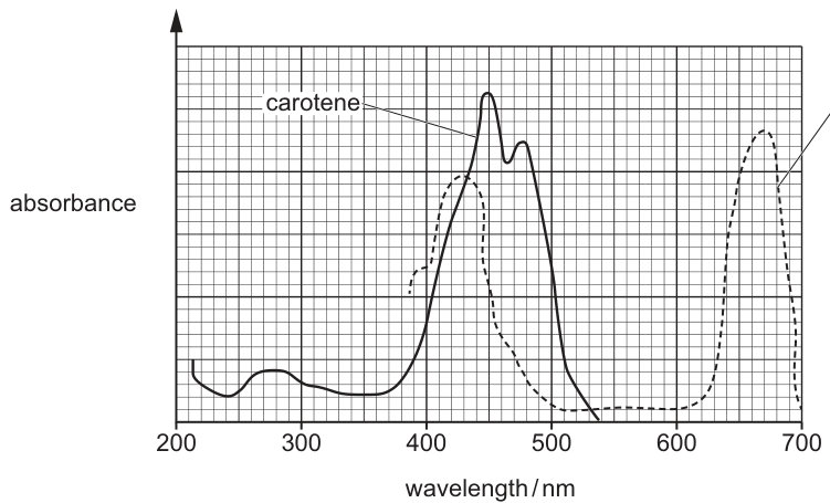
With reference to Fig. 7.1, describe and explain the role of carotene in photosynthesis.
Answer
- Carotene absorbs light between 400 and 500 nm, with a peak at about 450 nm.
- Carotene is an accessory pigment.
- It absorbs wavelengths of light that are not absorbed by chlorophyll a, so it extends the range of wavelengths that can be used in photosynthesis.
- The energy absorbed by carotene is transferred to chlorophyll a in the reaction centre, improving the efficiency of the light-dependent stage.
Carotene absorbs light at 400-500 nm (peak ~450 nm); it is an accessory pigment that absorbs wavelengths chlorophyll a cannot, transfers the energy to chlorophyll a and so extends the range of wavelengths used in the light-dependent stage, improving its efficiency.
Background Concept
Photosynthetic pigments absorb photons of particular wavelengths and use the energy to drive the light-dependent reactions. Chlorophyll a is the primary pigment located in the reaction centres of photosystems I and II. Other pigments — chlorophyll b, xanthophylls and carotenes — are accessory pigments. They are bound in light-harvesting complexes that surround the reaction centres. When an accessory pigment absorbs a photon, the excitation energy is passed by resonance to chlorophyll a in the reaction centre, where it is used to excite an electron that enters the electron transport chain.
Understanding the Question
Fig. 7.1 is an absorption spectrum showing absorbance (y-axis) against wavelength in nm (x-axis, 200–700) for two pigments: carotene (solid line) and chlorophyll a (dashed line). Carotene peaks strongly between 400 and 500 nm with a maximum around 450 nm, and shows essentially zero absorbance above 550 nm. Chlorophyll a peaks in the blue (~430 nm) and the red (~660 nm) and is very weakly absorbing in the green window around 500–600 nm.
The question asks the candidate to describe AND explain the role of carotene, with explicit reference to the figure. "Describe" means read the figure; "explain" means link the data to the function.
Approach
Read the two curves, identify the wavelengths at which carotene absorbs, compare them with the wavelengths at which chlorophyll a absorbs, and then convert this comparison into the biological function of an accessory pigment.
Step-by-Step Reasoning
- From the figure, carotene absorbs light between 400 and 500 nm, with a peak at approximately 450 nm.
- Carotene is an accessory pigment rather than a primary pigment.
- Carotene absorbs strongly in the blue-green window where chlorophyll a absorbs poorly, so it extends the range of wavelengths the plant can harvest.
- The absorbed energy is not used directly by carotene — instead, the excitation energy is transferred by resonance to chlorophyll a in the reaction centre.
- More wavelengths captured → more photons available → higher rate of light-dependent reactions → improved efficiency of the light-dependent stage.
Key Takeaways
Accessory pigments broaden the spectrum of light a plant can use and funnel the absorbed energy into chlorophyll a in the reaction centre. Reading an absorption spectrum correctly is essential to identifying which wavelengths a pigment can contribute.
Common Mistakes
Stating that carotene is the primary pigment (it is accessory). Saying that carotene "produces energy" rather than absorbing light and passing the energy on. Failing to describe the figure (i.e. giving the peak/region of absorbance) and instead giving a generic answer that could apply to any accessory pigment.
Things to Be Careful About
The command word is "describe and explain" — both are required. To describe, the candidate must quote wavelengths from the figure (400–500 nm / peak 450 nm). To explain, the candidate must say why carotene is useful (extends range, transfers energy, improves efficiency). Do not say carotene "carries out photosynthesis" or "makes ATP directly".
The Boelen’s python, Simalia boeleni, is a non-venomous snake found only on the island of Papua New Guinea.
Fig. 8.1 shows a Boelen’s python.
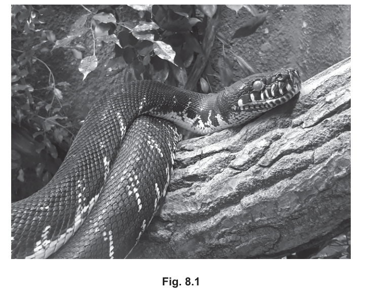
The International Union for Conservation of Nature (IUCN) has not assessed the conservation status of S. boeleni because the python is very hard to detect and locate.
S. boeleni is listed in one of the appendices of the Convention on International Trade in Endangered Species of Wild Fauna and Flora (CITES).
Suggest the advantages of S. boeleni being listed in CITES, even though it does not have an IUCN conservation status.
Answer
- CITES prevents illegal international trade / poaching / hunting of S. boeleni.
- Any international trade in S. boeleni requires permits, so trade is regulated and monitored.
- Listing in CITES raises awareness / educates the public and governments that S. boeleni is a protected species.
Prevents illegal trade, regulates trade via permits, and raises awareness/education.
Background Concept
CITES (Convention on International Trade in Endangered Species of Wild Fauna and Flora) is an international agreement between governments. Its aim is to ensure that international trade in specimens of wild animals and plants does not threaten their survival. Species are placed in one of three CITES Appendices depending on how threatened they are by trade:
- Appendix I – trade is prohibited (species threatened with extinction).
- Appendix II – trade is permitted only with permits/certificates.
- Appendix III – trade is regulated in cooperating countries.
The IUCN Red List is a separate assessment of a species' conservation status (e.g. Critically Endangered, Vulnerable), based on population data. The IUCN has not assessed Simalia boeleni because the python is very hard to detect and locate, so the population cannot be evaluated properly. A lack of an IUCN Red List status, however, does not mean the species is safe — and this is precisely where CITES becomes useful.
Understanding the Question
The question asks the student to suggest advantages of S. boeleni being listed in a CITES appendix, given that the IUCN has not assessed it. The command word "suggest" means the answer needs reasoned points, not pure recall — but the reasoning is built directly on what CITES does. Three marks are available, so three distinct advantages are expected.
Approach
Think about the practical effects of being on a CITES list. The list does not protect the species directly on the ground (that is the role of national parks, anti-poaching units, etc.) — instead, it controls what happens when animals or their parts cross international borders. The advantages therefore concern:
- the regulation of trade itself,
- the deterrent effect on illegal trade,
- the wider knock-on benefits (awareness, monitoring, research funding).
Step-by-Step Reasoning
-
Prevents illegal trade / poaching / hunting — When a species is in any CITES appendix, customs officers in signatory countries can intercept shipments and check for permits. This makes smuggling the animal (or skins, body parts, etc.) across borders far harder, which directly reduces the incentive to poach it.
-
Trade is regulated — permits required — Legal, documented trade is only possible if the exporter and importer can show that the specimen was obtained sustainably and that the trade is not detrimental to the species. Permits therefore channel trade through a controlled, recorded route, and the data gathered can be used to monitor population pressures.
-
Raises awareness / education — Simply being listed gives the species a public profile. Conservation NGOs, customs officers, scientists, and local people become aware that the species is protected. This often leads to further research, funding, and education campaigns, all of which benefit the species.
A useful extra point the examiner may accept: even without an IUCN Red List assessment, CITES still triggers a level of protection until proper data can be gathered, so the species is not unprotected in the meantime.
Key Takeaways
- CITES and the IUCN are different tools: IUCN assesses status, CITES controls trade.
- A species can be protected by CITES even when its IUCN status is "Not Assessed".
- The core benefits of CITES listing are: regulation of trade via permits, deterrence of illegal trade, and raised public/scientific awareness.
Common Mistakes
- Writing about IUCN functions (assessing population size, classifying as Endangered, etc.) rather than CITES functions. The question is specifically about the advantage of being in CITES.
- Saying that CITES "saves the species" or "stops extinction" — CITES only controls trade; it does not directly prevent extinction.
- Repeating the same idea in different words (e.g. "prevents poaching" and "stops illegal hunting") rather than offering three distinct advantages. The mark scheme explicitly lists three different ideas.
- Suggesting that CITES protects habitat — it does not; it only regulates trade in specimens.
Things to Be Careful About
- The marking point says "prevents illegal trading/poaching/hunting"; the word illegal is key — CITES does not stop all trade, just illegal trade.
- "Permits" is a precise term worth using; "licences" is acceptable but the more general "rules" is too vague.
- Three marks require three separate points. Do not bundle two advantages into one sentence.
S. boeleni is a member of the kingdom Animalia.
Outline the characteristic features of the kingdom Animalia.
Answer
Any four from:
- Multicellular.
- Eukaryotic — cells contain a nucleus (and membrane-bound organelles).
- Cells are specialised, forming tissues, organs and organ systems.
- Heterotrophic nutrition (obtain food by ingesting / consuming other organisms).
- Possess a nervous system.
- Some cells possess cilia or flagella (e.g. sperm cells).
- Motile / capable of locomotion (at least at some stage of the life cycle).
Multicellular eukaryotes with specialised cells/tissues/organs, heterotrophic nutrition, a nervous system, some cilia/flagella, and motility.
Background Concept
Living organisms are classified into five kingdoms in the traditional five-kingdom system used by CIE Biology: Prokaryotae (Monera), Protoctista, Fungi, Plantae and Animalia. Each kingdom is defined by a suite of features, not just one. The features of Kingdom Animalia can be grouped into three themes:
- Cell-level features — eukaryotic cells, usually lacking a cell wall and chloroplasts, so they cannot photosynthesise. Cells may carry cilia or flagella at some life stage.
- Body-plan features — multicellular bodies built from specialised cells organised into tissues → organs → organ systems, allowing division of labour.
- Lifestyle features — heterotrophic (consume other organisms for food), motile (can move from place to place at some point in the life cycle), and possessing a nervous system to coordinate responses to stimuli.
Understanding the Question
The question asks the student to outline the characteristic features of the kingdom Animalia. "Outline" means give the main points in a summary form — a short bulleted list is ideal. Four marks are available, so four distinct features should be given. The mark scheme accepts any four of seven possible points, so the student can choose the four they know best.
Approach
Work through the characteristic features of animals in a logical order: cell structure → body organisation → nutrition → coordination → movement. Pick any four; do not waste time trying to cover all seven.
Step-by-Step Reasoning
-
Multicellular — all animals are made of many cells (unlike Protoctists, which include many unicellular forms, and Prokaryotes, which are all unicellular).
-
Eukaryotic / cells contain a nucleus — animal cells have membrane-bound organelles including a true nucleus containing linear chromosomes. This separates them from Prokaryotae.
-
Specialised cells / tissues / organs — animal cells differentiate to perform specific roles (e.g. nerve cells conduct impulses, muscle cells contract), and these are arranged into tissues (e.g. muscle tissue), then organs (e.g. the heart), then organ systems (e.g. the circulatory system). This is a feature shared with the more complex plants and most multicellular fungi, but combined with the other features it is diagnostic for Animalia.
-
Heterotrophic nutrition — animals cannot make their own food; they ingest other organisms (or organic matter) and digest it internally. This contrasts with plants (autotrophic, photosynthetic) and fungi (saprotrophic, absorbing digested food externally).
-
Nervous system — a defining feature of animals: a network of neurones that detect stimuli, transmit impulses, and coordinate responses (including movement). Plants and fungi do not have a true nervous system.
-
Cilia / flagella (in some cells) — many animal cells (e.g. spermatozoa, ciliated epithelium of the trachea) have cilia or flagella made of microtubules in a 9+2 arrangement. This is not true of all animal cells but is a diagnostic feature of the kingdom.
-
Motile / mobile — most animals can move from place to place at some stage in their life cycle, and most are motile as adults (larvae especially are often free-swimming). Sessile adult forms such as sponges and some molluscs are exceptions, so the mark scheme allows "at some life stage".
The mark scheme accepts any four of these, so a candidate who confidently remembers "multicellular, eukaryotic, heterotrophic, nervous system" will score full marks.
Key Takeaways
- Kingdom Animalia is defined by a combination of features, not just one.
- The classic combination to remember is: multicellular, eukaryotic, heterotrophic, with a nervous system and motility.
- The features distinguish Animalia from the other four kingdoms: from Prokaryotae (prokaryotic), from Protoctista (often unicellular, no true tissues), from Fungi (saprotrophic, no nervous system, usually non-motile), and from Plantae (autotrophic, cell walls, sessile).
Common Mistakes
- Saying "autotrophic" instead of "heterotrophic" — animals are consumers, not producers.
- Confusing multicellular with eukaryotic — these are separate marks. Unicellular eukaryotes (e.g. Amoeba) are not animals.
- Writing "have a backbone" or "have four limbs" — these are features of specific animal groups (vertebrates, tetrapods), not of the whole kingdom. The mark scheme would reject this.
- Writing "cells have a cell wall" — animal cells do not.
- Omitting the nervous system — this is one of the strongest distinguishing features and should be included if known.
Things to Be Careful About
- "Motile" is the precise term; "can move" is acceptable but "move by themselves" risks being too vague. The mark scheme also accepts "locomotion".
- "Cilia/flagella" should be qualified with "some cells" — not all animal cells have them. Saying "all animal cells have cilia" would be wrong.
- "Heterotrophic" can be described ("feed on other organisms") if the technical word is forgotten, but the precise term is preferred.
- Four marks = four distinct points. Do not pad one point with extra words; write a short list of four clear features.
Most carnivorous mammals need to move to hunt their prey.
Outline why a carnivorous mammal makes more use of its nervous system, rather than its endocrine system, when it hunts.
Answer
- (Named) sense organs, e.g. eyes/ears, detect the prey.
- The nervous system gives faster transmission of impulses (than hormones travel in the blood).
- This produces a faster reaction to stimuli.
- The brain / CNS acts as a control centre, making decisions rapidly.
- The response is faster, e.g. via a reflex arc.
- The effectors (muscles) contract rapidly to capture the prey.
Sense organs detect prey; nervous transmission is faster than hormonal, giving faster reactions, faster CNS decision-making and faster (reflex) responses by muscles — ideal for hunting.
Background Concept
Animals have two coordination systems. The nervous system uses neurones to transmit electrical impulses, and the endocrine system uses hormones carried in the blood. Their properties differ sharply:
- Nervous: very fast (impulses travel at up to ~120 m s⁻¹ along myelinated axons); short-lived; acts on precise, localised targets; suited to rapid responses to a changing environment.
- Endocrine: slow (hormones may take seconds to minutes to arrive); long-lasting; acts on widespread targets; suited to slower, sustained regulation such as growth, metabolism, water balance and reproduction.
A carnivorous mammal hunting depends on split-second timing: detecting moving prey, deciding whether to pursue, and triggering coordinated muscle contractions to chase, pounce and subdue it.
Understanding the Question
The question uses the verb outline — the candidate should give the main features of an explanation, here in the context of hunting. The marks scheme offers six valid points, of which any four will score full marks. The keyword faster appears three times, highlighting that speed is the central contrast.
The student must therefore contrast the speed of the two systems AND show why that speed is critical for hunting.
Approach
The best answers thread the comparison through the whole reflex/response pathway:
- Detection — sense organs (receptors) need to pick up the prey.
- Transmission — impulses travel much faster than hormones in the blood.
- Decision / coordination — the CNS integrates the information quickly.
- Response — a rapid (often reflex) response, executed by muscles (effectors).
These are the same four steps a reflex arc covers, so structuring the answer around the reflex pathway gives a clean, mark-rich response.
Step-by-Step Reasoning
- Receptors in the eyes and ears (and nose) detect the sight, sound or scent of prey. The mark scheme allows any named sense organ.
- Nervous transmission is faster than hormonal — electrical impulses along axons outpace any hormone moving in the bloodstream. This is the key comparison.
- A faster reaction to stimuli follows automatically from faster transmission.
- The CNS / brain acts as the control centre, making decisions about whether to pursue or attack.
- The faster response (often via a reflex arc) closes the loop; reflexes are particularly important because the response can occur before conscious awareness.
- Muscles are the effectors that actually move the mammal towards the prey, or the jaws/claws to capture it.
A top-scoring answer mentions at least four of the six and emphasises speed in at least three of them.
Key Takeaways
- Nervous system = fast, precise, short-lived; endocrine = slow, widespread, long-lasting.
- The reflex arc (receptor → sensory neurone → CNS → motor neurone → effector) is the prototype of a fast nervous response.
- Always link the comparison to the biological context the question specifies — here, hunting requires rapid, well-timed movements.
Common Mistakes
- Vague wording: "the nervous system is faster" without saying what is faster (transmission of impulses, reaction, response) — credit is given only when what is fast is named.
- Confusing stimuli and responses: a stimulus is detected; a response is made.
- Writing that the endocrine system is "not used at all" — it still performs slower background roles (e.g. adrenaline), but is not the main one for hunting movements.
- Forgetting to name the effectors (muscles).
Things to Be Careful About
- Bold-marked words in the mark scheme (faster) must appear in the answer.
- The sense organ does not have to be specifically the eye/ear/nose — any named sense organ is acceptable.
- A reflex is a specific type of response, not a separate pathway; mention it where the mark scheme allows it as an alternative to "faster response".
Fig. 9.1 is a diagram of a motor neurone.
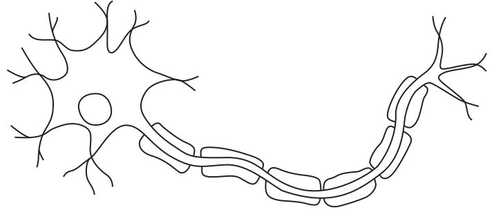
On Fig. 9.1, add label lines and the letters R, S and T to label a part of the neurone that:
- can become depolarised – use the letter R
- contains many mitochondria – use the letter S
- acts as an insulator – use the letter T.
Answer
- R — placed on a dendrite (or the cell body, or the axon plasma membrane): the membrane of these regions can become depolarised.
- S — placed on a synaptic terminal (axon ending): this region contains many mitochondria to provide ATP for synthesising neurotransmitter and for active transport.
- T — placed on a myelin sheath (Schwann cell): the myelin acts as an electrical insulator around the axon.
R on dendrites/cell body/axon membrane; S on synaptic terminals; T on the myelin sheath.
Background Concept
A motor neurone has four recognisable regions, each with a distinct structure and function:
- Dendrites — branched extensions from the cell body that receive impulses from other neurones. Their membranes carry ligand-gated and voltage-gated channels and can depolarise.
- Cell body (soma) — contains the nucleus and a high density of organelles, including many mitochondria (to supply ATP for protein synthesis and active transport) and Nissl bodies (rough ER).
- Axon — a single long process that conducts the action potential. Its plasma membrane is excitable (it has many voltage-gated Na⁺/K⁺ channels) and can depolarise.
- Myelin sheath — formed by Schwann cells wrapping around the axon in vertebrates. The lipid-rich myelin acts as an electrical insulator, increasing the speed of conduction. Gaps between adjacent Schwann cells are the nodes of Ranvier, where the membrane is exposed and the action potential is regenerated (saltatory conduction).
- Synaptic terminals (axon terminals / synaptic knobs) — the swollen ends of the axon. They are packed with synaptic vesicles and large numbers of mitochondria to power the ATP-requiring steps of neurotransmitter synthesis, vesicle recycling and the Na⁺/K⁺-ATPase that maintains the membrane potential.
Understanding the Question
The candidate is given a printed drawing of a motor neurone (Fig. 9.1) and must add three label lines terminating in the letters R, S and T. Each letter must point to a region whose function matches the description in the bullet list. The mark scheme adds the warning: if a letter is used more than once, all instances must be correct. This means a single letter can legitimately point to two regions (e.g. S used at both ends of the neurone), provided each chosen region is correct.
Approach
Read each description and identify the function the structure must perform:
| Description | Function | Where on the neurone? |
|---|---|---|
| can become depolarised | excitable membrane (voltage-gated ion channels) | dendrites, cell body, axon |
| contains many mitochondria | high ATP demand | synaptic terminals, cell body |
| acts as an insulator | electrically isolates the axon | myelin sheath (Schwann cell) |
Then place the corresponding label on the diagram.
Step-by-Step Reasoning
- R = can become depolarised. The plasma membrane of the dendrites, cell body and axon all contain voltage-gated Na⁺ channels and can generate an action potential. The myelin sheath itself cannot depolarise (it is a Schwann-cell wrapping), so do not place R there. The marking scheme's image shows R used twice — on a dendrite and on the axon — both of which are correct.
- S = contains many mitochondria. The synaptic terminals are the busiest ATP-consuming region of the neurone (vesicle recycling, Na⁺/K⁺-ATPase, transmitter synthesis), so they are densely packed with mitochondria. The cell body also has many mitochondria, so S can be placed there too.
- T = acts as an insulator. The myelin sheath, formed by Schwann cells, is the only structure on the diagram that fits. Do not place T on a node of Ranvier — the node is precisely the gap in the insulation.
Key Takeaways
- Structure–function matching is core to A-level biology: always know which membrane property or organelle fits the function asked for.
- Dendrites, cell body and axon are all excitable; only the myelin sheath is not.
- Synaptic terminals and cell body are the regions of highest mitochondrial density.
- The mark scheme permits using the same letter twice only if every placement of that letter is correct.
Common Mistakes
- Placing R on the myelin sheath — myelin does not depolarise, so this scores zero.
- Placing T on a node of Ranvier — the node is the un-insulated gap, the opposite of what the question asks.
- Confusing dendrites (receive) with axon (transmit); both are excitable, but only the axon has myelin.
- Drawing the label line through another structure so it is unclear which region is being indicated.
Things to Be Careful About
- The label line must end on the structure, with no ambiguity about which region is intended.
- If the same letter is used twice, both placements must be correct (a single wrong placement costs the mark for that letter entirely).
- The Schwann cell nucleus sits outside the myelin wrapping — placing the label there is acceptable as a way of indicating the myelin sheath, but the line should still clearly point to the myelin / Schwann-cell wrapping rather than to the bare axon between segments.
Fig. 9.2 summarises changes that occur during the contraction of a sarcomere.
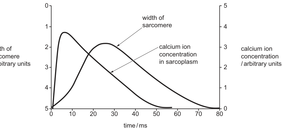
Suggest an explanation for the shape of the curve that shows changes in the width of the sarcomere.
Answer
Where the curve rises (curve goes up, sarcomere width decreases — contraction, 0 to ~25 ms):
- Cross bridges form: myosin heads bind to actin.
- The power stroke occurs, so actin filaments slide over myosin filaments.
- The sarcomere gets shorter (width decreases).
Where the curve falls (curve goes down, sarcomere width increases — relaxation, ~25 ms to 80 ms):
- Cross bridges break: myosin heads detach from actin.
- The sarcomere gets longer (width increases).
Cross-bridge formation and the power stroke shorten the sarcomere (contraction phase); cross-bridge breakage lengthens the sarcomere again (relaxation phase).
Background Concept
The sliding filament model explains how a sarcomere shortens during muscle contraction:
- A sarcomere is the unit between two Z-lines and contains thin (actin) filaments anchored at the Z-lines and thick (myosin) filaments lying in the middle. Pulling the actin filaments inwards along the myosin shortens the sarcomere; the filaments themselves do not contract.
- Myosin heads are ATPases that bind to actin, perform a power stroke, then detach. The cycle needs ATP for detachment and for re-cocking the head.
- Troponin and tropomyosin regulate the binding sites on actin: when Ca²⁺ binds to troponin, tropomyosin moves out of the binding groove and myosin heads can attach.
- The sarcoplasmic reticulum (SR) is a modified endoplasmic reticulum that stores Ca²⁺ at high concentration. An action potential in the sarcolemma / T-tubule causes Ca²⁺ to be released into the sarcoplasm, triggering contraction; relaxation occurs when Ca²⁺ is pumped back into the SR.
Understanding the Question
Fig. 9.2 is a graph with two curves sharing a time axis. The width-of-sarcomere axis is inverted (0 at the top, 5 at the bottom). This means:
- Curve visually moving up = width value decreasing from ~5 toward ~2 = the sarcomere is getting shorter (contraction).
- Curve visually moving down = width value increasing back toward 5 = the sarcomere is getting longer (relaxation).
The mark scheme labels the two phases "increase in curve" (visual rise = contraction) and "decrease in curve" (visual fall = relaxation). The candidate must explain both phases.
Approach
- Identify which phase of the curve corresponds to contraction and which to relaxation, using the inverted axis.
- For each phase, write the molecular events that produce the change in sarcomere width.
- Pair the change in width (observation) with the molecular cause (explanation) — both are required for full marks.
Step-by-Step Reasoning
Phase 1 — curve rises (0 → ~25 ms); sarcomere width DECREASES (contraction).
- Ca²⁺ (released earlier — see part ii) has bound to troponin, exposing the myosin-binding sites on actin.
- Cross bridges form as myosin heads bind to actin.
- The power stroke pulls the actin filaments towards the centre of the sarcomere; actin filaments slide over the myosin filaments.
- The I-band and H-zone narrow and the sarcomere width decreases — exactly what the curve shows as it rises from 5 toward 2.
Phase 2 — curve falls (~25 ms → 80 ms); sarcomere width INCREASES (relaxation).
- Ca²⁺ has been pumped back into the SR (see part ii), so tropomyosin once again blocks the myosin-binding sites on actin.
- Cross bridges break as myosin heads detach from actin (this step also requires ATP binding to myosin).
- With no further pulling force, the sarcomere is pulled back to its resting length by elastic elements in the muscle (e.g. titin) and by opposing muscles / antagonists.
- The sarcomere width increases back toward 5 — exactly what the curve shows as it falls from 2 toward 5.
Key Takeaways
- The sliding filament model does not involve the filaments themselves contracting — they slide past one another.
- Cross-bridge formation drives contraction; cross-bridge breakage allows relaxation.
- A graph with an inverted y-axis is common in Cambridge papers — always read the axis label and the order of numbers before interpreting the curve's direction.
- "Suggest an explanation" still demands a molecular explanation, not just a description of the curve's shape.
Common Mistakes
- Confusing the direction of the curve with the direction of the change in width because the y-axis is inverted.
- Naming only "myosin and actin" without describing the cross-bridge cycle.
- Saying "the muscle contracts" without specifying that the sarcomere is the unit that shortens.
- Forgetting to explain the relaxation phase (the rising part of the curve after the minimum) — both halves are required.
Things to Be Careful About
- Read both axes (left and right) and the units before describing what the curve is doing.
- The mark scheme offers alternatives: "power stroke" OR "actin filaments slide over myosin filaments" — either is acceptable; pairing it with "cross bridges form" gives the strongest answer.
- The widening of the sarcomere is passive (elastic recoil / antagonist muscles) once cross-bridge cycling stops — say so if asked for completeness, but it is not required here.
Answer
Where the Ca²⁺ curve rises (0 → ~10 ms):
- Calcium ions are released from the sarcoplasmic reticulum into the sarcoplasm.
- Calcium ions bind to troponin.
Where the Ca²⁺ curve falls (~10 → 50 ms):
- Calcium ions are pumped back into the sarcoplasmic reticulum (by active transport).
Ca²⁺ is released from the sarcoplasmic reticulum and binds troponin (rise); Ca²⁺ is then pumped back into the SR (fall).
Background Concept
Calcium ions are the trigger that switches a sarcomere from the relaxed to the contracted state:
- The sarcoplasmic reticulum (SR) is a specialised endoplasmic reticulum surrounding each myofibril. It actively pumps Ca²⁺ into its lumen, so the resting sarcoplasmic [Ca²⁺] is very low.
- When an action potential travels along the sarcolemma and into the T-tubules, voltage-sensitive DHP receptors in the T-tubule membrane open ryanodine receptors on the SR membrane, releasing Ca²⁺ into the sarcoplasm (a process called excitation–contention coupling).
- Ca²⁺ binds to troponin, which moves tropomyosin away from the myosin-binding sites on actin, allowing cross-bridge formation and contraction.
- Relaxation requires Ca²⁺ to be removed from the sarcoplasm. The SERCA pump (a Ca²⁺-ATPase) actively transports Ca²⁺ back into the SR lumen, using ATP. As sarcoplasmic [Ca²⁺] falls, Ca²⁺ dissociates from troponin, tropomyosin blocks the binding sites again, and the sarcomere relaxes.
Understanding the Question
The Ca²⁺ curve on the right-hand axis of Fig. 9.2 shows a fast rise (peak around 10 ms) followed by a slower fall (back to baseline by ~50 ms). The candidate must suggest an explanation for both the rise and the fall. The mark scheme gives three creditable points: release from SR, binding to troponin (both on the rising phase), and reuptake into SR (falling phase).
Approach
- Rising phase (0 → ~10 ms): Ca²⁺ enters the sarcoplasm — this requires (i) a source (the SR) and (ii) something for it to do once it is there (bind to troponin).
- Falling phase (~10 → ~50 ms): Ca²⁺ leaves the sarcoplasm — it is pumped back into the SR by active transport.
Step-by-Step Reasoning
Why the curve rises.
- The action potential in the sarcolemma / T-tubule triggers the SR to release stored Ca²⁺ into the sarcoplasm through ryanodine receptors. Sarcoplasmic [Ca²⁺] therefore rises rapidly.
- The released Ca²⁺ binds to troponin on the thin filament. This is the key molecular consequence of the rise in free Ca²⁺ — it explains why the rise in Ca²⁺ is followed (with a small lag) by the contraction seen in part (i).
Why the curve falls.
- Ca²⁺ is removed from the sarcoplasm by being actively transported back into the lumen of the SR by SERCA pumps (Ca²⁺-ATPases), using ATP.
- As sarcoplasmic [Ca²⁺] falls, Ca²⁺ dissociates from troponin; tropomyosin re-covers the myosin-binding sites on actin; cross-bridge cycling stops and the sarcomere relaxes (the widening seen in part i).
Why the fall is slower than the rise.
- Release from the SR is via opening of ion channels — fast, passive movement down the steep concentration gradient between SR lumen and sarcoplasm.
- Reuptake is via ATP-driven pumps — slower, because each pump cycle takes a finite time and the rate is limited by the number of pumps and the available ATP.
This explains the asymmetric shape of the Ca²⁺ curve (sharp rise, gentler fall).
Key Takeaways
- The SR is the source (release) and the sink (reuptake) for sarcoplasmic Ca²⁺.
- Ca²⁺ binding to troponin is the molecular link between the electrical signal and the mechanical contraction.
- Reuptake is active, so it requires ATP and is intrinsically slower than release.
- The lag between the Ca²⁺ peak (~10 ms) and the minimum sarcomere width (~25 ms) reflects the time taken for cross-bridge cycling to pull the filaments together.
Common Mistakes
- Saying Ca²⁺ "comes from the blood" or "from the extracellular fluid" — at this scale, the Ca²⁺ is exclusively from the SR.
- Saying "Ca²⁺ is used up" or "broken down" — Ca²⁺ is an ion, not a metabolite; it is recycled, not consumed.
- Forgetting to mention troponin (or naming tropomyosin instead) — the mark scheme requires troponin specifically.
- Saying reuptake is by diffusion — it is by active transport and requires ATP.
- Confusing the SR with the smooth endoplasmic reticulum of other cell types — in muscle the SR is a highly specialised, Ca²⁺-storing organelle wrapped around each myofibril.
Things to Be Careful About
- The mark scheme allows either "calcium ions" or the symbol Ca²⁺ — using the symbol is cleaner and is preferred at A-level.
- Always state direction and destination when describing reuptake (Ca²⁺ is pumped back into the SR).
- Notice the temporal sequence in the figure: Ca²⁺ peak → sarcomere minimum → Ca²⁺ baseline → sarcomere baseline. The biological order (Ca²⁺ rise causes contraction; Ca²⁺ fall allows relaxation) is preserved, with the contraction/relaxation lagging the Ca²⁺ change by a few ms.
The golden poison dart frog, Phyllobates terribilis, lives in the Colombian rainforest ecosystem.
Fig. 10.1 shows golden poison dart frogs.
Answer
An ecosystem is a self-contained unit consisting of a community of organisms (biotic factors) interacting with each other and with the abiotic (non-living) factors of their environment, with flow of energy and cycling of nutrients / minerals through it.
A self-contained unit comprising a community of organisms interacting with each other and with the abiotic factors of their environment, with flow of energy and cycling of nutrients.
Background Concept
An ecosystem is one of the most fundamental concepts in ecology. It describes a unit of the living world that contains both living and non-living components and the interactions between them.
The biotic component includes all the living organisms — plants, animals, fungi, bacteria — that form a community of several different species living in the same area at the same time.
The abiotic component includes the non-living features of the environment: temperature, light intensity, water availability, soil pH, mineral ions, wind, etc. These determine which organisms can survive in a particular place.
A key feature of an ecosystem is that the biotic and abiotic components interact: organisms are affected by their environment and, in turn, change it (e.g. plants take up mineral ions, decomposers return nutrients to the soil). These interactions drive the flow of energy through feeding relationships and the cycling of nutrients (e.g. the carbon cycle, the nitrogen cycle).
The mark scheme awards credit for any three of the following:
- A self-contained / discrete unit
- A community of organisms (or the idea of several species)
- Biotic and abiotic factors (a named example is acceptable)
- Reference to interaction
- A valid additional point — most commonly the flow of energy and cycling of minerals/nutrients
Understanding the Question
This is a 'define' question worth 3 marks, so the answer needs to be precise and cover several distinct points. The term to define is 'ecosystem' — not 'habitat' (which is just the place where an organism lives) and not 'community' (which only refers to the living organisms, not the abiotic environment).
The golden poison dart frog, Phyllobates terribilis, is given as the example: it lives in the Colombian rainforest alongside other organisms and the rainforest's abiotic environment (rain, soil, temperature, etc.), and interacts with all of them. The photograph in Fig. 10.1 simply provides visual context — the marks are awarded for the definition itself.
Approach
Combine the required marking points into a single, well-structured definition:
- Start with what the unit is (a self-contained unit)
- State what it contains (a community of organisms + abiotic factors)
- Say what the components do (interact)
- Add a relevant further point about energy flow / nutrient cycling — this is the classic A-level addition that frequently earns the third mark
Step-by-Step Reasoning
The five (or more) marking points the candidate can include are:
-
Self-contained unit — an ecosystem is, by definition, a unit that can be studied as a system. The rainforest pond, a single rotting log, and the entire Colombian rainforest can each be called ecosystems because each is a self-contained system in terms of its energy flow and matter cycles.
-
Community of organisms — the biotic part: many species of plants, animals, decomposers, etc. all living together. A community always involves more than one species (the mark scheme accepts this phrasing).
-
Biotic and abiotic factors (or a named example) — the non-living part: e.g. light, temperature, water, soil pH, mineral ions, humidity. The mark scheme says 'named' is acceptable — so a single example such as 'water' or 'temperature' is fine.
-
Reference to interaction — the living organisms interact with each other (e.g. predator–prey, competition) and with the abiotic environment (e.g. plants using light, animals drinking water, soil organisms respiring).
-
Additional valid point (AVP) — typically the flow of energy (e.g. through feeding relationships) and the cycling of nutrients/minerals (e.g. carbon, nitrogen).
A full, 3-mark definition that covers all the key points reads:
'An ecosystem is a self-contained unit comprising a community of organisms (biotic factors) interacting with each other and with the abiotic factors of their environment, with flow of energy and cycling of nutrients/minerals.'
Key Takeaways
- An ecosystem = a community of organisms + the abiotic environment + their interactions.
- The two classic additions that lift a definition from 2 to 3 marks are: (a) the idea of an ecosystem as a self-contained unit, and (b) the flow of energy / cycling of nutrients.
- Do not confuse 'ecosystem' (community + environment + interactions) with 'habitat' (just the place an organism lives) or 'community' (just the organisms).
Common Mistakes
- Defining 'ecosystem' as simply 'a community of organisms' or 'a habitat' — this misses the abiotic component and the interaction / energy-flow ideas that the mark scheme requires.
- Writing 'an ecosystem is a place where organisms live' — this is a definition of habitat, not ecosystem.
- Omitting the self-contained / discrete-unit idea.
- Failing to include at least one example of a biotic OR abiotic component to anchor the abstract definition.
Things to Be Careful About
- The command word is 'define', not 'state' or 'describe'. A 'define' answer should be a single, clear, paragraph-style definition (or a tight list) that names the term and captures all the required ideas. Avoid padding with examples that do not score extra marks.
- Make sure at least one marking point mentions both biotic AND abiotic (or gives named examples of each).
- The 'AVP' mark can be earned by including the flow of energy and cycling of minerals — this is the most credit-worthy extra point.
One way of estimating the size of a population of golden poison dart frogs is to use the mark-release-recapture method.
Suggest the assumptions that must be made for the mark-release-recapture method to be valid.
Answer
Any three from:
- The frogs are mobile (so they can move around freely and re-mix with the rest of the population after release).
- The marking method is not harmful to the frogs and the marks cannot be removed / fall off (so marked frogs behave normally and are still recognisable as marked on recapture).
- Sufficient time is allowed for the marked individuals to mix randomly back into the rest of the population before the second sample is taken.
- There are no births or deaths, and no immigration or emigration, so the population size is constant between the two samples.
Assumptions: (1) the frogs are mobile, (2) the marking is not harmful and the marks stay on, (3) the marked frogs have had time to mix with the rest of the population, and (4) the population size is constant (no births, deaths, immigration or emigration).
Background Concept
The mark-release-recapture method is a standard ecological technique for estimating the size of an animal population that is too large or too mobile to count directly. The procedure is:
- Capture a first sample of individuals, mark them in a way that does not harm them, and release them back into the population.
- After sufficient time, capture a second sample of individuals and count how many of them () already carry marks.
- Apply the Lincoln index (Petersen estimate):
The underlying logic is that the proportion of marked individuals in the second sample should equal the proportion of marked individuals in the whole population:
which rearranges to the formula above. For this equality to give a valid estimate, several assumptions must be true. The mark scheme lists four such assumptions; the candidate needs to give any three for full marks.
Understanding the Question
The question is specifically about the mark-release-recapture method being used to estimate the size of a population of golden poison dart frogs in the Colombian rainforest. The command word is 'suggest', so the candidate is expected to think through what conditions must hold for the method to work, rather than recall a list verbatim.
The marks reward candidates who identify three distinct assumptions. The four credit-worthy answers are:
- Mobility: frogs move around freely, so that a marked frog released at one point can later be captured at any other point in the same area.
- Marking has no effect / is durable: the mark is not harmful (does not increase the frog's chance of being caught by a predator, does not affect its behaviour) and the mark does not fall off or wear away.
- Mixing: there has been enough time between the release of the marked frogs and the second sample for the marked individuals to redistribute themselves randomly among the rest of the population.
- Closed population: the population size is constant — no births, no deaths, no immigration of new frogs into the area, and no emigration of frogs out of the area, between the two samples.
Approach
The Lincoln index rests on a single equality: the fraction of marked animals in the second sample equals the fraction of marked animals in the whole population. A useful way to generate the assumptions is therefore to ask, 'What could break this equality?'
- If the frogs are not mobile, the marked individuals will all stay in one place and the second sample will be biased.
- If the marking is harmful or the marks fall off, the marked individuals will not be recaptured in the expected proportion.
- If there has not been time for mixing, the second sample will not represent the population as a whole.
- If the population is not closed (births, deaths, immigration, emigration), the second sample will not represent the population as it was when the first sample was taken.
Step-by-Step Reasoning
Marking point 1 — mobility
The golden poison dart frog is a small, active amphibian. For the method to work, marked frogs released into the rainforest must be able to move freely around the study area so that they can later be captured at random in the second sample. If the frogs were sessile (e.g. a barnacle) or had very small home ranges, the marked individuals would cluster in the area where they were released and the second sample would catch an unrepresentative number of them. The mark scheme credits the simple statement that '(frogs are) mobile'.
Marking point 2 — marking has no effect / mark is durable
For the method to work, the mark must remain on the frog and must not change its behaviour, its chance of being caught, or its chance of survival. If marking causes the frog to behave abnormally (e.g. hide more), it will be under-represented in the second sample. If marks fall off, recaptured 'marked' frogs will appear to be unmarked. The mark scheme credits 'marking, not harmful / cannot be removed'.
Marking point 3 — time for mixing
After the first sample is released, the marked frogs must have time to redistribute themselves among the rest of the population. If the second sample is taken too soon, the marked individuals will still be clustered in the area where they were released and the ratio of marked to unmarked frogs in the second sample will not reflect the true ratio in the whole population. The mark scheme credits '(sufficient time for) marked individuals to mix with rest of population'.
Marking point 4 — closed / constant population
The Lincoln index assumes that the population size is the same at the time of the first capture and at the time of the second capture. Any change in (through births, deaths, immigration, or emigration) will bias the estimate. The mark scheme accepts any of: 'no births/deaths', 'no immigration/emigration', or 'constant population size'.
Key Takeaways
- The Lincoln index rests on the assumption that the proportion of marked animals in the second sample equals the proportion of marked animals in the whole population.
- Any of the following would invalidate the estimate: lack of mobility, harmful or removable marks, insufficient time for mixing, births, deaths, immigration, or emigration.
- For a population of mobile animals, the most important practical assumptions to verify are that the marks are durable and that enough time has been allowed for mixing.
Common Mistakes
- Vague answers such as 'the method is accurate' or 'the frogs are healthy' do not score: the mark scheme wants specific assumptions about mobility, marking, mixing, and population constancy.
- Listing 'no migration' only — the mark scheme wants 'no immigration AND no emigration' (or the equivalent statement that the population is constant).
- Confusing 'mark-release-recapture' assumptions with those of quadrat-based plant sampling (e.g. suggesting 'random sampling', which is a feature of good experimental design but not one of the mark-scheme-listed assumptions for this method).
- Repeating the same idea in different words — only scores once.
Things to Be Careful About
- The command word is 'suggest', so the candidate is expected to reason out the assumptions rather than recall a list. The mark scheme nevertheless provides a fixed list, so phrasing each assumption clearly (e.g. 'the marks do not fall off') is what earns the credit.
- Three distinct assumptions are required for the three marks — repeating the same idea in different words only scores once.
- 'No immigration AND no emigration' is required — the mark scheme rejects 'no migration' on its own.
A first sample of 27 golden poison dart frogs was captured, marked and released. When a second sample of 33 frogs was captured, 13 had marks on them.
Use the Lincoln index to estimate the population size of the frogs.
= number of individuals captured in first sample
= number of individuals (both marked and unmarked) captured in second sample
= number of marked individuals recaptured in second sample
answer ______
Working
Given:
frogs (first sample, marked and released)
frogs (second sample)
marked frogs recaptured
The estimate must be expressed as a whole number.
Answer
69
69
Background Concept
The Lincoln index (also called the Petersen estimate) is the formula that converts a mark-release-recapture experiment into a numerical estimate of the population size . It assumes that the proportion of marked individuals in the second sample equals the proportion of marked individuals in the whole population:
Rearranging for :
where:
- = number of individuals captured, marked and released in the first sample
- = number of individuals (both marked and unmarked) captured in the second sample
- = number of marked individuals recaptured in the second sample
Because estimates the number of individual animals in a population, the result must be a whole number. If the calculation gives a non-integer value (which it almost always does), it is rounded to the nearest whole number.
Understanding the Question
The question supplies all the necessary data and the formula itself. The candidate only has to substitute the values and round to a whole number:
- (first sample size)
- (second sample size)
- (marked individuals recaptured)
The question is worth 2 marks: one for the correct working (substitution) and one for the correct final answer. The mark scheme is unusually strict here: it states that the answer 'must be a whole number, one mark only if not whole number'. A correct calculation that gives 68.5 or 68.54 instead of 69 therefore scores only 1 of the 2 marks.
Approach
Substitute the three values into the formula, evaluate the numerator first (this gives a clear, easy-to-mark number), divide by the denominator, and round to the nearest whole number.
Step-by-Step Reasoning
Step 1 — Substitute the values into the formula
Step 2 — Calculate the numerator
So:
Step 3 — Divide
Step 4 — Round to a whole number
The unrounded value is , which rounds to (using the standard 'round half up' rule). The mark scheme explicitly states: 'must be a whole number, one mark only if not whole number'. The candidate therefore loses one mark if they write 68.5 or 68.54, even though the calculation is correct.
Step 5 — Final answer
Key Takeaways
- The Lincoln index formula is: .
- The final answer is the estimated number of individuals in the population, so it must be a whole number. If the calculation gives a non-integer, round it.
- The mark scheme for this question is unusually strict: 1 mark for correct working (substitution), 1 mark for the rounded whole-number final answer. A non-rounded answer therefore scores at most 1 of the 2 marks, even if the calculation is correct.
Common Mistakes
- Failing to round — the most common error. Writing 68.5 or 68.54 instead of 69 loses one mark, as the mark scheme explicitly states that the answer must be a whole number.
- Inverting the formula or swapping and — would give a tiny number (e.g. ), which is obviously not a population estimate.
- Forgetting to use all three numbers from the question (e.g. using only and , or and ).
- Using as the second sample size instead of as the number of recaptured marked frogs. Reading the question carefully is essential.
Things to Be Careful About
- The mark scheme awards 'credit working if wrong answer', so showing the substitution and the division step is enough to earn 1 mark even if the final rounded answer is incorrect.
- The units are 'individuals' or 'frogs' — the candidate should make it clear that the answer is a number of frogs in the population.
- The result is an estimate, not an exact count, and assumes the four assumptions from part (b)(i) hold true.