Biology 9700/41 — May/June 2025
Cambridge A-Level · A Level Structured Questions · worked solutions for every part, with the mark scheme
Topics Energy and Respiration · Inheritance · Selection and Evolution · Classification, Biodiversity and Conservation · Control and Coordination · Genetic Technology · +2 more
Organisms need a source of energy for many cellular processes. Respiration involves the release of energy from energy-rich molecules for the synthesis of ATP molecules. ATP is described as the energy currency of cells.
Answer
- Glucose (phosphate)
- Triose phosphate
(Any two from: glucose (phosphate); fructose (phosphate / bisphosphate); triose phosphate; a named fatty acid / keto-acid; glycerol; an amino acid.)
Glucose (phosphate) and triose phosphate.
Background Concept
Respiration is the controlled release of energy from energy-rich organic molecules. Many different substrates can be respired, but the most common entry points into the pathway are the carbohydrates glucose and the three-carbon sugar phosphates (triose phosphates and fructose phosphates) produced during glycolysis. Lipid breakdown releases glycerol and fatty acids; fatty acids are broken down by β-oxidation into acetyl (2-carbon) units that enter as acetyl coenzyme A. Amino acids, after deamination, also feed carbon skeletons into the pathway.
Understanding the Question
The command word is name, so a short, accurate response is all that is required — no description is needed. Two molecules must be supplied, each worth one mark, and any two from the mark scheme's accepted list are acceptable. The parent stem makes clear we are looking for molecules that enter the respiration pathway (i.e. substrates that feed in), not for every molecule produced along the way.
Approach
Recall which substrates connect directly to glycolysis or the link reaction. Glucose and its phosphorylated derivatives (glucose phosphate, fructose bisphosphate, triose phosphate) are the most fundamental and form the basis of glycolysis. For two marks, the simplest, most secure answer is the substrate (glucose) and the immediate glycolytic intermediate (triose phosphate).
Step-by-Step Reasoning
- Glucose (phosphate) is the starting substrate for glycolysis; in some textbooks it is shown entering the cell as glucose and being immediately phosphorylated to glucose 6-phosphate.
- Triose phosphate is the three-carbon sugar phosphate produced when fructose 1,6-bisphosphate is split, and it is the substrate that donates a high-energy phosphate group directly to ADP (substrate-level phosphorylation) to form ATP.
- Other acceptable answers include fructose bisphosphate, glycerol (from triglycerides), fatty acids, and amino acids, all of which can be channelled into the pathway at various points.
Key Takeaways
- Carbohydrate substrates enter glycolysis; triose phosphate is the immediate ATP-yielding intermediate.
- Lipid and protein breakdown products (fatty acids, glycerol, amino acids) can also be respired once converted into appropriate entry-point molecules (e.g. acetyl CoA, pyruvate).
Common Mistakes
- Listing products of respiration (e.g. pyruvate, CO₂) rather than substrates that enter the pathway. Pyruvate is produced by glycolysis; it is not a substrate that "enters" the pathway in the sense of the question.
- Writing only "glucose" without the phosphate option — fine in itself, but two distinct entries are required for two marks.
- Naming ATP, ADP or Pi as energy-rich substrates. They are intermediates/energy carriers, not the energy-rich molecules the question is about.
Things to Be Careful About
- The mark scheme accepts glucose, fructose, triose phosphate, glycerol, fatty acids and amino acids. Anything outside this list (e.g. sucrose, starch, triglycerides) is too far upstream and would not be credited as a molecule that directly enters the pathway.
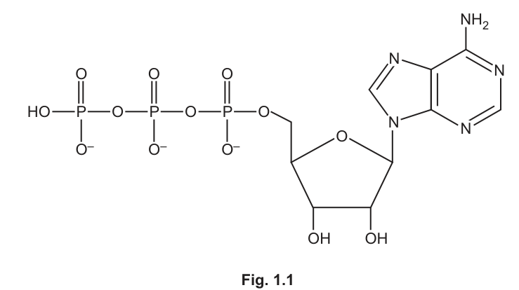
Fig. 1.1 shows a molecule of ATP.
Explain the features of ATP that make it suitable to be the universal energy currency of cells.
Answer
- ATP is hydrolysed to ADP and Pi, releasing a small but useful packet of energy (~30.5 kJ mol⁻¹) that can be coupled to energy-requiring reactions.
- The reaction is readily reversible, so ATP can be regenerated from ADP and Pi, giving a high turnover and constant supply.
- ATP is a small, soluble molecule that can diffuse freely to wherever energy is required in the cell.
- AVP — e.g. the negatively charged phosphate groups repel one another, making the phosphoanhydride bonds high-energy / easily broken; ATP can phosphorylate other molecules, activating them for further reactions.
Hydrolysis releases ~30.5 kJ mol⁻¹; reaction is reversible (high turnover); small and soluble so diffuses freely; activates other molecules by phosphorylation.
Background Concept
ATP (adenosine triphosphate) consists of three parts visible in Fig. 1.1: an adenine base (a double-ring nitrogenous base with an –NH₂ group), a ribose sugar (a five-carbon sugar with two –OH groups on the ring), and a chain of three phosphate groups attached to carbon 5 of the ribose. Note the negative charges shown on the oxygens of each phosphate group — these like charges repel one another, making the bonds between adjacent phosphates (the phosphoanhydride bonds) unstable and therefore high in energy.
ATP is called the energy currency of the cell because it stores energy in a form that is portable, transferable, and immediately usable. When the terminal (γ) phosphate bond is hydrolysed, energy is released that can be coupled to energy-requiring processes such as active transport, biosynthesis, and muscle contraction.
Understanding the Question
The command word is explain, which means each marking point must be a feature plus the reason it matters. Four independent points are required. The mark scheme lists five possibilities (plus AVP), so any four earn full marks. The parent stem reminds us that ATP is the energy currency of the cell, so the answer must explain why ATP is suitable to play this role universally.
Approach
Think about what any energy currency needs: a way of releasing energy on demand, a way of being recharged, portability, and the ability to interact with the reactions that spend energy. Map each of these ideas to a specific property of ATP shown in the diagram.
Step-by-Step Reasoning
- Hydrolysis releases a usable amount of energy. Removing the terminal phosphate to form ADP + Pi releases about — small enough to avoid damaging the cell but large enough to drive many reactions.
- The reaction is reversible. Because the energy released on hydrolysis is modest, the reaction has a small energy of activation in both directions, and ATP can be resynthesised from ADP and Pi during respiration. This makes it a currency rather than a fuel — it circulates and is reused.
- Small and soluble. The molecule is small enough to diffuse freely through the cytosol to wherever it is needed, so it can be supplied rapidly to energy-consuming processes in any part of the cell.
- AVP — coupling to other reactions. The phosphate group can be transferred to another molecule (phosphorylation), raising the energy of that molecule and allowing it to take part in a reaction that would otherwise be energetically unfavourable. The repulsion between the negatively charged phosphates (visible on Fig. 1.1) is what makes the terminal bond so reactive.
Key Takeaways
- ATP is the universal energy currency because of four key properties: modest energy release, reversibility, solubility, and ability to phosphorylate other molecules.
- The negative charges on the phosphates are crucial — they create the unstable, high-energy bonds that make ATP reactive.
- Being a currency (recyclable) is just as important as being an energy source (fuels are consumed, currency is reused).
Common Mistakes
- Stating only that "ATP releases energy" without saying how much, or that it is "high energy" without describing what this means for the cell.
- Confusing ATP (an energy currency) with glucose (an energy store or fuel). Glucose must be broken down over many steps; ATP releases energy in a single, coupled step.
- Saying ATP "contains energy" without explaining how it releases it (through hydrolysis of a phosphate bond).
- Writing about the structure of ATP without linking each feature to its functional consequence — "it has three phosphates" alone is not an explanation.
Things to Be Careful About
- The mark scheme uses both "" and the value 30.5. Quoting the energy value is the cleanest way to score that marking point.
- "Reversible" must be qualified: the mark scheme accepts either "reversible reaction", "high turnover" or "can be regenerated" — any of these is fine.
- The question awards 4 marks for any four from five options, so there is no need to include all five.
Identify the type of phosphorylation reaction to synthesise ATP that occurs during glycolysis and the Krebs cycle.
Answer
Substrate-level (phosphorylation).
Substrate-level phosphorylation.
Background Concept
There are two ways in which ATP is made during respiration:
- Substrate-level phosphorylation: a phosphate group is transferred directly from a phosphorylated substrate (an intermediate in the pathway) onto ADP, forming ATP. The energy has already been "stored" in that intermediate during an earlier step.
- Oxidative phosphorylation (chemiosmosis): ATP is made by ATP synthase, driven by a proton gradient across the inner mitochondrial membrane that is generated by the electron transport chain.
In a single turn of glycolysis, ATP is made twice by substrate-level phosphorylation (at two different steps). In each turn of the Krebs cycle, one ATP (or GTP, which is then converted to ATP) is also made by substrate-level phosphorylation.
Understanding the Question
The command word is identify; the question asks for the type of phosphorylation that occurs in both glycolysis and the Krebs cycle. Since both stages make ATP directly from a phosphorylated intermediate, the answer is the same for both. The contrast the question is hinting at is with oxidative phosphorylation, which happens on the inner mitochondrial membrane (not in the cytosol or matrix at the named steps).
Approach
Recall that in glycolysis the steps that produce ATP involve 1,3-bisphosphoglycerate and phosphoenolpyruvate; in the Krebs cycle succinyl CoA → succinate is the equivalent step. In every case, a phosphate is transferred from a substrate to ADP. That is, by definition, substrate-level phosphorylation.
Step-by-Step Reasoning
- The term to remember is substrate-level phosphorylation (also written "substrate-linked phosphorylation" by the mark scheme).
- It is specifically not oxidative phosphorylation, which occurs at the inner mitochondrial membrane and depends on oxygen.
- One mark for the correct term; do not add extra, uncreditable material.
Key Takeaways
- Substrate-level phosphorylation produces a small number of ATP directly from pathway intermediates; oxidative phosphorylation produces the much larger bulk of ATP and depends on the electron transport chain and a proton gradient.
- Glycolysis and the Krebs cycle both yield ATP by substrate-level phosphorylation; only the electron transport chain uses oxidative phosphorylation.
Common Mistakes
- Writing "oxidative phosphorylation" — this is wrong for the stages named in the question and loses the mark.
- Writing "photophosphorylation" — this is the photosynthesis equivalent, not the respiration one.
- Writing "substrate phosphorylation" without the "-level" or "-linked" qualifier — accepted in conversation, but the mark scheme requires the full term.
Things to Be Careful About
- The mark scheme accepts "substrate-level" or "substrate-linked". Either is fine; "substrate phosphorylation" on its own is risky.
Pyruvate moves into the matrix of the mitochondrion only when a particular inorganic molecule is present.
Name the inorganic molecule that must be present in the cell for pyruvate to enter the matrix of the mitochondrion.
Answer
Oxygen.
Oxygen.
Background Concept
Pyruvate, the three-carbon end-product of glycolysis in the cytosol, can only continue through aerobic respiration if it crosses both mitochondrial membranes into the matrix, where the link reaction and Krebs cycle take place. This requires the cell to be respiring aerobically, i.e. the electron transport chain is functional and reduced NAD is being reoxidised.
The link reaction itself is catalysed by the pyruvate dehydrogenase complex:
For the cell to keep producing reduced NAD and acetyl CoA at a useful rate, the reduced NAD must be reoxidised by the electron transport chain — and that requires the inorganic molecule that acts as the final electron acceptor.
Understanding the Question
The part is a one-mark name question. The stem hints that pyruvate can enter the matrix only "when a particular inorganic molecule is present", and asks the candidate to name it. The word "inorganic" is the key clue: organic cofactors (CoA, NAD⁺) are not the answer.
Approach
Think of what is consumed during aerobic respiration and is therefore essential for the pathway to keep running. Carbon dioxide is a product, not a requirement. Water is plentiful. The inorganic molecule that is required and that disappears during aerobic respiration is oxygen.
Step-by-Step Reasoning
- Without oxygen, the electron transport chain stalls because there is no acceptor for the electrons at the end of the chain.
- Reduced NAD therefore accumulates, and the link reaction (and Krebs cycle, and glycolysis via the triose phosphate dehydrogenase step) slows or stops because NAD⁺ is not regenerated.
- Pyruvate therefore cannot meaningfully enter the mitochondrial matrix for the link reaction.
- The single mark is awarded for the term oxygen (O₂).
Key Takeaways
- Aerobic respiration requires O₂ as the terminal electron acceptor; without it, the link reaction and Krebs cycle halt.
- The question's word "inorganic" rules out all the organic cofactors (CoA, NAD⁺) and points unambiguously to oxygen.
Common Mistakes
- Writing "carbon dioxide" — this is a product of the link reaction, not a requirement.
- Writing "water" — water is produced in oxidative phosphorylation, not required for the link reaction.
- Writing "NAD" or "coenzyme A" — these are organic cofactors and do not satisfy "inorganic".
Things to Be Careful About
- The question awards only one mark; the answer must be a single, accurate term. "Oxygen" is the mark-scheme answer. "O₂" is also accepted.
Explain how the presence of the inorganic molecule named in (d)(i) affects the ATP yield from respiration.
Answer
- The ATP yield increases when oxygen is present.
- Oxygen acts as the final electron (and proton) acceptor at the end of the electron transport chain.
- This allows oxidative phosphorylation / chemiosmosis to occur, in which energy released as electrons pass along the carriers is used to pump H⁺ into the intermembrane space, creating a proton gradient that drives ATP synthesis by ATP synthase.
ATP yield increases; oxygen is the final electron/proton acceptor, allowing oxidative phosphorylation and chemiosmosis to occur.
Background Concept
The yield of ATP from one molecule of glucose depends critically on whether the electron transport chain (ETC) is operating. In aerobic respiration:
- Glycolysis yields 2 ATP by substrate-level phosphorylation and 2 reduced NAD.
- The link reaction (×2, per glucose) yields 2 reduced NAD.
- The Krebs cycle (×2, per glucose) yields 6 reduced NAD, 2 reduced FAD, and 2 ATP by substrate-level phosphorylation.
- The electron transport chain reoxidises 10 reduced NAD and 2 reduced FAD; the energy released pumps H⁺ from the matrix into the intermembrane space, creating an electrochemical gradient. H⁺ flowing back into the matrix through ATP synthase drives the synthesis of ~34 ATP.
So the great majority of ATP — the bulk of the ~38 ATP per glucose — comes from oxidative phosphorylation, which only runs if oxygen is present to accept electrons at the end of the chain.
Understanding the Question
Part (d)(ii) follows from (d)(i). The question asks the candidate to explain how the presence of oxygen (named in (d)(i)) increases the ATP yield. Three marks are available: the first for the direction of the effect, the next two for two of the listed explanations (the role of oxygen, oxidative phosphorylation / chemiosmosis, energy from reduced coenzymes, the proton gradient, or ATP made in the Krebs cycle).
Approach
Build a logical chain: oxygen is the final electron acceptor → the ETC can run → reduced NAD/FAD is reoxidised → the energy released pumps H⁺ into the intermembrane space → H⁺ flowing back through ATP synthase makes ATP. Three discrete points are needed from this chain.
Step-by-Step Reasoning
- The ATP yield is higher when oxygen is present. Without oxygen, only the 2 ATP from glycolysis are made (anaerobic respiration in many cells regenerates NAD⁺ by reducing pyruvate to lactate or ethanol, but no further ATP is produced). With oxygen, the ETC runs and the total yield rises to ~38 ATP per glucose.
- Oxygen is the final electron (and proton) acceptor. At the end of the ETC, electrons are passed to oxygen, which combines with H⁺ to form water. This keeps the chain "pulling" electrons from reduced NAD and reduced FAD, regenerating NAD⁺ and FAD so that the link reaction and Krebs cycle can continue.
- Oxidative phosphorylation / chemiosmosis occurs. Energy released as electrons pass along the ETC carriers is used to actively transport H⁺ from the matrix into the intermembrane space, creating an electrochemical (proton) gradient. H⁺ diffuses back into the matrix through ATP synthase, and the energy released drives the synthesis of ATP from ADP and Pi.
- (Alternative marking point) ATP is also made in the Krebs cycle by substrate-level phosphorylation, but only when oxygen is available, because the Krebs cycle requires NAD⁺ and FAD (regenerated by the ETC) to keep turning.
Key Takeaways
- Oxygen dramatically increases the ATP yield of respiration because it allows oxidative phosphorylation to run, generating the great majority of ATP per glucose.
- Without oxygen, the electron transport chain stalls, reduced NAD cannot be reoxidised, the link reaction and Krebs cycle halt, and only the 2 ATP from glycolysis are made.
- The role of oxygen is specifically as the terminal electron acceptor; this is why it is described as an inorganic molecule — it is the only non-organic participant in the chain.
Common Mistakes
- Saying "oxygen is needed for Krebs cycle" without explaining why — the mark requires the role as the final electron acceptor, which is the link to oxidative phosphorylation.
- Conflating substrate-level phosphorylation (in the Krebs cycle) with oxidative phosphorylation (on the inner membrane); the question is about the latter.
- Writing only "more ATP is made" without explaining the mechanism. "Explain" demands a reason, not just an observation.
- Stating that "oxygen is used to make water" without linking this to the electron transport chain and ATP yield.
Things to Be Careful About
- The mark scheme's point 6 ("ATP made in Krebs cycle") is a consequence of oxygen being present (because NAD⁺ and FAD can be regenerated), but the bulk of the extra ATP comes from oxidative phosphorylation — the candidate should make this clear.
- " into intermembrane space" is the precise wording the mark scheme uses for the proton gradient; mentioning the matrix → intermembrane direction is what earns the mark.
A respirometer can be used to investigate how temperature affects the rate of respiration of woodlice.
Woodlice are small invertebrate animals. Fig. 2.1 shows a single woodlouse.
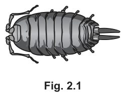
Fig. 2.2 shows a simple respirometer.
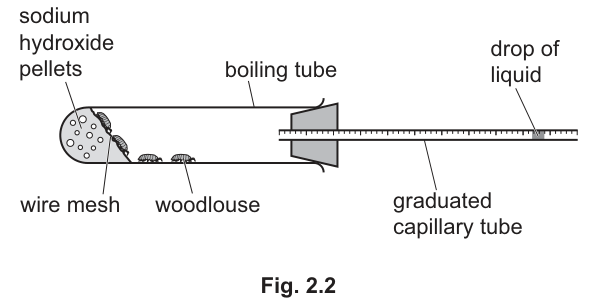
Explain how the experimental set-up in Fig. 2.2 allows the rate of respiration of woodlice to be determined.
Answer
- Sodium hydroxide pellets absorb the released by the respiring woodlice.
- Uptake of by the woodlice (with the produced being absorbed by the NaOH) reduces the pressure (and gas volume) inside the boiling tube.
- The drop of liquid in the capillary tube moves towards the boiling tube (towards the woodlice).
- The distance moved by the drop in a set time is measured, so:
- The volume of used can be calculated from the cross-sectional area of the capillary tube multiplied by the distance moved by the drop:
Rate of respiration (volume of O₂ taken up per unit time) is obtained from the distance the drop moves along the capillary tube in a set time, because the NaOH absorbs CO₂ so the only net gas change is the O₂ consumed.
Background Concept
A respirometer is a simple piece of apparatus used to measure the rate at which a small organism takes up oxygen during aerobic respiration. It relies on the fact that in aerobic respiration, an organism consumes and releases an equal volume of .
If both gases were released and absorbed equally, there would be no net change in the volume of gas inside the sealed tube, and so no way to detect respiration. Sodium hydroxide (or another absorbent such as soda lime) removes the as soon as it is produced, leaving only the decrease in volume due to uptake. This change in gas volume produces a tiny decrease in pressure inside the tube, which is amplified and visualised by a coloured drop of liquid in a narrow horizontal capillary tube.
Understanding the Question
Part (a) asks the candidate to explain — point by point — how the apparatus shown in Fig. 2.2 actually produces a measurement of the rate of respiration. The apparatus consists of a horizontal boiling tube containing woodlice, separated by wire mesh from sodium hydroxide pellets. The right-hand end of the boiling tube is closed by a rubber bung through which a graduated capillary tube passes; a coloured drop of liquid sits in the capillary tube. The question wants the chain of reasoning that links "respiring woodlice" → "drop moving" → "rate of respiration".
Approach
Work through the chain in the order the events actually happen:
- Respiration: in, out.
- The is absorbed by the NaOH, so the only net change is loss of .
- The gas (and therefore pressure) inside the sealed tube falls.
- Atmospheric pressure pushes the drop of liquid inwards (towards the woodlice).
- The distance moved in a known time gives a rate; the volume of used = cross-sectional area of capillary × distance moved.
Step-by-Step Reasoning
- NaOH absorbs the produced by respiration. The wire mesh keeps the woodlice away from the pellets, preventing them from touching the corrosive NaOH while still allowing the gas in the tube to circulate freely.
- consumed by aerobic respiration is no longer replaced, so the number of gas molecules inside the sealed boiling tube decreases.
- This produces a small reduction in pressure inside the tube relative to atmospheric pressure outside.
- Atmospheric pressure pushes the drop of liquid in the capillary tube towards the boiling tube (i.e. to the left in the figure). The narrower the capillary, the greater the distance moved for a given volume change, so the apparatus is sensitive.
- The distance the drop moves in a set time is recorded, allowing calculation of a rate:
- To convert this to a true volume of used per unit time, multiply the distance moved by the cross-sectional area of the capillary tube (, where is the distance moved). This volume is the consumed in that time interval, i.e. the rate of aerobic respiration.
Key Takeaways
- A respirometer measures uptake, not release, because the is removed by an alkali.
- The liquid drop in the capillary tube is a sensitive manometer: it amplifies the small pressure/volume change into a measurable distance.
- A valid rate must always be expressed as volume of per unit time (often per unit mass of organism, e.g. ).
- Standard controls: a parallel tube set up identically but with dead woodlice (or no woodlice) — to compensate for any physical effects of temperature or pressure change that are unrelated to respiration.
Common Mistakes
- Stating that the drop moves because the woodlice "give off heat" or "use energy" — this is wrong; it is the fall in gas pressure, not temperature change, that moves the drop.
- Saying the drop moves "outwards" or "to the right" — because gas is being consumed (not produced), the drop must move towards the woodlice.
- Failing to state the role of NaOH; without it, uptake and release would approximately cancel and the drop would not move.
- Writing "rate = distance" without dividing by time.
- Not converting distance to a volume — to compare experiments using different capillary tubes, a true volume is required.
Things to Be Careful About
- In a Cambridge practical question you may be expected to give a worked equation for the volume of gas used.
- Woodlice must be given an equilibration period after being placed in the tube so that handling (and any change in temperature/activity) does not affect the first reading.
- The tube must be airtight (well-greased bung, no gaps around the capillary) for the pressure change to drive the drop predictably.
- Always run a control tube without woodlice to allow for any change in atmospheric pressure or temperature during the experiment.
A student determined the rate of respiration of woodlice at a temperature of and at a temperature of .
Predict and explain how the rate of respiration of woodlice differs at compared to .
Answer
- The rate of respiration at is lower / slower than at because the lower temperature gives the enzymes and substrates less kinetic energy, so there are fewer successful enzyme–substrate collisions per unit time and the enzyme-catalysed reactions of respiration (glycolysis, link reaction, Krebs cycle) proceed more slowly.
Rate at 5 °C is lower than at 20 °C because the enzymes have less kinetic energy, so fewer enzyme–substrate collisions occur per unit time.
Background Concept
Aerobic respiration is catalysed by a long sequence of enzymes — those of glycolysis in the cytoplasm and those of the link reaction, Krebs cycle and oxidative phosphorylation in the mitochondria. Like all enzymes, these catalysts have an optimum temperature (around 35–40 °C for many mammalian enzymes) and their activity depends on the kinetic energy of the molecules involved. Below the optimum, the rate of an enzyme-catalysed reaction falls because the molecules have less kinetic energy and collide less frequently, and because a smaller fraction of collisions have enough energy to exceed the activation energy of the reaction.
Understanding the Question
Part (b) asks the candidate to compare the rate of respiration of woodlice at with that at and to explain the difference. The mark scheme demands both a prediction (which temperature gives the higher rate) and a reason linked to enzymes or kinetic energy.
Approach
Identify that woodlice are poikilothermic (their body temperature follows the surrounding temperature), so a change in environmental temperature directly alters the kinetic energy of the enzymes and substrates inside their cells. Apply the general rule for the effect of temperature on enzyme activity below the optimum.
Step-by-Step Reasoning
- At the lower temperature (), the kinetic energy of the enzyme and substrate molecules is reduced.
- Molecules therefore move more slowly and collide less often.
- A smaller proportion of those collisions have sufficient energy to overcome the activation energy of the enzyme-catalysed reaction.
- The rate of each step of respiration (glycolysis, link reaction, Krebs cycle) — and therefore the overall rate of oxygen uptake — is lower.
- The drop of liquid in the respirometer would therefore move a shorter distance in a given time at than at .
Key Takeaways
- For any enzyme-controlled process, a temperature below the optimum gives a slower rate because of reduced kinetic energy and fewer effective collisions.
- A "predict and explain" question requires both parts: a clear comparative statement and a biological reason.
- Woodlice are ectotherms, so the temperature of their environment directly determines the temperature inside their cells.
Common Mistakes
- Saying the rate is higher at (incorrect — colder temperatures slow enzyme activity below the optimum).
- Giving only the prediction without an explanation: the mark scheme requires a linked cause (kinetic energy, collisions, or named respiration steps).
- Stating that "the enzymes are denatured" at — denaturation requires much higher temperatures; cold simply reduces activity, it does not destroy the enzyme.
- Vague answers such as "the woodlice are cold and so are less active" — this is not biological reasoning; the question demands an enzyme-based explanation.
Things to Be Careful About
- The mark scheme is strict: it wants the keyword "kinetic energy" or "enzyme–substrate collisions" (or an explicit reference to a named respiration step). A general phrase such as "the enzymes don't work as well" will not score.
- Note the temperature range used ( versus ): both are well below the typical denaturation temperature, so the comparison is about kinetic energy only, not about denaturation.
Porphyria is a group of rare genetic diseases in which molecules called porphyrins accumulate in the body.
Three examples of porphyria are:
- X-linked protoporphyria
- variegate porphyria
- congenital erythropoietic porphyria (CEP).
X-linked protoporphyria is caused by a mutant allele located on the X chromosome.
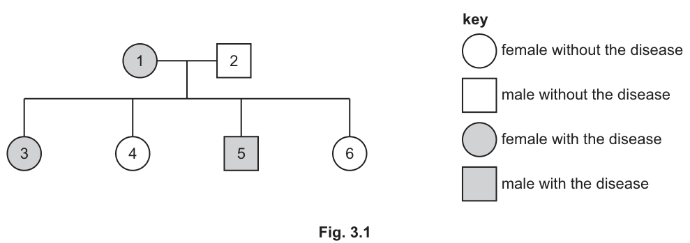
Fig. 3.1 shows the pattern of inheritance of X-linked protoporphyria in one family.
X-linked protoporphyria is described as a sex-linked disease.
Use the information in Fig. 3.1 to make one other conclusion about the pattern of inheritance of X-linked protoporphyria.
Give evidence to support your conclusion.
conclusion _____
evidence _____
Answer
Conclusion: the (mutant) allele is dominant.
Evidence: individual 3 (affected daughter) has the disease, but her father (individual 2) does not. If the allele were recessive, the affected daughter would need to have inherited the recessive allele on the X chromosome from her father, which would mean the father would also have the disease. Therefore, the allele must be dominant (so the daughter has inherited a dominant mutant allele from her affected mother).
Dominant; individual 3 (affected daughter) has the disease but her father (individual 2) does not, which would not be possible if the allele were recessive (as the daughter would have to have inherited the recessive allele on an X chromosome from her father).
Background Concept
Sex-linked (X-linked) inheritance describes genes carried on the X chromosome. A recessive X-linked allele, such as the one for haemophilia, is normally only expressed in males (who have just one X chromosome) or in homozygous females. For a female to express a recessive X-linked trait, she must inherit the recessive allele on both of her X chromosomes — one from her mother and one from her father. The father must therefore also show the trait (or be hemizygous for the recessive allele, in which case he will express it because he has no second X chromosome to carry a dominant normal allele).
A dominant X-linked allele is different: only one copy is needed for the trait to be expressed. An affected heterozygous female (X^D X^d) will pass the dominant allele to roughly half her offspring regardless of the father's genotype, and the daughter can be affected even though her father is unaffected.
Understanding the Question
Fig. 3.1 shows a two-generation pedigree. In generation 1 the mother (1) is affected and the father (2) is unaffected. In generation 2 they have four children: an affected daughter (3), an unaffected daughter (4), an affected son (5), and an unaffected daughter (6). The question is already given the information that the allele is X-linked, and is asking for one other conclusion about the inheritance pattern, supported by evidence from the pedigree.
The command words are "conclusion" and "evidence" — the mark scheme rewards a single clear statement of the conclusion (the word "dominant") plus the supporting logic drawn from the pedigree.
Approach
Test the two possibilities (dominant vs. recessive) against the pedigree:
- If the X-linked allele were recessive, the affected daughter (3) would need X^p X^p. She must have received one X^p from each parent. From the father (2), who is unaffected, the only X chromosome he can pass on carries the normal allele (X^P). So he could not pass on X^p. This contradicts the pedigree — therefore the allele cannot be recessive.
- If the X-linked allele were dominant, the affected daughter only needs one copy of the mutant allele. She can have received the mutant X from her affected mother and a normal X from her unaffected father. This is consistent with the pedigree.
Step-by-Step Reasoning
- The mother (1) is affected, so her genotype is X^A X^? (at least one mutant allele, written as A assuming dominant).
- The father (2) is unaffected, so his genotype is X^? Y (one normal X chromosome and a Y).
- Daughter 3 is affected. She has genotype X^A X^? — she received one X from each parent.
- From the father, she must have received the X chromosome, which carries the normal allele (since the father has no disease).
- The mutant allele she shows must therefore have come from the mother.
- With one mutant allele from the mother and one normal allele from the father, she is heterozygous, yet she expresses the disease. This is only possible if the mutant allele is dominant to the normal allele.
- If the allele were recessive, daughter 3 would need two copies of the mutant allele (one from each parent), and so the father would have to be affected — which he is not.
Key Takeaways
- A dominant X-linked trait can pass from an affected mother to a daughter whose father is unaffected, because the daughter needs only one copy of the mutant allele.
- For a recessive X-linked trait, an affected daughter must have an affected father, because she must have received the recessive allele on his X chromosome.
- A single pedigree can therefore often rule out one of the two possibilities (dominant vs. recessive) just by looking at father–daughter combinations.
Common Mistakes
- Saying the allele is recessive because the disease is rare — incorrect; dominance and rarity are independent.
- Stating the conclusion but not providing the supporting evidence drawn from the pedigree (e.g. simply saying "the allele is dominant because the disease is X-linked").
- Failing to explain why the recessive possibility is ruled out (i.e. the father would have to be affected).
Things to Be Careful About
- The conclusion must be a single, clear genetic statement ("dominant"). Avoid vague answers like "the disease is inherited".
- The evidence must refer explicitly to specific individuals in the pedigree (individual 2 and individual 3).
- The mark scheme accepts either the affirmative argument (daughter affected, father not) or the negative argument (if recessive, daughter could not be affected).
Variegate porphyria occurs in 1 in 100000 people in Europe.
In the 1600s, approximately 100 Dutch people migrated from Europe to South Africa.
The population of people of Dutch descent in South Africa is now 2.5 million. Variegate porphyria occurs in 1 in 1000 people in this population.
Suggest and explain why variegate porphyria is more common among people of Dutch descent in South Africa than among people in Europe.
Answer
- Founder effect — the South African Dutch population was founded by only ~100 Dutch migrants, so the allele frequencies in this small group may not have been representative of the European population as a whole. (1 mark)
- The mutant allele for variegate porphyria was carried by some of these migrant founders at a higher frequency than in Europe, so the founding population contained the allele. (1 mark)
- Because the population that established itself was small, genetic drift had a large effect on allele frequencies, and inbreeding between members of the same small group maintained the high frequency of the mutant allele over many generations. (1 mark)
Founder effect: the small group of Dutch migrants to South Africa carried a higher frequency of the variegate porphyria allele than the European population as a whole; genetic drift and inbreeding within this small isolated population then maintained the high allele frequency over many generations.
Background Concept
The founder effect is a special case of genetic drift. When a new population is established by a very small number of individuals drawn from a larger parent population, the allele frequencies in the new population are determined mainly by the genotypes of those few founders — not by the frequencies in the parent population as a whole. By chance, some alleles that were rare in the parent population may be present in the founders at much higher frequency (or vice versa).
Once established, a small population is also strongly affected by genetic drift — random fluctuations in allele frequency from one generation to the next — and by inbreeding, which does not change allele frequency by itself but keeps rare alleles (in homozygous form) circulating in the population.
Understanding the Question
The question gives two pieces of data:
- In Europe as a whole, variegate porphyria occurs in 1 in 100 000 people.
- In South Africans of Dutch descent, it occurs in 1 in 1000 people — about 100 times more often.
The historical context is that this South African Dutch population was founded in the 1600s by approximately 100 migrants. The question asks you to suggest and explain why the disease is more common in this descendant population.
Approach
The key concept to reach for is the founder effect. Then layer in additional reasoning:
- How did the higher frequency arise in the founders? (The allele must have been carried by some of the 100 founders at a higher-than-European frequency.)
- Why has the high frequency been maintained over ~400 years? (Small population → genetic drift; inbreeding within the group.)
Step-by-Step Reasoning
- The original European population is large, and the disease allele is rare (1/100 000).
- A small group of ~100 Dutch people migrated to South Africa; the allele frequencies in this group are determined by chance (which alleles the founders happened to carry).
- By chance, the founders included at least one (probably more) carrier(s) of the variegate porphyria allele, so the allele frequency in the founding population was higher than in Europe overall.
- This is the founder effect: a new population's allele frequencies are not representative of the source population.
- After establishment, the new South African Dutch population remained relatively small and isolated. In a small gene pool, genetic drift has a disproportionately large effect on allele frequencies, and random fluctuations are amplified.
- The group was also highly inbred (people tended to marry within their own community), so the mutant allele was maintained in the gene pool over many generations rather than being diluted out by mixing with populations carrying the normal allele.
- The result is that today, the descendant population of 2.5 million still shows a much higher frequency of the allele (1/1000) than the European source population (1/100 000).
Key Takeaways
- The founder effect is a genetic-drift phenomenon that operates on a small founding population; the allele frequencies in the descendants reflect those of the founders, not the source population.
- Founder effect plus a long period of relative isolation (with inbreeding) is the classic explanation for high frequencies of otherwise rare alleles in particular human populations (e.g. the high frequency of certain alleles in the Afrikaner population, the Old Order Amish, or isolated island populations).
- "Suggest and explain" questions on evolution want the mechanism named and the specific link to the data given.
Common Mistakes
- Saying only "founder effect" without explaining how the higher frequency arose or how it was maintained.
- Confusing founder effect with bottleneck effect: a bottleneck reduces population size suddenly and randomly; a founder effect establishes a new population from a small number of individuals. Both are forms of genetic drift, but the timing and mechanism differ.
- Suggesting natural selection favoured the allele — there is no evidence for any selective advantage of variegate porphyria, and the mark scheme is asking for a neutral, drift-based explanation.
- Suggesting a higher mutation rate in the South African Dutch population — not supported by the data.
Things to Be Careful About
- The mark scheme wants specific points tied to the data: small founding population, allele present in the founders, frequency higher than in the source population, genetic drift in a small gene pool, and inbreeding.
- Any 3 of these (out of the 7 listed in the mark scheme) will earn full marks, so give the strongest, most specific points.
Microarrays can be used to detect various forms of porphyria caused by mutant alleles.
Describe how microarrays can be used to detect a disease by analysing gene expression.
Answer
- mRNA is extracted from the patient's cells and used as a template, with reverse transcriptase, to make cDNA (single-stranded DNA). (1 mark)
- Fluorescent labels (dyes / tags / markers) are attached to this cDNA. (1 mark)
- The fluorescently labelled cDNA is added to the microarray, where it hybridises (by complementary base pairing) to DNA probes on the array that are complementary to the gene / allele of interest. (1 mark)
- The location and intensity of fluorescence on the microarray shows whether the disease / mutant allele is being expressed (transcribed) — a fluorescent spot indicates that the gene is being expressed. (1 mark)
mRNA is extracted from the patient's cells and reverse-transcribed into fluorescently labelled cDNA; the cDNA binds by complementary base pairing to probes on the microarray, and the pattern of fluorescence shows whether the mutant allele is being expressed.
Background Concept
A DNA microarray is a small chip carrying thousands of different single-stranded DNA probes, each representing a known gene or allele, fixed at a specific position on the chip. By exposing the chip to a sample of DNA (or cDNA), complementary sequences hybridise (form base pairs) with their matching probe. Any hybridised sample can be detected because it has been labelled, usually with a fluorescent dye.
A microarray can be used in two distinct ways:
- Genotyping / allele detection — to test whether a person carries a particular mutant allele. The probes on the chip are designed to match either the normal or the mutant sequence, and the patient's DNA (or cDNA) is tested against both.
- Gene expression profiling — to measure how actively a gene is being transcribed. mRNA is extracted from the patient's cells; the more mRNA a cell makes, the more actively that gene is being expressed.
This part of the question is specifically about gene expression (the wording is "detecting disease by analysing gene expression"), so the procedure must include extracting mRNA and converting it to cDNA.
Understanding the Question
The question is worth 4 marks and asks for a description of how a microarray detects a disease by analysing gene expression. The mark scheme rewards four distinct steps: making cDNA from mRNA, labelling the DNA, hybridising to probes, and reading fluorescence as expression.
Approach
The key is to follow the biological chain in order:
mRNA (from the cell) → cDNA (by reverse transcription) → labelled cDNA → hybridisation to probe → fluorescence = expression of the gene.
Step-by-Step Reasoning
- mRNA extraction. Cells from the patient are broken open and the mRNA is isolated. mRNA is chosen because it is a direct read-out of which genes are being transcribed. Genes that are not expressed will produce little or no mRNA.
- Reverse transcription. The enzyme reverse transcriptase uses the mRNA as a template to make a complementary strand of DNA, called cDNA (complementary DNA), which is single-stranded.
- Fluorescent labelling. A fluorescent dye (often Cy3, which fluoresces green, or Cy5, which fluoresces red) is incorporated into the cDNA, either during the reverse-transcription step or afterwards.
- Hybridisation. The fluorescently labelled cDNA is washed over the microarray. The cDNA molecules base-pair (hybridise) only with probes whose sequence is complementary. If the patient is expressing the mutant allele, cDNA corresponding to that allele will bind to its matching probe.
- Detection. A laser scans the array; the spot(s) where cDNA has hybridised fluoresce, and the position on the array identifies which gene/allele is being expressed, while the intensity of the fluorescence indicates how strongly it is being expressed. A fluorescence pattern different from that of a healthy control indicates disease-associated expression.
Key Takeaways
- Microarrays for gene expression start with mRNA, not genomic DNA. The conversion of mRNA to cDNA via reverse transcriptase is the key step.
- The fluorescent label does not "find" the gene on its own — hybridisation by complementary base pairing is the matching step, and the label is what makes that match visible.
- A spot's position on the array identifies the gene; the intensity of fluorescence indicates how strongly the gene is being expressed.
- Microarrays for allele detection (genotyping) work slightly differently — they use short allele-specific oligonucleotide probes and test genomic DNA, not mRNA. The question specifies expression, so the mRNA → cDNA route is required.
Common Mistakes
- Skipping the reverse-transcription step and saying "DNA is added directly to the microarray". Without the mRNA → cDNA step, you are not measuring expression.
- Saying the microarray "reads the DNA sequence" or "sequences the gene". That is sequencing, not microarray analysis.
- Saying the fluorescence "detects the disease" without specifying that it is detecting expression of the disease allele. Microarrays do not detect the presence of an allele directly unless used in genotyping mode.
- Forgetting the role of complementary base pairing: the cDNA does not "stick" non-specifically to the chip; it hybridises to its complementary probe.
Things to Be Careful About
- Mark scheme explicitly credits the use of mRNA → cDNA (or ssDNA), the fluorescent label, the hybridisation step, and the interpretation of fluorescence as expression.
- A fifth "AVP" mark is available, e.g. for mentioning that the position on the chip identifies which gene is being expressed, or for noting that comparing the patient's fluorescence pattern with a healthy control reveals the disease.
The nucleotide sequences of alleles that cause rare genetic diseases, such as the various forms of porphyria, are stored in a database.
State one benefit of having a database of nucleotide sequences for alleles that cause rare diseases.
Answer
A database of nucleotide sequences for alleles that cause rare diseases allows clinicians and researchers to identify / diagnose a person's disease (or to choose / develop the correct treatment). The database also acts as a global research resource that scientists anywhere can access. (Any one of these.)
Allows the disease to be diagnosed (so the correct treatment can be chosen) / provides a global research resource.
Background Concept
Many rare genetic diseases are caused by different mutations in the same gene, or by mutations in different genes that produce similar symptoms. Identifying the exact mutation in a patient — for example by sequencing the relevant gene and comparing the sequence against known pathogenic variants in a database — is often the first step in making a definitive diagnosis and in selecting the most appropriate treatment.
Sequence databases such as ClinVar, OMIM, the Human Gene Mutation Database (HGMD) and locus-specific databases (LSDBs) store the exact nucleotide changes recorded in patients with particular diseases. They are used by:
- Diagnostic laboratories, to interpret a patient's sequence (is the variant seen known to cause disease?).
- Clinicians, to choose a treatment that targets the specific mutation.
- Researchers, to find out what mutations are known, identify gaps, and plan new therapies.
Understanding the Question
The question is worth 1 mark and asks for one benefit of having a database of nucleotide sequences for alleles that cause rare diseases. The mark scheme accepts any of three benefits (diagnosis, treatment selection, or global research resource). The student should give one clear, specific benefit.
Approach
Pick the most direct benefit (usually diagnosis) and state it in one or two sentences. Avoid vague answers like "it is useful for science".
Step-by-Step Reasoning
- A patient with a suspected rare porphyria has their gene sequenced.
- The sequence is compared with the entries in the database.
- If a match is found, the disease is confirmed and its specific form identified — the patient can be diagnosed.
- Knowing the exact mutation allows the correct treatment to be chosen (or developed), and the data is available to researchers worldwide as a resource for further study.
Key Takeaways
- Sequence databases turn a patient's raw sequence into a clinically meaningful result by enabling comparison with known pathogenic variants.
- The benefit is not just "having the information" but the diagnostic, treatment and research uses to which it is put.
- For rare diseases, where a single clinician may never see a case, a global database is especially valuable.
Common Mistakes
- Vague answers: "it helps doctors" or "it is useful for research" without specifying the benefit.
- Saying the database "treats" the disease — it does not; it enables the right treatment to be chosen.
- Saying the database "prevents" the disease — sequence data does not, by itself, prevent disease.
Things to Be Careful About
- One mark is available for a single clear, specific benefit. Choose one and state it precisely.
Congenital erythropoietic porphyria (CEP) is caused by a substitution mutation in a gene coding for an enzyme.
The mutant CEP allele codes for an enzyme with little or no function.
Scientists used gene editing in the laboratory to correct the nucleotide sequence of the mutant allele in cultured human cells.
The scientists introduced three molecules to the cells:
- an enzyme called Cas9 that causes breaks in DNA strands
- guide RNA (gRNA), attached to Cas9, that is complementary to the mutant CEP allele
- a short length of DNA, known as template DNA, that can replace the section of the allele where the substitution mutation has occurred.
The repair mechanism within the cell allows the template DNA to be inserted so that the mutation is corrected.
Fig. 3.2 shows part of the nucleotide sequence in the mutated CEP allele before and after the gene editing procedure.

In the future, this gene editing procedure may be used in a person with CEP to prevent the accumulation of porphyrins in the body.
Suggest and explain how this gene editing procedure will prevent the accumulation of porphyrins in a person with CEP without damaging other genes.
Answer
- The gene editing procedure substitutes the mutant T with a C at the third position of the codon (changing TAT to TAC), correcting the nucleotide sequence back to the normal allele. (1 mark)
- The corrected allele now codes for a functional / normal enzyme, which can carry out its role in the porphyrin biosynthesis pathway and so prevent the accumulation of porphyrins in the body. (1 mark)
- The guide RNA (gRNA) is complementary only to the mutant CEP allele, so Cas9 is directed to cut only at that specific site; it does not bind to (or cut) other genes, so off-target damage is avoided. (1 mark)
The T is replaced by C in the mutant CEP allele, restoring the normal coding sequence so a functional enzyme is produced (preventing porphyrin accumulation); because the gRNA is complementary only to the mutant CEP allele, Cas9 cuts only at that site and other genes are not damaged.
Background Concept
Congenital erythropoietic porphyria (CEP) is an autosomal recessive condition in which a substitution mutation in the gene encoding the enzyme uroporphyrinogen III cosynthase (UROS) produces an enzyme with little or no activity. Without this enzyme, the haem / porphyrin biosynthesis pathway cannot proceed normally, and porphyrin intermediates accumulate. Porphyrins are light-sensitive, so their accumulation in the skin causes the characteristic photosensitivity and skin damage of CEP.
CRISPR–Cas9 gene editing uses two key components:
- Cas9, an endonuclease that cuts both strands of DNA at a precise location.
- A guide RNA (gRNA) that base-pairs with the target DNA sequence and directs Cas9 to that exact location. The gRNA is typically about 20 nucleotides long and must be complementary to the target site; this is what gives the system its specificity.
Once Cas9 has cut the DNA, the cell's own repair machinery can be exploited: if a template DNA with the correct sequence is provided, the cell's homology-directed repair (HDR) pathway will use it to repair the cut, replacing the faulty sequence with the correct one. The result is a precise, targeted change to a single gene.
Understanding the Question
Fig. 3.2 shows part of the CEP gene before and after editing. Before editing the sequence reads TATGGCTCG; after editing it reads TACGGCTCG. The change is a single nucleotide: the third base, a T, has been replaced by a C. This is the substitution that the question tells you produces a non-functional enzyme.
The question asks you to suggest and explain how the gene-editing procedure will:
- prevent the accumulation of porphyrins (the biological outcome), and
- do this without damaging other genes (the safety/specificity issue).
Three marks are available: one for the nucleotide change, one for the functional protein outcome, and one for the gRNA specificity (with an AVP mark available for a further valid point).
Approach
Read the question in two halves:
- How does the editing prevent porphyrin accumulation? The mutation is corrected → normal mRNA is transcribed → functional enzyme is translated → the porphyrin pathway works → no porphyrin accumulation.
- Why doesn't it damage other genes? The gRNA is complementary to the mutant CEP allele only, so Cas9 is targeted to that one site and does not cut elsewhere in the genome.
Step-by-Step Reasoning
- Identify the change. In Fig. 3.2, the mutant sequence contains T (third base) where the corrected sequence contains C. This single nucleotide change (T → C) restores the correct codon and hence the correct amino acid in the enzyme.
- Restored function. With the corrected sequence, the cell's transcription and translation machinery produce a normal, functional uroporphyrinogen III cosynthase enzyme.
- Metabolic outcome. The functional enzyme catalyses its step in the porphyrin / haem biosynthesis pathway, so porphyrin intermediates are no longer left unprocessed and no longer accumulate. The disease phenotype is therefore prevented.
- Specificity of gRNA. The guide RNA is a short single-stranded RNA designed to be complementary in base sequence to the mutant CEP allele. Because base pairing is sequence-specific, the gRNA will only bind to that exact target sequence and not to other regions of the genome. Cas9, which is carried to the DNA by the gRNA, will therefore only cut the CEP gene.
- Repair uses the template. Once Cas9 has cut the DNA at the CEP site, the cell's homology-directed repair mechanism uses the supplied template DNA to fill in the correct sequence, completing the correction.
- Off-target effects avoided. Because the gRNA does not bind to other genes, Cas9 does not cut them, so other genes are not damaged by the procedure.
Key Takeaways
- The biological principle of gene therapy for a loss-of-function disease is: correct the mutation → restore the normal protein → restore the biochemical pathway → prevent the build-up of the harmful intermediate.
- The specificity of CRISPR–Cas9 comes from the guide RNA's base sequence. A 20-nucleotide gRNA is long enough to be essentially unique in the human genome, so it binds only at its intended target.
- The cell's own repair machinery (homology-directed repair, HDR) is what actually inserts the new DNA; Cas9 only makes the cut.
Common Mistakes
- Saying only that "the gene is corrected" without explaining the downstream consequence (functional enzyme → working pathway → no porphyrin accumulation).
- Saying the gRNA "recognises the disease" or "finds the faulty gene" without explaining the molecular basis of this recognition (complementary base pairing to the mutant allele).
- Confusing Cas9 (the cutting enzyme) with the gRNA (the targeting molecule) and saying Cas9 itself "recognises" the target.
- Saying the procedure "removes the mutant gene" rather than correcting it — gene editing in this protocol alters the sequence in place, it does not delete the gene.
Things to Be Careful About
- Three distinct ideas are needed: the T → C substitution, the functional enzyme outcome, and the gRNA specificity. Watch for marks that are easy to leave on the table by being too vague.
- The AVP mark is for any further valid point — e.g. that HDR uses the supplied template DNA, or that the change is at a single specific site.
Scientists have invented a way of producing food by artificial photosynthesis.
- Solar panels convert sunlight energy into electricity.
- The electricity powers an electrolysis reaction between carbon dioxide gas and water to form the organic product acetate.
- The single-celled alga Chlamydomonas, a protoctist, can use acetate to grow and reproduce in the dark, instead of photosynthesising in the light.
- The algae can be processed to make a food product.
Fig. 4.1 shows an outline of the artificial photosynthesis process.
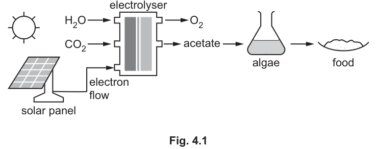
Identify three similarities between the artificial photosynthesis process shown in Fig. 4.1 and the normal process of photosynthesis.
Answer
Any three of:
- Both use/require .
- Both use/require (water).
- Both use/require (sun)light energy.
- Both make/produce an organic molecule/product.
- Both produce/release .
- Both involve the transduction of energy (from one form to another).
- Both involve electron flow.
Any three of: CO2; H2O; sunlight; organic product; O2; energy transduction; electron flow.
Background Concept
Natural photosynthesis, summarised by the overall equation
takes carbon dioxide and water and, using light energy captured by chlorophyll in chloroplasts, builds an organic molecule (a carbohydrate) and releases oxygen. The light-dependent stage, on the thylakoid membranes, uses light to split water, generate ATP and reduce NADP, with released as a by-product. The light-independent stage (Calvin cycle) in the stroma uses these to fix into triose phosphate. Throughout, energy is transduced from light into chemical form, and electrons flow from to NADP.
The artificial system in Fig. 4.1 uses a solar panel to convert sunlight into electricity, which then drives an electrolyser reacting with to give and acetate (an organic, two-carbon molecule). The acetate feeds Chlamydomonas, which is then processed into food.
Understanding the Question
The command word is "identify" — the candidate simply needs to name shared features, with three marks available, so three points are required. The question asks for similarities, i.e. features that BOTH processes have in common. It is easy to drift into differences, which score nothing here.
Approach
Compare the inputs and outputs of the two systems and look for overlap:
- Inputs to the artificial system: sunlight, , .
- Inputs to natural photosynthesis: sunlight, , .
- Outputs of the artificial system: , acetate (organic).
- Outputs of natural photosynthesis: , carbohydrate (organic).
- Energy pathway: light → electrical → chemical in the artificial system; light → chemical in natural photosynthesis.
- Electron pathway: electrons flow from the solar panel through the electrolyser; electrons flow from through the thylakoid electron transport chain to NADP in the natural system.
Any three matches earn the three marks.
Step-by-Step Reasoning
The mark scheme lists seven possible creditable points; any three are accepted:
- Both use .
- Both use .
- Both use sunlight energy.
- Both make an organic molecule/product.
- Both release .
- Both involve energy transduction (one form to another).
- Both involve electron flow.
A strong answer picks the three most concrete, most easily-verifiable points — usually , and either light or — to make sure all three marks are earned unambiguously.
Key Takeaways
- The overall equation of photosynthesis is the easiest reference point for any "compare photosynthesis with X" question.
- "Organic molecule/product" is broader than "carbohydrate" — the artificial system makes acetate, which is organic, so this wording covers both.
- Energy transduction and electron flow are deeper similarities that show the chemistries are fundamentally linked, even though the two systems use very different machinery.
Common Mistakes
- Confusing the algae's metabolism with photosynthesis. The algae in the dark are using acetate directly (heterotrophically), not photosynthesising. They contribute to the use of the organic product, not to making it.
- Listing features only one process has, e.g. "the artificial system uses electricity" — that is a difference, not a similarity.
- Vague answers such as "both involve chemical reactions" — the mark scheme rewards the named substance (, , ) or the named concept (light energy, electron flow), not generic statements.
Things to be Careful About
- Use the specific chemical species (, , ) — not "a gas" or "water".
- "Energy transduction" is the technical term; "energy change" is a weaker but still credit-worthy paraphrase.
- Do not list more than three unless time allows; the question only requires three, so extra points are wasted effort.
Fig. 4.2 shows the single-celled alga Chlamydomonas. It contains a large, cup-shaped chloroplast for photosynthesis.
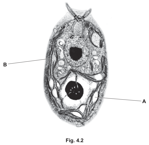
Structures A and B in Fig. 4.2 are found within the chloroplast.
Name structures A and B.
A _____
B _____
Answer
A: starch grain (granule) ;
B: thylakoid (membrane) / lamella / granum ;
A = starch grain/granule; B = thylakoid (membrane) / lamella / granum
Background Concept
A chloroplast is a double-membraned organelle specialised for photosynthesis. Inside, the thylakoid membrane system is folded into stacks called grana (singular: granum), connected by stromal lamellae. The thylakoid membrane carries chlorophyll and the protein complexes of the light-dependent reactions. The fluid stroma surrounding the thylakoids contains the enzymes of the Calvin cycle, starch grains (storage of carbohydrate product) and, in many algae, a pyrenoid — a proteinaceous body where the enzyme Rubisco is concentrated and around which starch is deposited.
In Chlamydomonas the single chloroplast is large and cup-shaped, occupying much of the cell. The pyrenoid is a prominent feature, embedded within the chloroplast and surrounded by starch plates/grains.
Understanding the Question
The question states explicitly that both A and B are found within the chloroplast, so the candidate is identifying sub-chloroplast structures on a transmission electron micrograph of Chlamydomonas. A points to a dense, dark, roughly circular deposit near the base of the cup-shaped chloroplast — the classic appearance of a starch granule in (or attached to) the pyrenoid. B points to the dark, layered, ribbon-like stacks running along the wall of the chloroplast — characteristic of thylakoid membranes (often arranged as grana in Chlamydomonas).
Approach
Match each label to the classic appearance of a chloroplast sub-structure and give the textbook name.
- A: dense, dark, rounded → starch grain (granule).
- B: layered, parallel, dark stripes → thylakoid membrane / lamella / granum.
Step-by-Step Reasoning
The mark scheme accepts:
- A: starch grain or starch granule (1 mark).
- B: thylakoid (membrane) / lamella / granum — any of these is acceptable (1 mark).
Both terms are simply named. The candidate does not need to add descriptions of function, but knowing the structure helps locate it on the unfamiliar micrograph.
Key Takeaways
- In a chloroplast TEM, thylakoids look like dark, stacked, ribbon-like layers; starch grains look like dense, dark, rounded deposits — often lying next to or within a pyrenoid in algae.
- "Thylakoid", "lamella" and "granum" are interchangeable in this context: a granum is a stack of thylakoid discs; a lamella is a single thylakoid disc; stromal lamellae connect grana.
- Starch grains in the chloroplast are temporary storage of the carbohydrate product of photosynthesis — they are not the same as the membrane stacks of thylakoids.
Common Mistakes
- Confusing A with the pyrenoid itself. The pyrenoid is the protein body; the dark deposit is usually a starch grain around the pyrenoid. "Starch grain" is the credit-worthy answer.
- Calling B "chloroplast" or "stroma" — too vague. The thylakoid membrane is a sub-chloroplast structure, and the question insists both A and B are within the chloroplast.
- Calling B "grana" (plural) when the diagram clearly shows a single stack — though "granum" or "grana" is credit-worthy, "grana" alone at this single spot is borderline.
Things to be Careful About
- Use the singular/plural form that matches the structure shown. "Granum" for a single stack; "thylakoid" for a single disc; "starch grain" for a single deposit.
- "Starch grain" and "starch granule" are interchangeable; either scores the mark.
- The question's image description notes the cell is a Chlamydomonas with a "cup-shaped chloroplast" — this is consistent with the structures named.
Thylakoid membranes are the site of the light-dependent stage of photosynthesis.
Answer
ATP and reduced NADP (and oxygen).
ATP and reduced NADP (and oxygen)
Background Concept
The light-dependent stage of photosynthesis takes place on the thylakoid membranes. Light energy absorbed by chlorophyll (in photosystems II and I) is used to: (1) photolyse water, releasing , electrons and protons; (2) drive an electron transport chain that pumps protons into the thylakoid lumen; (3) reduce NADP to reduced NADP using the electrons from water; and (4) drive chemiosmotic synthesis of ATP by ATP synthase as protons flow back out of the lumen. The overall products are therefore ATP, reduced NADP and .
Understanding the Question
The command word is "name", and only 1 mark is available, so a brief statement is needed. The mark scheme uses the wording "ATP and reduced NADP (and oxygen)" — the underscore on "and" is important: the candidate must name BOTH ATP AND reduced NADP for the mark. Oxygen is also a product, listed in brackets, and may be added without penalty.
Approach
Recall the three products of the light-dependent reactions and list them, ensuring both ATP and reduced NADP are named together.
Step-by-Step Reasoning
- Photolysis of water releases → oxygen is a product.
- Electron transport reduces NADP → reduced NADP is a product.
- Chemiosmosis via ATP synthase → ATP is a product.
The 1-mark answer must combine ATP AND reduced NADP, with oxygen as an acceptable extra.
Key Takeaways
- ATP and reduced NADP are the two products that link the light-dependent stage to the Calvin cycle; they are the immediate products most often tested.
- is a by-product of photolysis, not the main reason the light stage exists, but it is still a product.
- "Reduced NADP" is sometimes written as NADPH or NADPH + H; all are accepted at A-level.
Common Mistakes
- Naming only one of the two required products (e.g. "ATP only") — the mark scheme explicitly requires BOTH ATP AND reduced NADP.
- Calling reduced NADP "NADPH" — the modern, accepted CIE term is "reduced NADP" or "NADPH".
- Naming water as a product — water is a reactant, not a product.
Things to be Careful About
- Write "reduced NADP", not "NADP" alone (NADP is the oxidised form, a reactant).
- The wording "and" is essential — the answer needs both, not just one.
Some of the protein components of the thylakoid membrane have a role in the
light-dependent stage of photosynthesis.
Explain the roles of the different proteins that function in the light-dependent stage of photosynthesis.
Answer
- Electron carriers / electron transport chain proteins carry electrons between photosystem II and photosystem I.
- They release energy (as electrons pass along the chain) to pump protons () into the thylakoid space, generating a proton gradient across the thylakoid membrane.
- The oxygen-evolving complex (in photosystem II) catalyses the splitting / photolysis of water, releasing .
- ATP synthase uses the proton gradient (protons flowing back out of the thylakoid lumen through the enzyme) to phosphorylate ADP to ATP (chemiosmosis).
- Light-harvesting complex / antenna / photosystem proteins hold the photosynthetic pigments in the correct orientation so that absorbed light energy is funnelled to the reaction centre.
Electron carriers in the ETC carry electrons and pump protons to form a gradient; the oxygen-evolving complex catalyses photolysis of water; ATP synthase makes ATP using the gradient; LHC/photosystem proteins hold pigments.
Background Concept
The thylakoid membrane contains several distinct protein complexes that work together in the light-dependent stage:
- Photosystem II (PSII) — an antenna complex (light-harvesting complex, LHC) holds pigments (chlorophyll a, chlorophyll b, carotenoids) that absorb photons and pass the excitation energy to the reaction centre. The reaction centre uses this energy to excite electrons. The oxygen-evolving complex on the inner side of PSII catalyses the photolysis of , releasing , protons and electrons.
- Electron transport chain (ETC) — a series of electron carriers (plastoquinone, the cytochrome bf complex, plastocyanin) embedded in the membrane. Electrons from PSII pass along the chain to PSI, releasing energy that is used to pump protons from the stroma into the thylakoid lumen, generating a proton gradient.
- Photosystem I (PSI) — re-energises the electrons using more light, allowing them to reduce NADP to reduced NADP via the enzyme NADP reductase.
- ATP synthase — a transmembrane channel that allows protons to flow down their electrochemical gradient from the thylakoid lumen back to the stroma, using the energy released to phosphorylate ADP to ATP (chemiosmosis).
Understanding the Question
The question asks for the roles of the different proteins in the light-dependent stage. The mark scheme lists six possible creditable points, of which any four are needed for 4 marks. The candidate must therefore identify at least four distinct functions and explain them.
Approach
Mentally work through the thylakoid membrane from light absorption to ATP/NADPH output, and at each step name a protein (or protein complex) and what it does. Then check that four distinct functions have been covered.
Step-by-Step Reasoning
The mark scheme accepts any four of the following six points:
- Electron carriers / ETC proteins carry electrons between the photosystems. (1 mark)
- Electron transport releases energy that pumps protons () into the thylakoid space, generating a proton gradient. (1 mark)
- The oxygen-evolving complex is an enzyme (1 mark) that catalyses photolysis (splitting) of water to release (1 mark — often credited together).
- ATP synthase makes ATP using the proton gradient (chemiosmosis). (1 mark)
- LHC / antenna / photosystem proteins hold the pigments in the membrane, funnelling absorbed light energy to the reaction centre. (1 mark)
- (NADP reductase reduces NADP to reduced NADP — also a valid role for a protein, though not in the mark scheme as a separate line.)
A complete 4-mark answer would, for example, give: (i) electron carriers in the ETC carry electrons, (ii) ETC releases energy to pump protons and generate a gradient, (iii) oxygen-evolving complex catalyses photolysis of water, (iv) ATP synthase makes ATP using the gradient. The role of photosystem/LHC proteins in holding pigments is a strong alternative to (i) or (ii).
Key Takeaways
- The thylakoid membrane is essentially a protein-rich membrane: every functional step of the light-dependent stage is performed by a specific protein or protein complex.
- Chemiosmosis — the generation of a proton gradient and its use by ATP synthase — is a unifying principle connecting photosynthesis and oxidative phosphorylation in mitochondria.
- The "oxygen-evolving complex" is a specific name worth remembering; it is the cluster of manganese ions at the inner face of PSII that catalyses the splitting of water.
Common Mistakes
- Saying "the chlorophyll absorbs light" without naming the protein — the question asks about proteins, not pigments. The pigment is held by the protein.
- Confusing the direction of proton pumping — protons are pumped into the thylakoid lumen, not out of it.
- Saying "ATP synthase creates a proton gradient" — ATP synthase uses the gradient; the ETC creates it.
- Missing the photolysis role of the oxygen-evolving complex — students often say "the ETC releases " rather than locating it at PSII's oxygen-evolving complex.
- Vague "proteins make ATP" without naming ATP synthase.
Things to be Careful About
- Use the technical names: "electron transport chain", "oxygen-evolving complex", "ATP synthase", "light-harvesting complex / photosystem".
- Each marking point is a function — say what the protein does, not what it is.
- If you only have time for three functions, pick the three with the deepest mechanistic content (ETC + proton pumping; oxygen-evolving complex + photolysis; ATP synthase + ATP synthesis).
The products of the light-dependent stage of photosynthesis are used in the Calvin cycle. Calvin cycle intermediates are used to produce amino acids, carbohydrates and lipids.
Name the Calvin cycle intermediate that can be used to produce starch.
Answer
Triose phosphate (TP) ;
Triose phosphate (TP)
Background Concept
The Calvin cycle fixes onto the 5-carbon acceptor ribulose bisphosphate (RuBP) in a reaction catalysed by the enzyme Rubisco. The unstable 6-carbon intermediate splits immediately into two molecules of glycerate 3-phosphate (GP). GP is then reduced (using ATP and reduced NADP from the light-dependent stage) to glyceraldehyde 3-phosphate, more commonly called triose phosphate (TP). TP is the first carbohydrate produced by the Calvin cycle and is the branch point for almost all other photosynthetic products:
- Some TP leaves the cycle to form hexose phosphates (e.g. glucose, fructose), which can be made into sucrose for transport or starch for storage.
- Some TP is used to regenerate the acceptor RuBP (using ATP).
- Some TP is diverted into lipid synthesis (by forming acetyl-CoA) and into amino acid synthesis (by transamination of keto acids derived from TP).
Understanding the Question
The question asks for the Calvin cycle intermediate that is used to produce starch. The answer must be the 3-carbon sugar that is the direct precursor of all carbohydrates in the cycle, including starch. Only 1 mark.
Approach
Identify the immediate product of carbon fixation that is the precursor of all carbohydrates. The chain is: → GP → TP → hexose phosphate → starch. The intermediate that links the Calvin cycle to starch is therefore TP.
Step-by-Step Reasoning
- Starch is a polymer of glucose.
- Glucose is made in the Calvin cycle from two TP molecules combining to form a hexose phosphate.
- The Calvin cycle intermediate directly feeding into hexose synthesis is triose phosphate.
- Hence, triose phosphate is the answer.
Key Takeaways
- Triose phosphate (TP) is the first carbohydrate made by photosynthesis and the "gateway" to all the other major classes of biological molecule.
- Starch is built from TP, but only indirectly — TP must first be combined with another TP to make a hexose, and several further reactions are needed before starch.
- The other common Calvin cycle intermediates (GP, RuBP) are not direct starch precursors.
Common Mistakes
- Saying "glucose" or "glycerate 3-phosphate (GP)" — these are wrong. Glucose is the product of further reactions on TP, and GP is the immediate product of carbon fixation, not the starch precursor.
- Saying "RuBP" — RuBP is the acceptor that is regenerated, not the carbohydrate output.
- Spelling "triose phosphate" incorrectly (e.g. "triose-P", "TrioP") — at A-level, write the full name or the standard abbreviation TP.
Things to be Careful About
- The full name "triose phosphate" or the abbreviation "TP" are both credit-worthy. "GALP" (glyceraldehyde 3-phosphate) is also an accepted CIE synonym.
- Note that TP exists as a phosphate ester — "triose phosphate", not just "triose".
Scientists claim that the artificial photosynthesis process shown in Fig. 4.1 is more efficient at converting light energy into food than normal photosynthesis by crop plants.
Give reasons why this claim may or may not be true.
true _____
not true _____
Answer
True (reasons the claim may be correct):
- The solar panel can absorb/use a wider range of wavelengths/colours of light than chlorophyll in plant leaves, so more of the incident light energy is harvested.
- The algae can grow in the dark on acetate, so the system is not limited by light intensity, day length or seasonal variation — production can continue round the clock.
- (Alternative) Crop plants lose fixed carbon by respiration, but in the artificial system the only respiring organism is the alga, which is harvested at the end of a rapid growth cycle, so respiration losses are smaller.
Not true (reasons the claim may be incorrect):
- Solar panels themselves have a low efficiency (typically only ~15–20%) at converting light energy into electricity, so a large amount of light energy is lost at the very first step of the chain.
- Additional energy inputs are required (e.g. to drive the electrolysis, to mix and pump the algal culture), which further reduce the net efficiency.
- (Alternative) The food product is algae — potentially less palatable and less nutritionally complete than conventional crop food; or a large area of solar panels is required to capture enough light.
True: solar panel uses a wider spectrum; algae grow in the dark so are not limited by daylight. Not true: solar panel efficiency is low; additional energy inputs are needed; food quality may be lower.
Background Concept
Natural photosynthesis by crop plants is famously inefficient at converting incident sunlight into chemical energy stored in food — typical values are around 1–2% for the whole plant over a growing season, because:
- Plants can only use light of wavelengths absorbed by their pigments (mainly chlorophyll a and b — blue and red light).
- Photosynthesis is limited by the slowest of light intensity, concentration and temperature (Blackman's law of limiting factors).
- Plants lose fixed carbon by respiration, both day and night.
- Only a small fraction of the plant is edible (leaves, fruits, seeds, tubers — not roots, stems, woody tissue).
- Plants have a long generation time and are subject to pests, disease and weather.
The artificial system in Fig. 4.1 uses a solar panel (which is engineered to absorb a wide spectrum, although with low conversion efficiency) and an electrolyser (which can be optimised to operate continuously at a chosen partial pressure, temperature and pressure) to produce acetate, which is then fed to Chlamydomonas. The algae are grown heterotrophically in the dark.
Understanding the Question
The question asks the candidate to evaluate the claim — i.e. give reasons both for and against it. The mark scheme is phrased as "any two plus one opposing arguments", meaning the candidate must offer two reasons supporting the claim and at least one counter-argument (or vice versa) for a total of three marks. The candidate should structure the answer under "true" (reasons the claim may be correct) and "not true" (reasons the claim may be incorrect).
Approach
Think systematically about where each system gains or loses energy:
- Light capture efficiency
- Energy losses at each step of the conversion chain
- Losses to respiration
- Limiting factors that constrain the rate
- Quality of the food product
- Resources and space needed
Pick the strongest two points for one side, and one strong point for the other.
Step-by-Step Reasoning
The mark scheme lists 18 possible marking points, but only three are needed. The strongest biological arguments are:
In favour of the claim (true):
- The solar panel absorbs a wider range of wavelengths than chlorophyll, so a higher fraction of the light energy is captured (mark scheme point 2 — absorption spectrum / wavelengths used).
- The algae grow in the dark on acetate, so the system is not subject to the limiting factors of light intensity, day length or weather (mark scheme point 8 — night / dark process; also point 6/7 — limiting factors).
- (Optionally) Plants lose fixed carbon by respiration; algae grown heterotrophically in the dark and harvested at the end of a rapid growth cycle lose less to respiration (mark scheme point 3).
Against the claim (not true):
- Solar panels themselves have a low efficiency (typically 15–22% for converting sunlight to electricity) — much of the light energy is lost at the very first step (mark scheme point 4/5 — stage where energy is lost; also point 1 — percentage/amount of light absorbed).
- Additional energy inputs are required — e.g. the electrolysis reaction, mixing, pumping and maintaining the algae culture (mark scheme point 11/12).
- The food product is algae, which is less palatable and less familiar to consumers than conventional crop food; it may also be nutritionally less complete (mark scheme point 9 — quality/nutrition/palatability of food product).
- (Optionally) A large area of solar panels is required to capture enough light (point 16).
A strong answer gives two clear reasons for the claim and one clear reason against it, with the biological reasoning attached.
Key Takeaways
- A meaningful efficiency comparison must look at every step of the energy-conversion chain, not just one step in isolation.
- Natural photosynthesis by crop plants is limited by Blackman's law — the slowest of light, and temperature caps the rate.
- "Efficiency" depends on what is being measured — light-to-food energy conversion, edible biomass per hectare, or simply grams of food per unit area.
Common Mistakes
- Only giving arguments for one side. The question asks for reasons the claim may or may not be true — both sides are required.
- Vague answers like "it's better because it's more modern" or "it's not better because it's expensive". The mark scheme rewards specific biological reasons — wavelengths absorbed, named limiting factors, energy losses at each stage.
- Forgetting to mention the biological reasons. The candidate should anchor each argument in photosynthesis, respiration or ecology, not in pure engineering.
- Saying "plants use 100% of the light they absorb, but solar panels only 20%" — plants use a much smaller fraction of incident sunlight than this, because most wavelengths are not absorbed.
Things to be Careful About
- Quote a named biological concept (limiting factor, respiration, action spectrum) for each reason.
- Where possible, give a number or a comparison (e.g. "solar panel efficiency ~20% vs crop photosynthesis ~1–2%").
- Structure the answer clearly under "true" and "not true" so the examiner can find each marking point.
The Galápagos is a group of islands with a high biodiversity.
Geospiza fortis is one of the many species of finch that live on the Galápagos islands. On one of the Galápagos islands, Daphne Major, scientists measured the bill size (length, width and depth) of individuals in a population of G. fortis over several years.
Fig. 5.1 shows a G. fortis female.
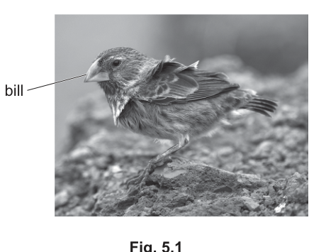
G. fortis feed on seeds. Seed size and bill size vary. It is easier for G. fortis with smaller bills to eat small seeds and for G. fortis with larger bills to eat large seeds.
In 1977, a drought occurred on Daphne Major, which resulted in a large decrease in the availability of seeds, particularly small seeds.
Scientists observed an increase in the mean bill size in the G. fortis population on Daphne Major after the drought.
State the type of natural selection that occurred in the G. fortis population on Daphne Major as a result of the drought.
Answer
Directional (selection).
Directional selection
Background Concept
Natural selection is the differential survival and reproduction of individuals due to differences in heritable traits. It has three main modes:
- Stabilising selection: selection acts against the extremes; the mean phenotype is unchanged but variance decreases. The classical example is human birth weight, where very small or very large babies have lower survival.
- Directional selection: selection acts against one extreme (e.g. small bills), shifting the mean phenotype towards the other extreme (e.g. large bills). Common after an environmental change that alters the selection pressure.
- Disruptive (diversifying) selection: selection acts against the mean (intermediate) phenotype, favouring both extremes and often producing a bimodal distribution.
A useful way to identify the mode is to ask what happens to the mean and the variance: directional shifts the mean, stabilising keeps the mean but reduces variance, disruptive keeps (or splits) the mean and increases variance.
Understanding the Question
G. fortis on Daphne Major feed on seeds. After the 1977 drought, small seeds became scarce. Individuals with larger bills could crack the remaining larger seeds, while individuals with small bills were disadvantaged. The observed result was an increase in the mean bill size of the population. The question asks for the named type of natural selection responsible.
Approach
- Identify the change in the mean: mean bill size has increased.
- Identify which extreme has been selected against: small bills (because their preferred food, small seeds, has become scarce).
- The phenotype has shifted in one direction → directional selection.
Step-by-Step Reasoning
- The drought removed most of the small seeds from Daphne Major. The new selection pressure was seed (food) size/availability.
- Birds with small bills lost their preferred food source, so they were less able to feed, had lower survival/reproductive success, and contributed fewer offspring to the next generation.
- Birds with larger bills were already able to crack large seeds and now had little competition for those seeds, so they had a selective advantage.
- The phenotype distribution of survivors therefore shifted to the right (larger bills), and the mean bill size increased.
- Selection acting against one extreme and shifting the mean in one direction is, by definition, directional selection.
Key Takeaways
- A shift in the mean phenotype, with one extreme being selected against, is directional selection.
- The Galápagos finches, studied by Peter and Rosemary Grant, are a classic textbook example — drought favours larger bills (as on Daphne Major in 1977) and a wet year can favour smaller bills.
Common Mistakes
- Writing 'natural selection' alone is not enough — the question asks for the type (directional/stabilising/disruptive).
- Confusing 'directional' with 'disruptive': disruptive selection produces two peaks (bimodal), not a single shifted peak.
Things to Be Careful About
- The single mark is for the precise term 'directional (selection)'. 'Directional evolution' or 'selection by drought' would not earn the mark.
Fig. 5.2 shows the distribution of bill size in the G. fortis population before the drought in 1977.
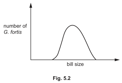
Sketch a new curve on Fig. 5.2 to show the distribution of bill size in the G. fortis population after the drought.
Answer
Draw a second bell-shaped curve on Fig. 5.2 whose starting point, peak and end point on the x-axis all lie to the right of the original curve's corresponding points (i.e. a normal-shaped curve of similar width centred on a larger bill size).
A new bell-shaped (normal) distribution curve, of similar width to the original, but shifted to the right so its start, peak and end all sit at larger bill sizes than the original.
Background Concept
Directional selection shifts the mean of a phenotype distribution towards the favoured extreme, while the shape of the distribution (its width / standard deviation) is approximately preserved. Graphically, the original normal curve is displaced sideways without becoming markedly narrower or broader, and without splitting into two peaks.
Understanding the Question
You are given Fig. 5.2, a normal (bell-shaped) distribution of bill size in the G. fortis population on Daphne Major before the 1977 drought. You must sketch a second curve on the same axes showing the distribution after the drought, when the mean bill size has increased.
Approach
- The new distribution must still look like a normal (bell-shaped) curve — directional selection does not change the shape, only the position.
- The whole curve (start, peak and end on the x-axis) must move to the right, towards larger bill size, because larger-billed birds survived the drought better.
- The width of the new curve should be similar to the original — selection has not narrowed or split the distribution.
Step-by-Step Reasoning
- After the drought, small-billed individuals had lower survival because their preferred small seeds were scarce.
- Large-billed individuals had higher survival and contributed more offspring.
- The mean bill size increased; the distribution did not become narrower (stabilising) and did not split into two peaks (disruptive).
- Therefore the correct sketch is a normal-shaped curve of similar width whose start, peak and end are all displaced to the right along the bill-size axis.
- The mark scheme rewards exactly this: the new curve's starting point, peak and end point must all sit to the right of the original's.
Key Takeaways
- Directional selection = a sideways shift of the entire distribution.
- The width is approximately unchanged, in contrast to stabilising selection (curve narrows) or disruptive selection (curve becomes bimodal).
Common Mistakes
- Drawing the new curve as taller and narrower — that would represent stabilising selection, not directional.
- Drawing only the right-hand half of the new curve, or raising only the right-hand side of the original — selection acts on the whole population's mean, not just one tail.
- Drawing a bimodal distribution — that would be disruptive selection, seen on Santa Cruz in part (b), not on Daphne Major after a drought.
Things to Be Careful About
- The mark scheme requires all three reference points (start, peak, end on the x-axis) to be to the right of the original. Moving only the peak is insufficient.
- Keep the new curve roughly the same width and shape as the original.
G. fortis also lives on Santa Cruz, another island in the Galápagos.
Most of the seeds available for G. fortis to eat on Santa Cruz are either small or large. There are few intermediate sizes of seed.
Fig. 5.3 shows the distribution of bill size in the G. fortis population on Santa Cruz.
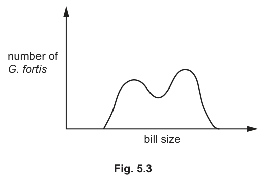
Explain how natural selection in the G. fortis population on Santa Cruz has resulted in the distribution in bill size shown in Fig. 5.3.
Answer
- The type of natural selection is disruptive (or diversifying) selection.
- The selection pressure is the availability of food (seeds) of mainly two sizes — small or large — with few intermediate-sized seeds on Santa Cruz.
- Birds with intermediate-sized bills cannot efficiently eat either small or large seeds; they are out-competed for food by both small-billed and large-billed birds.
- Therefore the extreme phenotypes (small-billed and large-billed birds) have a selective advantage: they can exploit the small or large seeds respectively, so they are more likely to survive and reproduce than intermediate-billed birds.
- Over generations, the allele frequencies for the genes controlling bill size change so that the alleles for small bills and for large bills become more common in the population, while alleles for intermediate bills become less common. This produces the bimodal distribution seen in Fig. 5.3.
Disruptive (diversifying) selection: birds with intermediate bills cannot eat either small or large seeds effectively, so small- and large-billed extremes are selected for; over time the alleles for small and large bills increase in frequency and the alleles for intermediate bills decrease, producing a bimodal distribution.
Background Concept
Disruptive (diversifying) selection occurs when individuals at both extremes of a phenotypic range have higher fitness than those near the mean. The intermediate phenotype is selected against. The classic graphical signature of disruptive selection is a bimodal distribution — two peaks with a trough at the mean — because the intermediate individuals survive and reproduce less successfully than the extremes.
This contrasts with:
- stabilising selection (mean unchanged, variance reduced) and
- directional selection (mean shifts to one side).
For disruptive selection to be sustained, the environment must offer two distinct resources that favour two different phenotypes — exactly the situation in this question, where seed sizes on Santa Cruz are bimodal.
Understanding the Question
G. fortis on Santa Cruz face a food supply that is bimodal: most available seeds are either small or large, with very few of intermediate size. The current population of G. fortis on Santa Cruz also shows a bimodal bill-size distribution (Fig. 5.3). The question asks you to explain how natural selection has produced this bimodal distribution from an originally unimodal one.
Approach
- Identify the type of selection: with two favoured extremes, this is disruptive selection.
- Identify the selection pressure: the bimodal seed-size distribution on Santa Cruz.
- Explain why the intermediate phenotype is at a disadvantage: intermediate-billed birds cannot crack large seeds (bill too small) and cannot pick up small seeds efficiently (or are out-competed by the better-adapted extremes).
- State which phenotypes are favoured: both extremes (small-billed and large-billed birds).
- Describe the genetic consequence: change in allele frequencies — alleles for small and large bills increase; alleles for intermediate bills decrease.
Step-by-Step Reasoning
- Selection pressure: food availability is bimodal (small and large seeds, few intermediates). So bill size has to match one of two resource sizes to be advantageous.
- Intermediate disadvantage: a bird with a mid-sized bill is poorly matched to both seed types — it cannot efficiently crack large seeds and it loses the small seeds to the more efficient small-billed birds. Competition for both seed types therefore reduces the fitness of intermediate-billed birds.
- Extreme advantage: small-billed birds efficiently eat small seeds; large-billed birds efficiently eat large seeds. Both extremes therefore have higher survival and reproductive success than the intermediate phenotype.
- Differential reproduction: extreme-billed birds pass more alleles to the next generation; intermediate-billed birds pass fewer. Over generations, the frequency of alleles producing small bills and the frequency of alleles producing large bills both rise, while the frequency of alleles producing intermediate bills falls.
- Outcome: the population's bill-size distribution becomes bimodal, exactly as shown in Fig. 5.3.
Key Takeaways
- A bimodal resource or habitat tends to drive disruptive selection.
- Intermediate phenotypes are at the greatest disadvantage when the resources themselves are bimodal — they are 'jacks of both trades and masters of none'.
- Disruptive selection can be a precursor to sympatric speciation if the two sub-populations become reproductively isolated.
Common Mistakes
- Naming the type of selection as 'directional' — that is a shift in one direction, not a split into two peaks.
- Naming it as 'stabilising' — the opposite; stabilising reduces variance.
- Saying only that 'birds with bigger bills survive' without also addressing the small-billed birds — disruptive selection favours both extremes.
- Failing to mention the intermediate disadvantage; this is the key feature that distinguishes disruptive from directional selection.
- Using vague terms like 'adapt' or 'evolve' without explaining the mechanism of differential survival and reproduction.
Things to Be Careful About
- Use the precise term 'disruptive (selection)' or 'diversifying (selection)'.
- The mark scheme credits any four of six points, but a complete answer hits all of them: type, selection pressure, competition, intermediate disadvantage, extreme advantage, change in allele frequency.
Scientists studied the effect of an increase in the number of tourists each year on the number of invasive alien species present in the Galápagos islands.
The scientists recorded the number of invasive alien species present each year in the Galápagos islands over many years. The scientists also recorded the number of tourists visiting the islands each year.
Fig. 5.4 shows a graph of the data for five different years during the study.
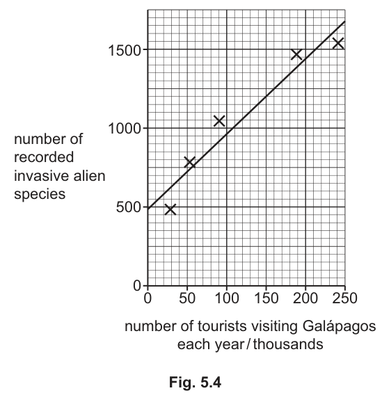
The scientists carried out a Pearson’s linear correlation calculation for the data in Fig. 5.4.
The scientists calculated an value of .
The scientists concluded that there is a significant correlation between the number of tourists visiting the Galápagos islands each year and the number of invasive alien species present.
Table 5.1 shows a probability table of critical values for Pearson’s linear correlation.
Table 5.1
| number of observations | probability level () 0.05 | probability level () 0.01 |
|---|---|---|
| 3 | 0.997 | 1.000 |
| 4 | 0.950 | 0.990 |
| 5 | 0.878 | 0.959 |
| 6 | 0.811 | 0.917 |
| 7 | 0.754 | 0.875 |
With reference to Table 5.1, explain why the value of indicates that there is a significant correlation between the number of tourists and the number of invasive alien species.
Answer
- The number of observations (years) is n = 5.
- From Table 5.1, the critical value of r for n = 5 at probability level p = 0.05 is 0.878.
- The calculated value r = 0.930 is greater than 0.878.
- Therefore the correlation is significant at the 5% probability level (i.e. there is less than a 5% probability that a correlation this strong would arise by chance).
r = 0.930 is greater than the critical value (0.878) for n = 5 at p = 0.05, so the correlation is significant at the 5% level.
Background Concept
Pearson's linear correlation measures the strength and direction of a linear relationship between two variables. The coefficient r lies between −1 (perfect negative correlation) and +1 (perfect positive correlation), with 0 meaning no linear correlation.
However, r calculated from a sample is an estimate; a small sample could give a moderately large |r| purely by chance. To decide whether an observed r is statistically significant, the calculated value is compared with a critical value from a probability table for the appropriate number of observations (n) and chosen probability level (usually p = 0.05 or p = 0.01).
- If |r (calculated)| > |r (critical)|, the correlation is significant at that p value (i.e. the probability that it arose by chance is less than p).
- If |r (calculated)| ≤ |r (critical)|, the correlation is not significant at that p value.
Degrees of freedom for Pearson's r is n − 2, but the critical-value table here is tabulated by n, so you read off the row matching the number of observations (years of data) directly.
Understanding the Question
The scientists have five data points (five years), giving n = 5. They have calculated r = 0.930 and want to know whether this provides evidence of a significant correlation between the number of tourists and the number of invasive alien species. You have to use Table 5.1 to justify the conclusion.
Approach
- Identify n from the scatter graph description (five plotted points → n = 5).
- Read the critical value of r for n = 5 at p = 0.05 from Table 5.1.
- Compare the calculated r with the critical value.
- State the conclusion using the standard phrasing ('greater than the critical value at p = 0.05' ⇒ significant).
Step-by-Step Reasoning
- n = 5 — there are five observations (one per year shown on Fig. 5.4).
- Reading Table 5.1, the critical value of r for n = 5 at p = 0.05 is 0.878.
- The calculated r is 0.930.
- 0.930 > 0.878, so the calculated r exceeds the critical value at the 5% level.
- The probability that a correlation this strong could occur by chance (if the true correlation were zero) is less than 0.05 (5%), so the correlation is statistically significant at p = 0.05.
- The same conclusion holds at p = 0.01 (critical value 0.959): here 0.930 < 0.959, so the correlation is not significant at the 1% level — but the mark scheme only requires the p = 0.05 justification.
Key Takeaways
- Significance is judged by comparing the calculated statistic with a critical value for the sample size and chosen probability level.
- p = 0.05 means 'there is a 5% probability that a result this extreme would occur by chance if there were truly no correlation'.
- A 'significant' correlation is not necessarily a strong or causal one — it just means unlikely to be due to chance, given the sample size.
Common Mistakes
- Using the wrong n (e.g. quoting the critical value for n = 4 or n = 6).
- Comparing r with the critical value for the wrong probability level (e.g. quoting 0.959 from the p = 0.01 column when justifying significance at p = 0.05).
- Stating that 'r is greater than 0.05' — that is comparing r to a probability, which is meaningless.
- Saying 'the correlation is significant' without quoting the critical value and the comparison.
Things to Be Careful About
- Quote both the critical value (0.878) and the probability level (p = 0.05) explicitly. The mark scheme awards one mark for each.
- A small sample (n = 5) means a very high threshold (0.878) for significance — even a strong-looking correlation can fail to reach it. Here r = 0.930 just exceeds it.
Invasive alien species are thought to have contributed to extinctions recorded in the Galápagos islands.
Discuss the negative effects of introducing invasive alien species to an ecosystem.
Answer
Invasive alien species can negatively affect an ecosystem by:
- Disrupting food chains / food webs — the alien species introduces new trophic links or removes existing ones, destabilising the ecosystem.
- Competing with native species for food, or for the same niche, so the native species has fewer resources and may decline.
- Predation / grazing on native species — the alien species eats native organisms that have no defence against it, reducing native populations.
- Introducing new diseases or parasites to which native species have no resistance, causing population decline or local extinction.
- Changing or damaging the habitat — e.g. altering vegetation, soil chemistry or water flow so that native species can no longer survive there.
- Being toxic or threatening human health (e.g. through allergens, toxins or as vectors of human disease).
- Damaging tourism or agriculture, with knock-on economic and social effects on the local human population.
Invasive alien species disrupt food webs, compete with native species, prey on or graze native species, introduce new diseases/parasites, change or damage the habitat, can be toxic / threaten human health, and can damage tourism and agriculture.
Background Concept
An invasive alien species is a non-native species that has been introduced outside its natural range and spreads aggressively, often causing ecological, economic or human-health harm. Native species have not co-evolved with the invader, so they typically lack behavioural, physiological or immunological defences against it. Common routes of introduction include ballast water, deliberate release, escapes from agriculture or horticulture, contaminated seed, and — relevant to the Galápagos — accidental transport by humans, including tourists.
The impacts can be grouped into broad categories:
- Trophic disruption (food webs broken or rewired)
- Competition (for food, space, light, nesting sites)
- Predation / herbivory (eating natives)
- Disease / parasitism (novel pathogens)
- Habitat alteration (e.g. changing fire regimes, soil chemistry, hydrology)
- Direct human effects (toxicity, allergens, disease vectors)
- Economic effects (losses to agriculture, fisheries, tourism)
Understanding the Question
You are given that invasive alien species are thought to have contributed to extinctions on the Galápagos and are positively correlated with tourist numbers (part c(i)). The question asks you to discuss the negative effects of introducing invasive alien species to an ecosystem — i.e. to give a structured, biological account of the ways in which such introductions cause harm. The mark scheme offers 4 marks from a list of 7 creditable points, so a strong answer will cover at least four distinct categories.
Approach
Aim for breadth, not depth — four clearly distinct points each scoring one mark. Use biological terminology (food web, niche, native species, habitat) rather than vague phrases like 'they damage nature'. Where possible, give a specific mechanism (how does the harm arise?) rather than a label (what is the harm?).
Step-by-Step Reasoning
- Food web disruption: an alien species introduces new feeding links or removes existing ones. For example, a plant-eating insect from elsewhere may consume a native plant that previously had no major herbivores, reducing the food supply for the native herbivores that depend on it.
- Competition with native species: by occupying the same niche or eating the same food, an invasive species reduces the resources available to natives, lowering their survival and reproductive rate. This is the classic mechanism of displacement.
- Predation / grazing on natives: native prey often lack anti-predator adaptations (camouflage, chemical defences, escape behaviour) against an unfamiliar predator. Examples include rats preying on ground-nesting birds' eggs and chicks on the Galápagos.
- Introduction of new diseases or parasites: native hosts have no evolved immunity, so an introduced pathogen can sweep through a naïve population, sometimes causing local or even global extinction.
- Habitat alteration: some invasives change the physical environment — e.g. by forming monocultures, altering fire frequency, fixing nitrogen or salinising soil — making the habitat unsuitable for species that previously lived there.
- Toxicity / human-health risk: some invasives produce toxins, allergens, or act as vectors of human disease (e.g. invasive mosquitoes carrying dengue or malaria).
- Damage to tourism / agriculture: economic and livelihood losses feed back into reduced capacity to manage conservation, compounding the ecological harm.
Key Takeaways
- A complete answer hits several different categories of impact, not just several versions of the same one.
- The Galápagos are a textbook example: tourism brings alien species on clothing, luggage and ships, and invasives such as rats, goats, cats and plants have devastated the islands' endemic fauna and flora.
- Invasive species are now considered one of the leading drivers of biodiversity loss worldwide, alongside habitat destruction and climate change.
Common Mistakes
- Repeating the same idea in different words (e.g. 'they eat native animals' and 'they hunt native species' both count as one point, not two).
- Vague statements such as 'they damage the environment' or 'they cause extinctions' — these earn no credit without an explanatory mechanism.
- Forgetting that 'competition' and 'predation' are different mechanisms and should be stated separately.
- Bringing in unrelated factors (climate change, habitat loss) that the question does not ask about.
Things to Be Careful About
- The question is about negative effects of introducing invasive alien species — keep the focus on the alien species as the cause.
- Four distinct, well-stated points will reliably reach 4 marks. The mark scheme allows seven, so you have plenty of options.
State two factors, other than competition and the introduction of invasive alien species, that can cause extinction.
1 _____
2 _____
Answer
- Climate change
- Hunting by humans (over-hunting / overexploitation)
(Other acceptable answers: habitat degradation / loss, named pollution such as pesticide or oil, disease, named natural disasters such as volcanic eruption or tsunami.)
Any two of: climate change; hunting by humans; habitat degradation/loss; (named) pollution; disease; (named) natural disaster.
Background Concept
Extinction is the permanent loss of a species from Earth (or, in conservation biology, locally from a region). The major anthropogenic drivers of extinction — sometimes remembered by the acronym HIPPO: Habitat loss, Invasive species, Pollution, Population growth / over-harvesting, Other (climate change, disease) — are well established. The question has already excluded invasive species and competition (a sub-component of over-exploitation and habitat change), so you must list two from the remaining categories.
Understanding the Question
This is a simple two-mark recall question: name any two factors (other than competition and invasive alien species) that can cause extinction. Each answer is worth one mark, so two clearly stated causes earn both marks.
Approach
Pick two of the strongest, most easily defensible causes from the standard list. Avoid the two that the question has already excluded (competition, invasive species). Use the specific term the mark scheme credits, not a vague paraphrase.
Step-by-Step Reasoning
The mark scheme accepts any two from:
- Climate change — shifting temperature and rainfall patterns push species beyond their tolerance ranges; corals bleached by warming seas are a current example.
- Hunting by humans (over-hunting, over-exploitation) — e.g. the dodo, passenger pigeon, many whale species reduced by commercial whaling.
- Degradation / loss of habitat — e.g. deforestation of tropical rainforests, drainage of wetlands, conversion of grassland to arable farmland.
- (Named) pollution — e.g. pesticide run-off, plastic pollution in oceans, oil spills, eutrophication, acid rain.
- Disease — e.g. chytridiomycosis in amphibians.
- (Named) natural disaster — e.g. volcanic eruption, tsunami, asteroid impact (the Cretaceous–Paleogene event ended the non-avian dinosaurs).
Key Takeaways
- Extinction has multiple, often interacting causes; the 'HIPPO' acronym is a useful mnemonic.
- The question excludes two common causes, so be ready to produce a list of others — climate change, habitat loss, hunting, pollution, disease, natural disasters.
Common Mistakes
- Writing 'competition' or 'invasive species' — these are explicitly excluded by the question.
- Writing 'humans' or 'human activity' — too vague to earn the mark; a specific human activity (e.g. hunting, deforestation, pollution) is needed.
- Writing 'global warming' instead of 'climate change' — usually accepted, but 'climate change' is the more precise term that the mark scheme uses.
Things to Be Careful About
- One factor per line; the mark scheme awards one mark per correctly stated cause.
- Avoid duplication: 'overhunting' and 'habitat loss' are not the same as 'competition' or 'invasive species', so they are fine.
In the kidney, various molecules move between the blood and the kidney nephron.
Table 6.1 describes the movements of molecules at three regions of the nephron, P, Q and R.
Table 6.1
| region of nephron | molecule(s) | direction of movement |
|---|---|---|
| P | amino acids, glucose and water | from nephron to blood |
| Q | water | from nephron to blood in the presence of antidiuretic hormone (ADH) |
| R | water and molecules with a molecular mass of less than 68,000 | from blood to nephron |
Name the processes, other than diffusion, osmosis and active transport, that are occurring at P, Q and R.
P _____
Q _____
R _____
No worked solution for this part yet — the official mark scheme is linked at the top of the page.
Identify the regions of the nephron represented by P, Q and R.
P _____
Q _____
R _____
No worked solution for this part yet — the official mark scheme is linked at the top of the page.
A student cut a fresh kidney lengthways and placed one half in the freezer. After 24 hours, the student examined the kidney section and tested its firmness with a mounted needle.
Sodium chloride concentration affects the freezing point of a solution.
The student drew the diagram in Fig. 6.1 to show an area that had frozen hard and an area that was softer and less frozen.
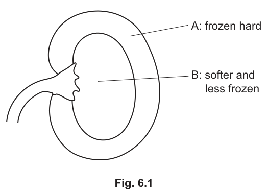
Suggest an explanation for the observations in Fig. 6.1 made by the student.
No worked solution for this part yet — the official mark scheme is linked at the top of the page.
Insulin is an example of a cell-signalling molecule of the endocrine system.
Outline why insulin can be described as an example of a cell-signalling molecule of the endocrine system.
Answer
- Insulin is a hormone (a chemical messenger) produced and secreted by an endocrine gland.
- It is secreted by the β (beta) cells of the islets of Langerhans in the pancreas in response to a stimulus (e.g. raised blood glucose concentration).
- Insulin is released into the blood and travels in the bloodstream around the body.
- It binds to specific receptors on the surface of target cells (e.g. liver, muscle and adipose cells), which recognise insulin as a signal.
- Binding triggers a response in the target cells — such as increased uptake of glucose from the blood — so insulin acts as a cell-signalling molecule of the endocrine system.
Insulin is a hormone secreted by the β cells of the pancreatic islets into the blood, where it travels to and binds receptors on target cells to trigger a response.
Background Concept
The endocrine system is one of the two major communication systems of the body (the other being the nervous system). Endocrine glands are ductless glands that secrete hormones — chemical messengers — directly into the blood. A hormone is defined as a chemical produced by an endocrine gland, transported in the blood, which acts on target cells carrying specific receptors, to produce a specific response. For a molecule to qualify as a cell-signalling molecule of the endocrine system it must therefore: (1) be made by an endocrine gland, (2) be released into the bloodstream, (3) be carried in the blood to distant target cells, (4) bind to specific receptors on those target cells, and (5) trigger a biological response.
Insulin is a peptide hormone produced by the β cells of the islets of Langerhans in the pancreas. The stimulus for its release is a rise in blood glucose concentration (e.g. after a carbohydrate-rich meal). Once released it travels in the blood and binds to insulin receptors on the plasma membrane of target cells — principally hepatocytes (liver), myocytes (muscle) and adipocytes (fat). Binding activates intracellular second-messenger cascades that increase glucose uptake (by inserting GLUT4 transporters into the membrane) and promote glycogenesis and lipogenesis, lowering blood glucose.
Understanding the Question
This is an "outline" question — the candidate must hit the key defining points of an endocrine cell-signalling molecule and apply them to insulin. The mark scheme (4 marks) allows any four from a list of creditable points. There is no graph to interpret; the answer comes from biological knowledge of how insulin works.
Approach
The most efficient way to score the four marks is to state, in order, the five classic defining features of a hormone and apply each to insulin. The examiner will accept any four, so the candidate should aim to cover as many as possible, naming the gland, the transport route, the target cells, the receptor interaction and the response.
Step-by-Step Reasoning
- State that insulin is a hormone — this is the umbrella term that the rest of the points flesh out.
- Name the source as an endocrine gland — the β cells of the islets of Langerhans in the pancreas. Naming the tissue shows understanding that endocrine glands are ductless.
- Note the stimulus for release (raised blood glucose), which demonstrates that secretion is regulated rather than continuous.
- State that insulin travels in the blood — this is what makes it endocrine rather than paracrine.
- State that it binds to specific receptors on target cells (liver, muscle, adipose) — the receptor concept is essential because it explains why only certain cells respond.
- State that binding triggers a response (e.g. increased glucose uptake) — closing the loop from signal to effect.
Key Takeaways
- A hormone is a chemical messenger secreted by an endocrine gland into the blood that acts on specific target cells with the appropriate receptor.
- Insulin fits every one of those criteria: β cells of pancreas → blood → insulin receptors on liver, muscle and adipose cells → increased glucose uptake.
- "Outline" means a brief statement of the main points, not a detailed essay.
Common Mistakes
- Writing "insulin is secreted by the pancreas" without specifying β cells or the islets of Langerhans — the mark scheme specifically credits the named tissue.
- Saying insulin "tells cells what to do" without mentioning a receptor — the receptor is the mark-bearing point.
- Confusing endocrine (blood-borne, distant target) with paracrine (local diffusion) signalling.
- Describing nervous-system features (e.g. "carried along neurones") — insulin is not a neurotransmitter.
Things to Be Careful About
- Use the precise term "hormone" rather than "chemical" or "messenger".
- Specify that transport is in the blood (plasma), not just "in the body".
- Receptors are on the target cell — not "in" or "near" — to make the mechanism clear.
Researchers compared the effect of two types of meal on the mean blood glucose concentration and the mean blood insulin concentration of twelve volunteers. The measurements were made for three hours after the meal.
Meal A contained 55% carbohydrate, 27% fat and 18% protein.
Meal B contained 3% carbohydrate, 90% fat and 7% protein.
Fig. 7.1 shows the results.
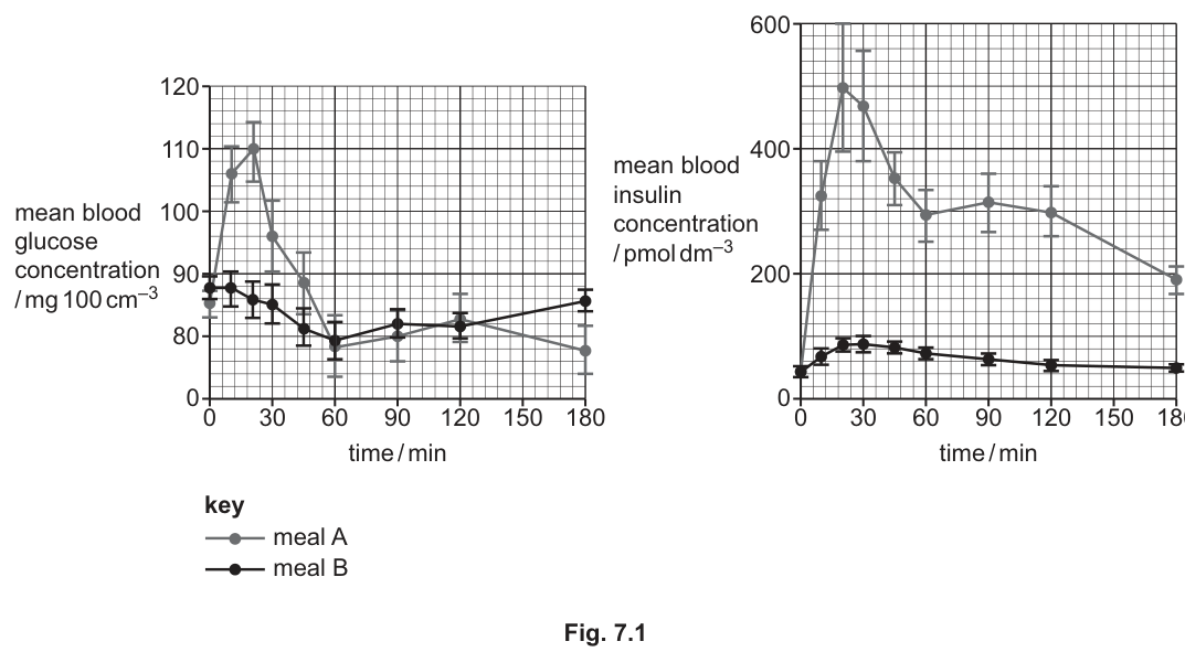
With reference to Fig. 7.1, explain how negative feedback operates to control mean blood glucose concentration in the volunteers who ate meal A.
Answer
- After meal A, mean blood glucose concentration rises to a peak of about at ~15–21 min.
- The rise in blood glucose is detected by the pancreas (β cells of the islets of Langerhans), which respond by secreting insulin; mean blood insulin concentration rises to a peak of about .
- Insulin increases the number of GLUT (glucose transporter) proteins in the cell-surface membranes of target cells (liver, muscle and adipose cells), so uptake of glucose from the blood into the cells increases.
- Insulin also stimulates glycogenesis (and lipogenesis), converting the excess glucose to glycogen (and fat) for storage.
- As a result of this increased uptake and storage, mean blood glucose concentration falls back to the normal range (~90 ), i.e. back towards the set point — this return to normal is the negative feedback that opposes the original rise.
Raised blood glucose after meal A is detected by the pancreas, which secretes insulin; insulin increases glucose uptake (via GLUT transporters) and glycogenesis in target cells, lowering blood glucose back to the set point.
Background Concept
Negative feedback is the mechanism by which a deviation from a set point triggers a response that reverses the deviation and restores the original level. Blood glucose control is a textbook example. When blood glucose rises above the set point, β cells of the pancreatic islets secrete insulin; insulin promotes glucose uptake, glycogenesis (glucose → glycogen) and lipogenesis (glucose → fat) in liver, muscle and adipose cells, lowering blood glucose. As blood glucose returns to normal, the stimulus for insulin secretion is removed and insulin output falls — completing the negative feedback loop. The opposite response (raised blood glucose) is mediated by glucagon from α cells.
Understanding the Question
The question supplies Fig. 7.1, two line graphs plotting mean blood glucose and mean blood insulin over 180 min after either meal A (55% carbohydrate) or meal B (3% carbohydrate, mostly fat). For part (b)(i) the candidate must focus on the Meal A curves and explain how the observed changes in blood glucose illustrate negative feedback. The mark scheme requires that the answer be tied to the figure — quoting a value from the graph is what earns one of the four marks.
Approach
A good way to structure the answer is to walk through the feedback loop in the order the graph shows it:
- Identify the rise in blood glucose (with a value from the graph).
- Identify the trigger and response of the pancreas (with a value from the insulin graph).
- Describe what insulin does at the target cells (mechanism of action).
- State the outcome — blood glucose returns to the set point — and link this to "negative feedback".
Step-by-Step Reasoning
- Reading the left-hand graph for Meal A, the blood glucose concentration rises from a baseline of ~88 to a peak of ~110 at about 15–21 min. This rise is the deviation from the set point.
- The rise in blood glucose is detected by the β cells of the islets of Langerhans in the pancreas (chemoreceptors respond to the change in plasma glucose). The β cells respond by secreting insulin, which appears in the blood (right-hand graph) at a peak of ~500 at about 15–21 min.
- Insulin travels in the blood and binds to insulin receptors on target cells (hepatocytes, myocytes, adipocytes). This increases the number of GLUT4 transporter proteins inserted into the plasma membrane, so these cells take up glucose from the blood more rapidly. Insulin also activates enzymes that increase the rate of glucose respiration and promotes glycogenesis (conversion of glucose to glycogen in liver and muscle) and lipogenesis (conversion to fat in adipose tissue).
- The combined effect — increased uptake, increased use, increased storage — lowers blood glucose. By ~60 min the mean concentration has fallen below the starting level (~80 ) and by 180 min it has returned to ~90 , the normal range. As the blood glucose approaches the set point, the stimulus for insulin secretion diminishes, and insulin output falls. This self-correcting response to the original rise is the hallmark of negative feedback.
Key Takeaways
- Negative feedback requires: a stimulus (raised blood glucose), a detector (β cells), a response (insulin secretion), an effector action (glucose uptake, glycogenesis), and a return to the set point.
- Graph-reading skills: identify peak values and times to anchor the answer in the data.
- Insulin's mechanism: GLUT4 insertion + glycogenesis + lipogenesis + increased respiration.
Common Mistakes
- Stating only that "insulin lowers blood glucose" without naming the cellular mechanism (GLUT proteins, glycogenesis, increased respiration). The mark scheme requires the mechanism to be stated.
- Failing to quote a value from the graph — one of the four marks is for a numerical reference (e.g. 110 or 500 ).
- Saying insulin "destroys" or "breaks down" glucose — it does not; it is taken up into cells and stored or respired.
- Describing the response as positive feedback because the insulin and glucose curves rise together — confusing correlation with causation; it is the fall in glucose that completes the negative feedback.
Things to Be Careful About
- The graph units are different on the two axes ( for glucose, for insulin) — make sure the value quoted matches the correct curve.
- The peak insulin (~500) appears earlier than the peak glucose appears to have fully resolved — make it clear that insulin is the response to the raised glucose, not the cause of the rise.
- "Negative feedback" must be linked to the return to the set point, not merely to the presence of insulin.
Suggest explanations for the changes in mean blood glucose concentration of the volunteers who ate meal B.
Answer
- Meal B is very low in carbohydrate, so very little glucose is absorbed from the gut; blood glucose concentration remains around the set point and even falls slightly as the body's cells continue to respire glucose to obtain ATP.
- With blood glucose concentration at or just below the set point, there is no stimulus to release insulin (and the right-hand graph confirms that insulin output remains low throughout).
- The fall in blood glucose is detected by the α cells of the islets of Langerhans in the pancreas, which secrete glucagon.
- Glucagon stimulates glycogenolysis — the breakdown of stored glycogen (mainly in the liver) to glucose, which is released into the blood.
- Glucagon also stimulates gluconeogenesis — the conversion of non-carbohydrate sources such as amino acids (from protein) and glycerol (from fat) into glucose — and promotes the use of fatty acids and amino acids in respiration. These combined actions raise blood glucose back towards the set point, so the curve returns towards ~85 by 180 min.
Little glucose is absorbed from the low-carb meal, so blood glucose stays near or slightly below the set point; insulin secretion is not stimulated, and glucagon triggers glycogenolysis and gluconeogenesis to restore blood glucose.
Background Concept
The second arm of blood-glucose homeostasis is mediated by glucagon, secreted by the α cells of the islets of Langerhans when blood glucose falls below the set point. Glucagon's actions are essentially the reverse of insulin's: it stimulates glycogenolysis (glycogen → glucose in liver), gluconeogenesis (amino acids, lactate, glycerol → glucose in liver), and the mobilisation and use of fatty acids and amino acids for respiration, sparing glucose. Together these raise blood glucose back towards the set point.
In a person who has eaten a meal that is very low in carbohydrate (such as the high-fat Meal B), the small intestine absorbs very little glucose. The body's cells continue to use glucose at the resting rate, so blood glucose tends to drift down. This fall is the trigger for the glucagon arm of the feedback loop.
Understanding the Question
The candidate is shown the Meal B curves on Fig. 7.1 (flat / slightly falling glucose, very low insulin) and asked to suggest explanations. The question is worth 3 marks, with the mark scheme offering a list of creditable points. The candidate must integrate the dietary information (3% carbohydrate, 90% fat) with the graphs and with knowledge of glucagon and its actions.
Approach
- Explain why blood glucose does not rise after this meal (lack of dietary carbohydrate).
- Explain why it falls slightly (continued respiration of glucose by the body's cells).
- Describe the response — glucagon release — and the actions of glucagon that restore blood glucose.
Step-by-Step Reasoning
- The left-hand graph shows that for Meal B, mean blood glucose concentration stays around 80–90 throughout, with a slight dip around 60 min. Because Meal B contains only 3% carbohydrate, very little glucose is absorbed from the small intestine into the blood, so there is no postprandial spike.
- The body's cells (especially the brain and red blood cells) continue to respire glucose to meet their ATP demand. With little new glucose entering the blood, the concentration drifts down slightly.
- The fall in blood glucose is detected by the α cells of the islets of Langerhans. Because blood glucose is not raised, there is no stimulus to release insulin — the right-hand graph confirms that insulin remains low (around 20–80 ) throughout.
- The α cells secrete glucagon. Glucagon binds to receptors on liver cells and stimulates:
- Glycogenolysis — breakdown of stored glycogen to glucose, which is released into the blood.
- Gluconeogenesis — synthesis of new glucose from non-carbohydrate precursors, particularly amino acids (from the digestion of the 7% protein in meal B) and glycerol (from the breakdown of triglyceride in the 90% fat).
- The mobilisation of fatty acids from adipose tissue and their use in respiration, sparing glucose.
- These actions raise blood glucose back towards the set point, which is why the curve returns to ~85 by 180 min. The homeostatic set point is therefore maintained even in the absence of a dietary carbohydrate load — but this time by the glucagon arm rather than the insulin arm of the loop.
Key Takeaways
- The endocrine control of blood glucose has two opposing arms: insulin (lowers glucose) and glucagon (raises glucose).
- A high-fat, low-carbohydrate meal produces no postprandial rise, and any slight fall in blood glucose triggers glucagon release.
- Glucagon's main actions are glycogenolysis, gluconeogenesis and the sparing of glucose by switching peripheral tissues to fatty-acid respiration.
- Negative feedback is preserved — the response opposes the deviation and restores the set point.
Common Mistakes
- Stating that "insulin is secreted after meal B" — the graph clearly shows insulin remains low. The candidate must recognise the absence of a stimulus for insulin.
- Saying "the body has no glucose" — wrong; blood glucose is maintained, and indeed the curve returns towards the set point, so the homeostatic system is working.
- Confusing gluconeogenesis (making new glucose) with glycogenolysis (releasing stored glucose) — the two are different processes, both stimulated by glucagon, and the mark scheme offers them as alternatives.
- Forgetting to link the response to a fall in blood glucose — the trigger must be stated to make the feedback loop clear.
Things to Be Careful About
- The question is about Meal B, not Meal A. Mixing the two descriptions is a common error.
- Glucagon is the hormone — not adrenaline, cortisol or growth hormone, all of which can also raise blood glucose but are not the immediate response here.
- The slight rise in glucose by 180 min should be attributed to the actions of glucagon (glycogenolysis / gluconeogenesis) and not dismissed as noise.
The presence of gibberellins in a plant cell leads to the expression of genes involved in stem elongation.
Describe how gibberellin causes stem elongation in plants.
Answer
- Gibberellin binds to a (gibberellin) receptor / GID1 receptor at the cell's interior, forming a receptor complex.
- This complex promotes the destruction / ubiquitination of DELLA (repressor) proteins.
- The transcription factor (PIF) is no longer bound to DELLA and is released / free in the nucleus.
- The free transcription factor / PIF (with RNA polymerase) binds to the promoter / DNA of target genes.
- The gene for xyloglucan endotransglucosylase (XET) / a wall-loosening enzyme is switched on / transcribed.
- XET loosens the cell wall / breaks bonds between cellulose microfibrils and hemicelluloses.
- The cell wall is more flexible, so water enters the cell by osmosis, the vacuole swells and the cell elongates, producing stem elongation.
Gibberellin binds its receptor → DELLA repressor destroyed → PIF transcription factor released → binds DNA → XET / wall-loosening gene expressed → cell wall loosened → water enters by osmosis → cell elongates.
Background Concept
Gibberellins are plant hormones that promote stem elongation, seed germination and other growth responses. Their action is a textbook example of how a small signalling molecule switches on specific genes by removing a molecular brake inside the cell.
In the absence of gibberellin, growth-promoting genes are kept switched OFF by a family of nuclear repressor proteins called DELLA proteins. DELLA proteins bind to and sequester transcription factors such as PIFs (Phytochrome Interacting Factors), preventing them from activating the genes that drive cell elongation. So DELLA = the brake, PIF = the accelerator pedal, and gibberellin = the hand that releases the brake.
When gibberellin is present, the brake is removed: gibberellin binds its intracellular receptor GID1, the GID1–gibberellin complex recruits DELLA, DELLA is tagged with ubiquitin and sent to the proteasome for destruction, and the PIF transcription factors are freed to bind DNA and switch on elongation genes. One key target gene encodes xyloglucan endotransglucosylase (XET), an enzyme that cuts and rejoins xyloglucan tethers between cellulose microfibrils, allowing the wall to stretch. Water then enters the elongating cell by osmosis, the vacuole enlarges, and the cell lengthens — producing stem elongation.
Understanding the Question
The question is worth 6 marks and uses the command word "describe", which on CIE mark schemes normally requires a sequenced, mechanistic account rather than a simple list of facts. The stem tells you that gibberellin ultimately switches on the genes for stem elongation, so your answer must explain the chain of events that gets from gibberellin being present to the cell physically getting longer. The marks will be awarded for each correct step in that chain, in logical order.
You are not asked to mention DELLA by name from the stem, but the mark scheme makes clear that the molecular detail (receptor, DELLA destruction, PIF release, gene switching, wall loosening, osmosis) is what scores the marks.
Approach
The strategy is to follow the signal from the outside (well, the cytoplasm — gibberellin is not perceived at the plasma membrane) all the way to the physical outcome (cell elongation). Six marks allows you to write the chain in roughly six linked points:
- Reception — gibberellin binds its receptor.
- Removal of the brake — DELLA destroyed.
- Release of the activator — PIF / transcription factor freed.
- Gene switching — target gene transcribed.
- Biochemical outcome — cell wall loosened.
- Physical outcome — water enters by osmosis, cell elongates.
Use the technical terms DELLA, PIF and XET — the mark scheme rewards each one specifically, and vague substitutes such as "a repressor" or "growth genes" lose marks.
Step-by-Step Reasoning
Point 1 — Reception. Gibberellin is a small, lipid-soluble molecule that crosses the plasma membrane passively. Inside the cell it binds to its receptor, GID1 (Gibberellin Insensitive Dwarf 1). The binding event is what changes the receptor's shape and lets it recruit the next player in the pathway. (Mark scheme point 1.)
Point 2 — Destruction of DELLA. When gibberellin sits in the GID1 pocket, the GID1–GA complex binds DELLA proteins. The complex then recruits an E3 ubiquitin ligase, which tags DELLA with ubiquitin. Polyubiquitinated DELLA is sent to the 26S proteasome and degraded. The "brake" is now removed. (Mark scheme point 2.)
Point 3 — Release of PIF. In the absence of gibberellin, DELLA was sitting on PIF transcription factors, holding them inactive. Once DELLA is destroyed, PIFs are released and free to act in the nucleus. (Mark scheme point 3.)
Point 4 — Binding to DNA. Free PIF, together with RNA polymerase, binds to specific promoter sequences in the DNA of target genes. This is the moment at which the genetic information is unlocked. (Mark scheme point 4.)
Point 5 — Transcription of growth genes. One important target gene encodes XET (xyloglucan endotransglucosylase), an enzyme that remodels the cell wall. The mark scheme accepts "XET" specifically, but also gives credit for any wall-loosening gene, or the general idea of growth/expansion genes being switched on. (Mark scheme point 5.)
Point 6 — Cell wall loosening. XET cleaves the xyloglucan cross-links that tether adjacent cellulose microfibrils in the cell wall. The wall becomes extensible rather than rigid, but the cell does not burst because the wall is still intact, just more flexible. (Mark scheme point 6.)
Point 7 — Water uptake and cell elongation. Because the loosened wall has a lower resistance to stretching, the cell's water potential is unchanged but the wall yields more easily. Water enters by osmosis down a potential gradient (the cell still has solutes in the vacuole), the vacuole swells and pushes the cytoplasm against the wall, and the cell lengthens in the direction of cellulose microfibril orientation. Stacks of elongating cells along the shoot produce visible stem elongation. (Mark scheme point 7.)
Key Takeaways
- Gibberellin is the signal, DELLA is the molecular brake, PIF is the accelerator, XET is the wall-loosening enzyme, water entry is the final physical driver.
- A "describe" question on a signalling pathway rewards mechanistic sequence — write the steps in the order in which they actually occur, not as a list of unconnected facts.
- The molecular details (GID1, DELLA, PIF, XET) are the precise terms the mark scheme credits; vague substitutes lose marks.
- Cell elongation is a physical event driven by turgor, not by gene expression alone — the gene-expression step has to be linked to wall loosening and osmosis to score the later marks.
Common Mistakes
- Skipping DELLA. Many candidates jump straight from "gibberellin switches on growth genes" without explaining how the brake is removed. This loses 2–3 marks (reception + DELLA destruction + PIF release).
- Writing "cell divides" instead of "cell elongates". Gibberellin-driven stem growth is primarily by cell elongation, not cell division. Auxin and cytokinin are more associated with division; gibberellin lengthens existing cells.
- Confusing XET with a digestive or wall-digesting enzyme. XET does not digest the wall — it cuts and rejoins xyloglucan cross-links, allowing the wall to be remodelled while remaining intact.
- Forgetting the osmosis step. Switching on XET only loosens the wall; the cell does not elongate until water enters by osmosis. Omitting this is a common reason to lose the last mark.
- Saying gibberellin "causes genes to be expressed" with no further detail. The mark scheme wants you to name a specific transcription factor (PIF) and ideally a specific target gene (XET).
- Confusing auxin and gibberellin pathways. Both promote elongation, but auxin works mainly via the acid-growth / expansin mechanism, whereas gibberellin works mainly via DELLA degradation and XET transcription.
Things to Be Careful About
- "Describe" does not mean "explain why it is useful to the plant" — stick to the mechanism.
- Six marks means you should aim for six clearly distinguished points, not three long ones.
- Use precise terminology: GID1 (receptor), DELLA (repressor), PIF (transcription factor), XET (enzyme), osmosis (water entry). The mark scheme's R (reject) and A (accept) annotations make clear that vague terms such as "receptor protein" or "wall enzyme" are weaker than the named forms but still acceptable; "growth gene" without any specific example is the weakest of all.
- Do not say gibberellin "directly causes elongation" — the mark scheme explicitly requires the intermediate molecular steps.
- The direction of the cell's water potential gradient matters: water enters down its potential gradient into a cell whose solutes (in the vacuole) make the inside more negative than the outside.
A species is classified into one of three domains.
Two of the domains contain only prokaryotic species.
With reference to the two prokaryotic domains, describe the features used to classify prokaryotic species into two different domains.
Answer
- The two prokaryotic domains are Archaea and Bacteria ;
- Bacteria have ester-linked membrane lipids, whereas Archaea have ether-linked (or non-ester) membrane lipids ;
- Bacterial cell walls contain peptidoglycan, whereas archaeal cell walls do not contain peptidoglycan / have a different composition ;
- Bacteria have a single type of rRNA, whereas Archaea have three types of rRNA (and their rRNA/ribosomal subunit is more similar to that of eukaryotes) .
Archaea and Bacteria are distinguished by: membrane lipid linkage (ester vs ether), rRNA type (one vs three) and the presence or absence of peptidoglycan in the cell wall.
Background Concept
All living organisms are placed in one of three domains: Archaea, Bacteria and Eukarya. The first two domains contain only prokaryotic organisms — single-celled organisms whose DNA is not enclosed within a membrane-bound nucleus. Although Archaea and Bacteria look superficially similar under the microscope (both are small, simple prokaryotes), molecular and biochemical studies have shown that they are profoundly different. Carl Woese proposed the three-domain classification in 1977, primarily on the basis of differences in ribosomal RNA (rRNA) sequences. The molecular distinctions between the two prokaryotic domains are now known to extend to their cell walls, their membrane lipids and many aspects of their metabolism.
Key distinguishing features:
- Membrane lipids — Bacterial phospholipids have fatty acids attached to glycerol via ester bonds (the same linkage as in eukaryotes). Archaeal membrane lipids are attached via ether bonds, and their isoprenoid chains can form a monolayer rather than a bilayer, giving Archaea greater stability in extreme environments.
- Ribosomal RNA — Bacteria have a single rRNA operon giving one type of ribosome. Archaea have three rRNA types and their ribosomes/proteins are more similar to those of eukaryotes (which is one reason Woese argued the three domains should be separate).
- Cell wall — Almost all Bacteria have a cell wall containing peptidoglycan (a polymer of sugars and amino acids). Archaea lack peptidoglycan; their walls are made of other polymers such as pseudopeptidoglycan, polysaccharides or proteins (S-layers).
Understanding the Question
The command word here is "describe" (worth 3 marks). The question asks for features that are used to classify prokaryotes into the two domains. The mark scheme explicitly says "with reference to the two prokaryotic domains", so candidates must name the two domains (Archaea and Bacteria) and then state the contrasting features. A common error is to give only one side of the comparison (e.g. "Archaea have ether-linked lipids") without giving the corresponding bacterial feature.
Approach
To earn all three marks, list the two domains first (this is the foundation of the comparison) and then pick the strongest three contrasting features from those on the mark scheme:
- Membrane lipid linkage (ester vs ether)
- rRNA type (one vs three) — or the alternative phrased as similarity to eukaryotes
- Cell wall composition (peptidoglycan vs not peptidoglycan)
State each as a paired contrast (Bacteria: …, Archaea: …) so both sides of the comparison are credited.
Step-by-Step Reasoning
- Mark point 1 (name the domains): The two prokaryotic domains are Archaea and Bacteria. This is worth one mark and must come first to anchor the comparison.
- Mark point 2 (membrane lipids): Bacteria have ester-linked lipids, while Archaea have ether-linked (or non-ester) lipids. The mark scheme accepts "not ester-linked" as an alternative for the Archaea side.
- Mark point 3 (rRNA): Bacteria have a single type of rRNA, while Archaea have three types of rRNA. The mark scheme also accepts the alternative formulation that archaeal rRNA/ribosomes are more similar to eukaryotic ones.
- Mark point 4 (cell wall): Bacterial cell walls contain peptidoglycan; archaeal cell walls do not. "Different" or "absent" is accepted for Archaea.
Because the question is out of 3 marks, only three of the above are required. A complete answer might give all four; the strongest three to learn are membrane lipids, rRNA and peptidoglycan.
Key Takeaways
- The three domains are Archaea, Bacteria and Eukarya; only the first two are prokaryotic.
- Archaea and Bacteria are biochemically distinct: ester vs ether membrane lipids, 1 vs 3 rRNA types, presence vs absence of peptidoglycan in the cell wall.
- Always give both sides of a comparison when describing a difference between two named groups.
Common Mistakes
- Stating only the Archaea feature (e.g. "Archaea have ether-linked lipids") without the corresponding bacterial side. The mark scheme requires the contrast.
- Confusing peptidoglycan with cellulose or chitin. Cellulose is a plant cell-wall polysaccharide, chitin is in fungal cell walls — only peptidoglycan is the bacterial marker.
- Saying Archaea have no cell wall — they do, but it lacks peptidoglycan.
- Including eukaryotic features (nucleus, membrane-bound organelles) — the question explicitly restricts the answer to prokaryotic differences.
Things to Be Careful About
- Use the precise terms: "peptidoglycan", "ester-linked", "ether-linked", "rRNA".
- The mark scheme allows error-carried-forward style alternatives, so "Archaea rRNA similar to eukaryotic" also scores on the rRNA point.
- An AVP (additional valid point) is credited — e.g. differences in RNA polymerase, in flagellin structure, or in the initiator tRNA. Only mention these if you are sure of the detail.
- "Describe" does not require reasons; it requires a clear, structured statement of the features.
Tigers are large mammals that live in many parts of Asia.
Fig. 9.1 shows a tiger, Panthera tigris.
Complete Table 9.1 by writing the domain, kingdom and genus in which tigers are classified.
Table 9.1
| taxon | name |
|---|---|
| domain | |
| kingdom | |
| phylum | Chordata |
| class | Mammalia |
| order | Carnivora |
| family | Felidae |
| genus |
Answer
| taxon | name |
|---|---|
| domain | Eukarya |
| kingdom | Animalia |
| phylum | Chordata |
| class | Mammalia |
| order | Carnivora |
| family | Felidae |
| genus | Panthera |
Domain: Eukarya; Kingdom: Animalia; Genus: Panthera
Background Concept
The taxonomic hierarchy places every named organism into a series of nested ranks. For CIE 9700, the principal ranks to know are:
The binomial scientific name of a species has two parts written in italics: the genus (capital letter) followed by the specific epithet (lower-case). For example, Panthera tigris — the genus is Panthera and the specific epithet is tigris. Both parts are needed to identify the species uniquely, but only the first part of the binomial gives the genus.
The three domains are Archaea, Bacteria and Eukarya. All animals — including the tiger — are eukaryotes, so the domain is Eukarya. Within Eukarya, the five kingdoms are commonly listed as Protoctista, Fungi, Plantae, Animalia and (sometimes) Monera, with multicellular heterotrophs placed in Animalia.
Understanding the Question
The candidate is shown a photograph of a tiger and given its scientific name Panthera tigris. Table 9.1 is partially completed: phylum, class, order and family are filled in. Three entries are blank: domain, kingdom and genus. The mark scheme awards 1 mark for each correct entry, giving 3 marks total.
The genus is read directly from the binomial (Panthera). The domain (Eukarya) and kingdom (Animalia) must be recalled from the classification of an animal.
Approach
- Genus — read the first word of the binomial Panthera tigris: it is Panthera. Note that the species epithet tigris is not the genus; only the first word is.
- Kingdom — the tiger is a multicellular, heterotrophic, motile organism with no cell wall, no chlorophyll and no true tissues other than the standard animal tissues. This places it in Animalia.
- Domain — any organism with membrane-bound organelles and a true nucleus (i.e. any animal, plant, fungus or protoctistan) belongs to Eukarya.
Step-by-Step Reasoning
- Panthera tigris: a tiger is a large, carnivorous mammal native to Asia. The image confirms it is a mammal (fur, four limbs, tail).
- The genus is the first word of the binomial, Panthera.
- The kingdom Animalia contains all multicellular heterotrophs that develop from a blastula and lack cell walls.
- The domain Eukarya contains all organisms whose cells have a true membrane-bound nucleus, distinguishing them from the prokaryotic Archaea and Bacteria.
Key Takeaways
- The genus is always the first word of a binomial; the second word is the specific epithet, which by itself does not give the genus.
- All animals belong to the kingdom Animalia and the domain Eukarya.
- The full hierarchy for Panthera tigris is: Eukarya > Animalia > Chordata > Mammalia > Carnivora > Felidae > Panthera > tigris.
Common Mistakes
- Writing the species epithet (e.g. tigris) in the genus box — a very common error that loses the mark. The genus is the first word of the binomial only.
- Omitting italics in the genus — the binomial must always be italicised, with a capital on the genus and a lower-case specific epithet.
- Putting "Animal" or "Mammalia" as the kingdom — the kingdom rank for a tiger is Animala (the taxonomic rank), not the common description.
- Writing "Eukaryota" or "Eukarya" — both forms are accepted, but consistency with the syllabus (Eukarya) avoids ambiguity.
Things to Be Careful About
- The genus name must be italicised and begin with a capital letter.
- The species epithet alone does not identify the genus; several species share the epithet across different genera (e.g. Felis tigris is a different, outdated classification of the tiger).
- When the question gives a binomial, read the first word for the genus; do not be distracted by the rest.
- The phylum Chordata is already supplied — do not change it. Only the blank cells should be filled in.
During the course of an action potential, the potential difference across a neurone membrane changes.
Table 10.1 shows the potential difference across the neurone membrane at three sequential time points, X, Y and Z, just before and during an action potential.
Table 10.1
| time point | potential difference / |
|---|---|
| X | –70 |
| Y | +40 |
| Z | –90 |
Answer
Point X (–70 mV) — resting potential — any three from:
- The sodium–potassium (Na⁺/K⁺) pump actively transports ions across the membrane.
- It moves 3 Na⁺ out of the axon and 2 K⁺ into the axon (against their concentration gradients, using ATP).
- More K⁺ diffuses out of the axon than Na⁺ diffuses in (because the membrane is more permeable to K⁺ at rest and the membrane is less permeable to Na⁺).
- This makes the outside of the membrane relatively more positive than the inside, establishing/maintaining the resting potential.
Point Y (+40 mV) — depolarisation — any two from:
- The membrane is depolarised.
- Voltage-gated Na⁺ channels open.
- Na⁺ ions move/diffuse into the axon down their electrochemical gradient, reversing the membrane potential to a positive value.
Point Z (–90 mV) — hyperpolarisation — any two from:
- The membrane is hyperpolarised.
- Voltage-gated K⁺ channels open.
- K⁺ ions move/diffuse out of the axon, making the inside of the membrane more negative than the resting potential.
X (–70 mV): resting potential maintained by the Na⁺/K⁺ pump (3 Na⁺ out, 2 K⁺ in) with more K⁺ diffusing out than Na⁺ in, leaving the outside positive. Y (+40 mV): depolarisation — voltage-gated Na⁺ channels open and Na⁺ diffuses in. Z (–90 mV): hyperpolarisation — voltage-gated K⁺ channels open and K⁺ diffuses out.
Background Concept
A neurone's membrane potential is the potential difference (pd) across its plasma membrane, measured in millivolts (mV), with the inside taken as the reference. Three named states appear in this question:
- Resting potential (~ –70 mV): the membrane is polarised — outside positive, inside negative. It is set up and continually maintained by the sodium–potassium pump (an active-transport carrier that uses ATP to move 3 Na⁺ out and 2 K⁺ in per cycle) and by the membrane's greater permeability to K⁺ than to Na⁺ at rest, so K⁺ leaks out faster than Na⁺ leaks in.
- Depolarisation (rising to +40 mV): a stimulus above threshold opens voltage-gated Na⁺ channels; Na⁺ rushes in down its electrochemical gradient, reversing the polarity so the inside becomes positive relative to the outside.
- Hyperpolarisation (overshoot to –90 mV): voltage-gated Na⁺ channels inactivate and voltage-gated K⁺ channels open more slowly; K⁺ continues to leave, driving the pd even more negative than the resting value before the K⁺ channels close and the Na⁺/K⁺ pump restores –70 mV.
The terminology in the mark scheme is precise: candidates must distinguish diffusion (passive, down a gradient) from active transport / pump (against a gradient, ATP-using), and must say which ion moves and in which direction.
Understanding the Question
Table 10.1 lists three pd values, and part (a) asks you to describe the events that cause each potential difference. The command word "describe" here is being used in an extended sense — examiners want you to identify the named state (resting, depolarisation, hyperpolarisation) and then explain the underlying ion movements, channels and pump activity that produce it. The 7 marks are split 3 (X) + 2 (Y) + 2 (Z) — you do not need to give an equal amount for each.
Approach
For each point, work in the same order:
- Name the state (resting, depolarised, hyperpolarised).
- Identify which ion channels/pumps are active.
- State the direction of ion movement.
- Explain the consequence for the membrane potential.
Use precise ion symbols (Na⁺, K⁺) and the words "diffuses" (for passive movement) and "active transport / pump" (for ATP-driven movement). Avoid vague phrases like "ions move".
Step-by-Step Reasoning
Point X (–70 mV):
- The Na⁺/K⁺ pump uses ATP to move 3 Na⁺ out per 2 K⁺ in. Because 3 positive charges leave for every 2 that enter, the outside accumulates positive charge relative to the inside — this is the basis of the resting potential.
- In addition, the resting membrane is more permeable to K⁺ than to Na⁺, so K⁺ diffuses out faster than Na⁺ diffuses in, reinforcing the inside-negative state.
- Net result: outside positive, inside negative, pd ≈ –70 mV. The pump does not create –70 mV alone — it maintains a steep Na⁺/K⁺ gradient that, combined with selective permeability, produces the resting pd.
Point Y (+40 mV):
- A stimulus that exceeds the threshold opens voltage-gated Na⁺ channels in the membrane.
- Na⁺ ions diffuse into the axon down both their concentration gradient (high outside) and electrical gradient (attracted to the negative inside).
- This inward positive charge reverses the membrane polarity, producing a pd of about +40 mV — this rising phase is depolarisation.
Point Z (–90 mV):
- Voltage-gated Na⁺ channels inactivate (close and become refractory), while voltage-gated K⁺ channels open (they are slower to open, so they peak as Na⁺ channels are closing).
- K⁺ ions diffuse out of the axon, carrying positive charge out faster than Na⁺ is moving in.
- The inside of the membrane becomes more negative than the resting value (–90 mV rather than –70 mV) — this is hyperpolarisation (the "undershoot").
- The K⁺ channels then close, the Na⁺/K⁺ pump restores the ion gradients, and the membrane returns to –70 mV.
Key Takeaways
- Resting potential = Na⁺/K⁺ pump + greater K⁺ permeability → inside negative (~ –70 mV).
- Depolarisation = voltage-gated Na⁺ channels open, Na⁺ diffuses in → inside positive (~ +40 mV).
- Hyperpolarisation = voltage-gated K⁺ channels open, K⁺ diffuses out → inside more negative than rest (~ –90 mV).
- The mark scheme rewards specific ion names, direction of movement and the distinction between diffusion and active transport.
Common Mistakes
- Writing "ions move" without naming the ion (Na⁺ or K⁺) or the direction — both are required for the mark.
- Saying the pump "creates" the resting potential on its own — it sets up the gradients, but selective permeability (K⁺ leak > Na⁺ leak) is what actually produces –70 mV.
- Confusing depolarisation with hyperpolarisation, or with the falling phase (repolarisation back to –70 mV).
- Writing "K⁺ enters" or "Na⁺ leaves" at any of the three points — these are wrong directions and score 0 for that point.
- Calling the –90 mV point "repolarisation" — repolarisation is the fall from +40 mV back towards –70 mV; the –90 mV value is past the resting line and is hyperpolarisation.
Things to Be Careful About
- Use the correct symbols: Na⁺, K⁺, with the plus sign as a superscript.
- State both the channel and the ion direction for full credit at Y and Z.
- The pump ratio (3 Na⁺ out : 2 K⁺ in) is a specific marking point — quoting it earns the second point at X, and missing the "out" / "in" direction is a common way to lose it.
- Hyperpolarisation is a separate mark point from depolarisation; do not combine them into a single confused statement.
- "Voltage-gated" is not strictly required at Y/Z by the mark scheme, but using it shows precise knowledge and is a safe wording.
Suggest how one action potential causes an action potential in an adjacent section of the axon of an unmyelinated neurone.
Answer
- When one section of the axon is depolarised (inside positive), local circuit currents flow in the cytoplasm/axoplasm and across the membrane to the adjacent resting section of the axon (where the inside is still negative).
- This local current depolarises the adjacent section of the membrane to threshold, opening its voltage-gated Na⁺ channels and generating a new action potential in that section. The depolarised region then enters its refractory period while the impulse moves on to the next section.
Local circuit currents flow from the depolarised (positive) region into the adjacent resting (negative) region, depolarising that next section of membrane to threshold and triggering a new action potential there.
Background Concept
An action potential is not "pushed" down the axon like electricity in a wire — it is regenerated at every successive patch of membrane. The driving force for this regeneration is the difference in potential between the currently depolarised patch (inside positive) and the neighbouring resting patch (inside negative). Ions flow between these two regions through the axoplasm and the extracellular fluid, and this movement of charge is called a local circuit current (or local current).
Where the local current crosses the membrane of the next resting patch, it depolarises that membrane. If the depolarisation reaches threshold (~ –55 mV), voltage-gated Na⁺ channels in that next patch open and an action potential is fired there. The previous patch is in its refractory period, so the impulse can only travel in one direction.
In an unmyelinated axon there are no nodes of Ranvier; voltage-gated Na⁺ channels line the entire length of the membrane, and the action potential is regenerated at every adjacent patch in turn — a relatively slow, continuous process.
Understanding the Question
Part (b) follows directly from part (a): once one section of the membrane has fired an action potential, how is the next section triggered? The question specifically says "unmyelinated neurone" and "adjacent section of the axon", so the answer must be about local circuits between neighbouring patches of membrane, not saltatory conduction (which belongs to part (c)).
The command word is suggest — examiners want a concise, mechanistically correct explanation, not a list of all possible ideas.
Approach
Two marking points are available:
- Name the mechanism: local circuit / local current.
- State the direction of current flow: from the depolarised (positive) region to the resting (negative) region.
The second point is what makes the next patch reach threshold. State both, in that order, for full marks.
Step-by-Step Reasoning
- After point Y, the membrane at that location has its pd reversed (inside ≈ +40 mV, outside negative). The membrane either side of it is still at resting potential (≈ –70 mV, outside positive, inside negative).
- Positive charge inside the axon flows away from the depolarised patch along the axoplasm toward the next resting patch, while in the extracellular fluid positive charge flows in the opposite direction toward the depolarised patch. This is the local circuit current.
- The current crossing the membrane of the next patch depolarises it. If the depolarisation is big enough to reach threshold, voltage-gated Na⁺ channels in that next patch open and a full action potential is generated there.
- Meanwhile, the previous patch is in its refractory period (Na⁺ channels inactivated, K⁺ channels still open), so the impulse cannot travel backwards — it propagates in one direction only.
Key Takeaways
- Action potentials are propagated, not conducted, along an axon — each new section of membrane regenerates the impulse.
- The trigger is a local circuit current from the depolarised region to the adjacent resting region.
- The previous section's refractory period enforces one-way travel.
Common Mistakes
- Saying "the electrical impulse travels along the axon" without mentioning local circuits or regeneration at the next patch — too vague for the mark.
- Confusing the direction: saying the current flows from the resting area to the depolarised area.
- Describing saltatory conduction / nodes of Ranvier here — the question is about an unmyelinated axon, so this earns no credit.
- Saying "ions diffuse along the axon" — diffusion of ions across the membrane is not the same as the local circuit current flowing in the axoplasm.
Things to Be Careful About
- "Local circuit" is the technical term; "local current" is accepted, but "electrical signal" or "nerve impulse moves" is too vague.
- Specify the polarity at each end of the current path: "from the depolarised (positive) area to the resting (negative) area".
- The question says "adjacent section of the axon" — make it clear the new action potential is generated in the next patch of membrane, not by the original one.
Vertebrate animals generally have a myelin sheath around the axons of motor neurones.
Explain why the presence of a myelin sheath around a motor neurone axon is an advantage.
Answer
Any three from:
- The myelin sheath (formed by Schwann cells) acts as an electrical insulator around the axon, preventing ion movement across the membrane in the myelinated regions.
- Voltage-gated Na⁺ channels are concentrated only at the nodes of Ranvier (gaps between adjacent Schwann cells).
- As a result, local circuit currents flow between nodes and the action potential is regenerated only at the nodes — this is called saltatory conduction.
- The action potential therefore "jumps" from node to node, greatly increasing the speed of impulse transmission along the axon compared with an unmyelinated axon.
- A faster impulse means a faster response (e.g. quicker muscle contraction), which is advantageous for a motor neurone controlling skeletal muscle.
The myelin sheath insulates the axon, restricting Na⁺ channel activity to the nodes of Ranvier; the action potential therefore jumps from node to node (saltatory conduction), greatly increasing the speed of impulse transmission and giving a faster response.
Background Concept
In a myelinated axon, specialised glial cells — Schwann cells in the peripheral nervous system (oligodendrocytes in the central nervous system) — wrap their plasma membranes around the axon in many tight layers, forming the myelin sheath. The sheath is a very good electrical insulator because the lipid-rich membrane layers prevent ion flow across the wrapped regions of the axolemma.
The sheath is not continuous: there are small gaps every 1–2 mm called the nodes of Ranvier, where the axolemma is exposed to the extracellular fluid and where the voltage-gated Na⁺ (and K⁺) channels are clustered at very high density (orders of magnitude higher than in unmyelinated axons). Because ion exchange can only occur at these nodes, the action potential cannot be regenerated along the myelinated stretches. Instead, the depolarisation at one node sets up a local circuit that travels passively (electrotonically) through the insulated axoplasm to the next node, where it depolarises that membrane to threshold and triggers a new action potential.
This node-to-node regeneration is called saltatory conduction (from Latin saltare, "to jump"). Because each node is depolarised almost instantaneously by the incoming local current — without having to regenerate the impulse at every micrometre of membrane — the overall speed of conduction is much greater than in an unmyelinated axon (up to ~120 m s⁻¹ vs ~1–2 m s⁻¹). A faster impulse means a faster reaction time and a faster muscle contraction, which is biologically advantageous for an animal responding to its environment.
Understanding the Question
The question stem reminds you that vertebrate motor neurones are myelinated and asks you to explain why this is an advantage — i.e. how does the myelin sheath improve the functioning of the motor neurone? The command word "explain" requires you to give a reason and a consequence, not just a label. Three marks are available, so two or three linked points are needed.
Approach
Build the answer as a causal chain:
- Myelin insulates the axon (structure).
- Therefore the action potential can only be regenerated at the nodes of Ranvier (consequence 1).
- The impulse effectively jumps from node to node — saltatory conduction (consequence 2, named phenomenon).
- This makes impulse transmission much faster than in an unmyelinated axon (consequence 3 — the advantage).
- A faster impulse gives a faster response / muscle contraction (biological benefit for a motor neurone).
The mark scheme credits any three of these. For full marks, name saltatory conduction explicitly.
Step-by-Step Reasoning
- Insulation: the lipid-rich, multilamellar myelin sheath prevents ion movement across the membrane in the wrapped regions, so no action potential can be generated under the sheath. The membrane behaves like the insulation around an electrical cable.
- Nodes of Ranvier: at the small gaps between adjacent Schwann cells, the axolemma is exposed and densely packed with voltage-gated Na⁺ channels. These are the only sites where the action potential can be regenerated.
- Local circuits between nodes: when one node fires, the depolarisation drives a local current through the axoplasm (and back through the extracellular fluid) to the next node. The high density of Na⁺ channels at the next node means that even a small depolarising current quickly brings the membrane to threshold.
- Saltatory conduction: because each node fires in turn, and the depolarisation "jumps" the insulated stretches between them, the action potential appears to leap along the axon. This is far faster than the continuous regeneration seen in unmyelinated axons.
- Functional advantage: for a motor neurone, faster impulse conduction means a shorter reaction time and more rapid, precisely timed muscle contraction. For a predator or prey animal this can be the difference between catching a meal and being caught; in any vertebrate it allows swift, coordinated movement.
Key Takeaways
- Myelin is an insulator; voltage-gated Na⁺ channels sit at the nodes of Ranvier.
- Action potentials "jump" between nodes — this is saltatory conduction.
- Saltatory conduction increases the speed of nerve impulses compared with unmyelinated axons.
- A faster impulse gives a faster biological response (e.g. quicker muscle contraction).
Common Mistakes
- Saying the myelin sheath "speeds up the impulse" without explaining how (insulation + nodes + jumping) — this is the consequence without the mechanism, and only earns one of the three marks.
- Omitting the word "saltatory conduction" — it is the named phenomenon the mark scheme explicitly rewards.
- Saying the impulse "travels along the myelin sheath" or "in the myelin" — the impulse is in the axon; the myelin is outside it.
- Confusing the function of myelin with the function of the synapse — myelin is about speed along the axon, not about transmitting the signal to the next neurone.
- Saying the myelin sheath "prevents the action potential" — it does not; it only prevents regeneration of the action potential under the myelinated regions, while allowing it at the nodes.
Things to Be Careful About
- The question specifies a motor neurone — credit goes to answers that connect the faster impulse to a faster muscle contraction or faster response, not just "faster thinking".
- "Insulator" is the precise word the mark scheme rewards; "protects" or "covers" is too vague.
- "Nodes of Ranvier" should be named explicitly, not just "gaps".
- "Saltatory conduction" is a technical term worth using in full — it is a discrete marking point (mark 4 in the scheme) and a single word that shows understanding.