Biology 9700/43 — May/June 2024
Cambridge A-Level · A Level Structured Questions · worked solutions for every part, with the mark scheme
Topics Inheritance · Homeostasis · Selection and Evolution · Genetic Technology · Photosynthesis · Energy and Respiration · +2 more
Fig. 1.1 outlines the effect of antidiuretic hormone (ADH) on the cells of the collecting duct. The cell-signalling mechanism of ADH is similar to that of glucagon on liver cells.
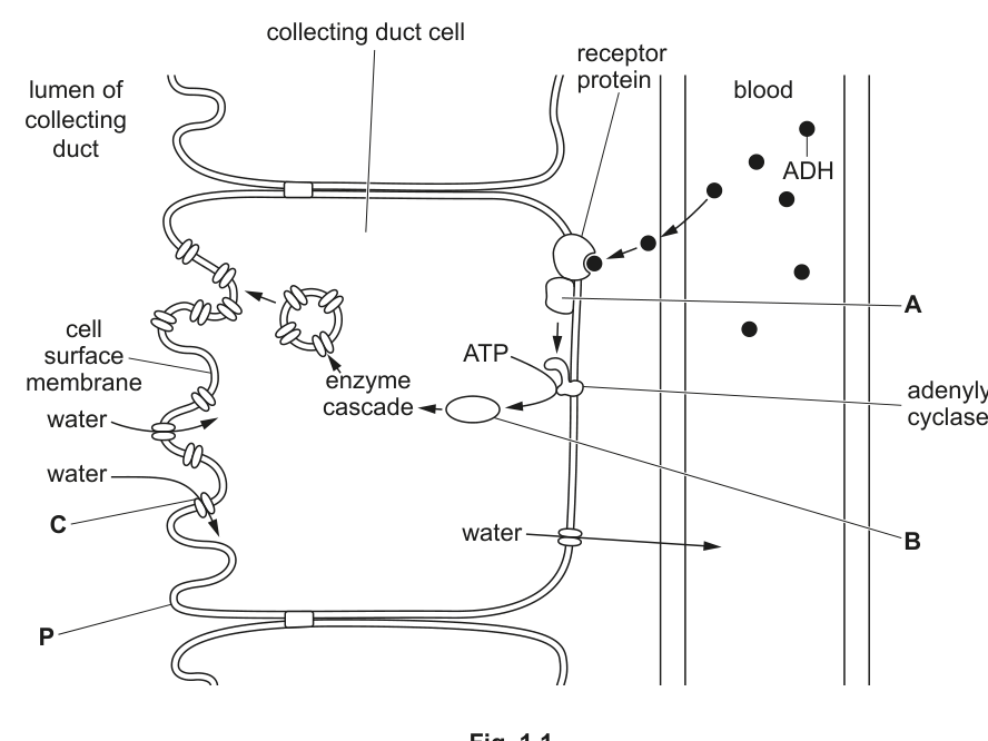
Fig. 1.1
Answer
A — G protein ;
B — cyclic AMP (cAMP) ;
C — aquaporin ;
A = G protein; B = cyclic AMP (cAMP); C = aquaporin
Background Concept
Antidiuretic hormone (ADH) is a small peptide hormone released from the posterior pituitary gland. Because ADH is too large and hydrophilic to cross the phospholipid bilayer, it binds to a specific receptor protein embedded in the cell surface membrane of its target cells (collecting-duct cells in the kidney, and similar receptors exist on hepatocytes for glucagon).
Binding of the hormone activates an associated G protein (a GTP-binding protein that acts as a molecular switch on the inner leaflet of the membrane). The activated G protein then stimulates the enzyme adenylyl cyclase, which catalyses the conversion of ATP into a small intracellular second messenger — cyclic AMP (cAMP). The rise in cAMP concentration triggers an enzyme cascade that ultimately causes vesicles containing aquaporin water-channel proteins to fuse with the cell surface membrane, increasing the permeability of the collecting-duct cell to water.
The structure C is therefore an aquaporin — a transmembrane protein that forms a pore allowing water to move down its water-potential gradient across the membrane.
Understanding the Question
The question provides Fig. 1.1, a labelled diagram of a collecting-duct cell showing the ADH cell-signalling pathway, and asks the candidate to name three structures: A, B and C. A is a structure inside the membrane associated with the receptor; B is produced from ATP; C is the channel through which water crosses the membrane on the lumen side.
Approach
Match each letter on the diagram to its position in the standard G-protein/adenylyl-cyclase/cAMP pathway. A sits on the inner face of the membrane next to the receptor — a G protein. B is generated from ATP by adenylyl cyclase — cyclic AMP. C is a transmembrane channel protein on the lumen-facing membrane through which water moves — an aquaporin.
Step-by-Step Reasoning
- A is shown attached to the inner side of the cell surface membrane, next to the ADH receptor. When ADH binds, this protein is activated — it is the G protein.
- B is produced by adenylyl cyclase from ATP. It is the second messenger that diffuses into the cytoplasm and triggers the enzyme cascade. This is cyclic AMP (cAMP).
- C is the water-permeable channel embedded in the cell surface membrane on the lumen side. The protein that forms this selective water pore is an aquaporin.
Key Takeaways
- The ADH signalling pathway uses a G protein, adenylyl cyclase, and cAMP as a second messenger — the same cascade used by glucagon in liver cells.
- Aquaporins are the water-selective channels inserted into the collecting-duct cell membrane in response to ADH.
- cAMP is generated from ATP; "cyclic" refers to the ring structure formed when the phosphate group loops back onto the ribose sugar.
Common Mistakes
- Writing "G-protein" is fine; avoid writing "G enzyme" or "GTP" for structure A.
- For B, students sometimes write "AMP" or "ATP" instead of "cyclic AMP / cAMP". The word cyclic (or the abbreviation cAMP) is the key marking point.
- For C, the mark scheme rejects "water channel" — the required term is the specific protein name aquaporin.
Things to Be Careful About
- The question stem notes that ADH's mechanism is similar to that of glucagon on liver cells, so identifying cAMP as the second messenger links this question to the glucagon/cAMP cascade students also learn.
- "Aquaporin" must be spelled correctly; "aquaporin-2" is the specific isoform inserted in the collecting duct but the name "aquaporin" alone is what the mark scheme accepts.
ADH is secreted by the posterior pituitary gland when the water potential of the blood decreases.
Suggest reasons why the water potential of the blood may decrease.
Answer
Any three of:
- dehydration / not drinking enough water ;
- ingesting salty food (high solute load in the blood) ;
- more, ions / glucose / amino acids / solute, in the blood ;
- sweating / perspiration / evaporation of water ;
- AVP e.g. disease where ADH is not, made / secreted ;
Any three valid reasons: dehydration, salty food, raised blood solutes, sweating, AVP.
Background Concept
Water potential () is a measure of the tendency of water to move by osmosis; pure water has and adding solutes makes more negative. The water potential of the blood depends on the balance between the water content of the plasma and the concentration of dissolved solutes (mainly Na, Cl, glucose, urea and plasma proteins). Anything that either removes water from the plasma or adds solute to it will lower (make more negative) the blood water potential. This fall is detected by osmoreceptors in the hypothalamus, which stimulate ADH release from the posterior pituitary.
Understanding the Question
The question gives a simple context: "ADH is secreted by the posterior pituitary gland when the water potential of the blood decreases." It then asks the candidate to suggest reasons why the water potential of the blood might decrease. The command word suggest means the answer does not have to be limited to textbook examples — any reasonable factor that would lower blood earns credit, up to the mark allowance of three.
Approach
There are two routes by which falls: (1) loss of water from the plasma, or (2) gain of solute by the plasma. Generate examples of each:
- Water loss — not drinking, sweating, vomiting, diarrhoea, evaporation, exercise.
- Solute gain — eating salty food, drinking sea water, high blood glucose (e.g. uncontrolled diabetes mellitus), high blood amino acids after a high-protein meal.
Any three distinct, sensible points gain the three marks.
Step-by-Step Reasoning
- Not drinking / dehydration reduces the water component of plasma, concentrating the remaining solutes — lower .
- Ingesting salty food introduces a large NaCl load into the blood; the additional solute lowers (and osmoreceptors respond before the kidney can excrete the excess).
- More ions, glucose or amino acids in the blood — any of these increases plasma osmolarity and so lowers ; this is why uncontrolled diabetes mellitus (high blood glucose) causes osmotic diuresis and thirst.
- Sweating / perspiration is evaporative water loss from the skin; the water leaves without solute, raising plasma concentration and lowering .
Key Takeaways
- Blood water potential is set by the ratio of water to solutes in plasma — either variable can change it.
- The hypothalamus detects small changes in plasma osmolarity (≈1%) and triggers ADH release and thirst — this is the afferent limb of an osmoregulatory negative-feedback loop.
- The question tests the application of water potential, not just rote learning.
Common Mistakes
- Saying "ADH is low" or "ADH is not secreted" — the question asks for causes of decreased water potential, not consequences.
- Listing only one type of cause (only water loss, or only solute gain) — the mark scheme rewards variety.
- Vague answers such as "hot weather" without explaining the mechanism (evaporative water loss → lower plasma ) — the link must be made.
Things to Be Careful About
- "Dehydration" is acceptable and worth a mark on its own; further detail (no water intake) is optional.
- "Salty food" is a single cause — no need to also list "more ions in the blood" as a separate point, as the mark scheme treats them as alternatives (separated by
;not\n). - Any creditable alternative (e.g. haemorrhage, severe burns, osmotic diuresis in diabetes mellitus) is accepted under the AVP line.
Diabetes insipidus is a condition affecting osmoregulation by the kidney. One form of diabetes insipidus is caused by a tumour in the pituitary gland, which results in a decreased secretion of ADH.
Suggest the symptoms that would occur in a person with diabetes insipidus.
Answer
Any two of:
- (large) increase in urine volume / polyuria / more frequent urination / dilute (less concentrated) urine ;
- fatigue / lethargy / weakness ;
- feelings of thirst / polydipsia / dry mouth ;
- AVP e.g. dehydration, change in blood pressure ;
Large volumes of dilute urine and thirst; fatigue and dehydration are also credited.
Background Concept
ADH increases the permeability of the collecting duct (and distal tubule) to water by triggering the insertion of aquaporin-2 channels into the luminal cell surface membrane. When ADH is low, the collecting duct remains relatively impermeable to water, so the filtrate that entered as plasma at the glomerulus passes through with little water reabsorption. The result is a large volume of dilute urine, and the body loses water it would normally have conserved. The drop in body water content raises plasma osmolarity and lowers blood volume (and pressure), which is sensed by osmoreceptors (thirst) and baroreceptors.
Understanding the Question
The stem tells the candidate that a pituitary tumour causes decreased ADH secretion (this is the central / neurogenic form of the condition). The candidate must therefore predict the symptoms: if ADH is low, water cannot be reabsorbed efficiently, so the body loses water and becomes dehydrated and thirsty.
Approach
Work forward from "less ADH" → "fewer aquaporins in collecting-duct membrane" → "less water reabsorbed" → "large volumes of dilute urine" + "net water loss from body" → "thirst, dehydration, fatigue".
Step-by-Step Reasoning
- With little ADH, the collecting-duct cells do not insert aquaporins into their luminal membrane; water is not reabsorbed and remains in the filtrate → large volume of dilute urine and frequent urination.
- The body therefore loses water faster than it is taken in → plasma becomes more concentrated and total body water falls → thirst / dry mouth (osmoreceptor response) and signs of dehydration.
- Loss of fluid from the circulation reduces blood volume and pressure; dehydration and electrolyte disturbance cause fatigue / lethargy.
Key Takeaways
- The triad of diabetes insipidus is polyuria (large urine volume), polydipsia (excess thirst) and dilute urine — these are the symptoms any candidate should aim to recall.
- This is mechanistically distinct from diabetes mellitus, where polyuria is caused by glucose-driven osmotic diuresis rather than ADH deficiency.
- The question illustrates how a single hormone deficiency produces a coherent set of symptoms through one disrupted pathway.
Common Mistakes
- Writing "less urine" — the opposite is true; without ADH the kidney cannot concentrate urine and produces a large volume of dilute urine.
- Saying "no aquaporins" — there is always a small baseline permeability; the right wording is fewer aquaporins or reduced water reabsorption.
- Confusing diabetes insipidus with diabetes mellitus and listing "high blood glucose" — the question specifies a pituitary tumour causing low ADH.
Things to Be Careful About
- The command word is suggest, so the marks reward any plausible symptom consistent with the mechanism. Aim for two clear, mechanism-based points.
- "Dilute urine" is one of the most distinctive features of diabetes insipidus and is worth a mark on its own.
Neurogenic diabetes insipidus (NDI) is another form of diabetes insipidus. In NDI, ADH molecules cannot bind to the receptor proteins located in the cell surface membranes of the cells of the collecting duct.
With reference to Fig. 1.1, explain the effect on the cell surface membrane labelled P if ADH cannot bind to the receptor proteins.
Answer
- (very) few / no, aquaporins added to membrane P ;
- membrane P has, reduced / low, permeability to water ;
- less water moves through, membrane P (into the cell from the lumen) ;
Few/no aquaporins are inserted into membrane P, so it stays of low water permeability and little water crosses it from the lumen.
Background Concept
The cell surface membrane of a collecting-duct cell is normally only sparingly permeable to water; its water permeability is regulated by the number of aquaporin-2 channels in the membrane. These channels are stored in vesicles just beneath the membrane and are inserted by exocytosis only when the cAMP cascade triggered by ADH is active. When ADH is removed, the channels are retrieved by endocytosis, and the membrane reverts to low water permeability.
In the figure, membrane P is the cell surface membrane on the lumen side of the collecting-duct cell — exactly where aquaporins are inserted to allow water to leave the tubular fluid and enter the cell (and then the blood).
Understanding the Question
The question sets up a different form of diabetes insipidus — neurogenic diabetes insipidus (NDI) — in which ADH molecules are present in the blood but cannot bind to the receptor proteins on the collecting-duct cells. The candidate must use Fig. 1.1 to explain the effect on membrane P.
Approach
Because the pathway is blocked at the very first step (no ADH–receptor binding), nothing downstream happens. Walk through Fig. 1.1 step by step and identify what fails to occur: no G-protein activation, no adenylyl cyclase activation, no cAMP produced, no enzyme cascade, and therefore no aquaporin vesicles fuse with membrane P. Then state the consequences: membrane P remains of low water permeability and little water crosses from the lumen.
Step-by-Step Reasoning
- ADH cannot bind the receptor → the G protein (A) is not activated → adenylyl cyclase is not stimulated → cAMP (B) is not produced from ATP → the enzyme cascade is not triggered.
- Without the cascade, vesicles carrying aquaporins (C) do not fuse with the cell surface membrane P, so few or no aquaporins are added to P.
- The permeability of membrane P to water therefore remains low, and less water moves through P from the lumen into the cell — which is exactly why NDI patients pass large volumes of dilute urine (linked to part (c)).
Key Takeaways
- The water permeability of the collecting-duct membrane is hormonally regulated, not fixed. It is a dynamic property set by the number of aquaporins in the membrane at any moment.
- Blocking any step of the cascade has the same end result as having no ADH: low membrane water permeability and dilute urine.
- The ecf note in the mark scheme is important: if the candidate called structure C a "water channel" in part (a), they can still score in (d) by saying "few/no water channels added to P".
Common Mistakes
- Saying the membrane becomes "impermeable to water" — the mark scheme rejects "impermeable"; the correct wording is reduced / low permeability.
- Describing only the biochemical consequences (no cAMP, no cascade) without linking them to the physical state of membrane P — the question specifically asks about the effect on P, so the answer must say something about its aquaporin content and water permeability.
- Forgetting to state the consequence in terms of water movement, which is what would actually be observed.
Things to Be Careful About
- The question says "with reference to Fig. 1.1", so at least one mark is essentially guaranteed by mentioning membrane P and the aquaporin (structure C).
- This part tests the link between cell signalling and a measurable physiological outcome — keep the chain of reasoning tight.
NDI is caused by a recessive allele of the gene coding for the receptor protein. The gene is located on the X chromosome.
Explain why a man with NDI could not have inherited the condition from his father.
Answer
- The (recessive) allele is on the X chromosome, which the man inherited from his mother ;
- He inherited the Y chromosome from his father (the Y chromosome does not carry the allele) ;
A man inherits his Y chromosome (not his X) from his father, so the X-linked recessive allele causing NDI must have come from his mother.
Background Concept
Sex-linked genes in humans are carried on the X chromosome; the Y chromosome is much smaller and carries very few genes that are not also present on the X. A male has the sex chromosomes XY — his single X comes from his mother (who contributes only an X to a son) and his Y comes from his father (who contributes either an X or a Y to each child). For a recessive X-linked allele to be expressed in a male, he needs only one copy (because he has no second X to carry a dominant allele that could mask it). Such a male must therefore have received the recessive allele on the X chromosome he got from his mother; the X chromosome cannot have come from his father because his father gave him a Y.
Understanding the Question
The stem states: "NDI is caused by a recessive allele of the gene coding for the receptor protein. The gene is located on the X chromosome." The question asks the candidate to explain why a man with NDI could not have inherited the condition from his father. This is a sex-linkage inheritance question framed in a clinical context.
Approach
Use the sex chromosome contributions of each parent:
- A son's X chromosome comes only from his mother.
- A son's Y chromosome comes only from his father.
- The recessive allele for NDI is on the X chromosome, so it can only be transmitted on an X. The father does not pass an X to a son; he passes a Y. Therefore the allele could not have come from the father.
Step-by-Step Reasoning
- The NDI allele is on the X chromosome; there is no corresponding allele on the Y chromosome.
- To inherit an X-linked allele, a person must receive an X chromosome. A son receives his single X from his mother (and his Y from his father).
- Therefore, if a man has the X-linked recessive allele, that X came from his mother. The Y he carries, which came from his father, does not carry the allele at all.
- Conclusion: a man with NDI cannot have inherited the condition from his father; he must have inherited it from his mother (who would be a carrier, if H is the dominant normal allele and h is the recessive NDI allele).
Key Takeaways
- A father's contribution to a son is the Y chromosome; the mother contributes the X.
- X-linked recessive conditions therefore appear in sons only via the maternal line; an affected father cannot pass an X-linked recessive allele to his son (he can only pass it to his daughters, who become carriers).
- Recognising which parent contributed which sex chromosome is the heart of any X-linked inheritance question.
Common Mistakes
- Saying "because the father is male he cannot pass the allele" — the father can carry and pass an X-linked allele, but only to his daughters (via his X); to a son he passes only the Y.
- Confusing X-linked with autosomal recessive inheritance and not invoking the sex chromosomes at all.
- Saying "the gene is only on the X chromosome, not on the Y" without connecting it to which parent gave which chromosome to the son.
Things to Be Careful About
- Use the words X chromosome and Y chromosome explicitly; vague statements like "from the mother because she's the one" will not earn both marks.
- A clear logical chain (X comes from mother, Y from father, allele is on X) is needed — the mark scheme awards one mark each for the X-from-mother and Y-from-father points.
- This question is a classic X-linkage reasoning item; practice with pedigree diagrams (aunts, uncles, grandfathers) reinforces the same logic.
Phenotypic variation exists in many forms.
Some examples of phenotypic variation in plants and animals are described in Table 2.1.
Complete Table 2.1 by stating whether the cause of variation for each described example is likely to be due to:
• genetic factors,
• environmental factors,
• a combination of genetic and environmental factors, .
Table 2.1
| description of phenotypic variation | cause of variation |
|---|---|
| Tomato plants grown in a glasshouse and grown outside vary in the yield of tomatoes they produce. Seventeen genes associated with tomato yield have been identified. | |
| New strawberry plants from the variety called Sweet Ann are made by asexual reproduction. The new plants grow to different sizes and produce different numbers of fruit. | |
| The domestic cat has a blood group system with three possible blood types: A, B and AB. The blood types are determined by antigens present on the cell surface membrane of red blood cells. | |
| Over 50 genes have variants that are associated with excessive weight gain in humans. Other risk factors for excessive weight gain include diet and exercise. | |
| Resting heart rate in humans varies between different individuals. Some factors that influence resting heart rate include: biological sex, family history of heart disease, number of cigarettes smoked, medication taken. |
Answer
| description of phenotypic variation | cause of variation |
|---|---|
| Tomato plants grown in a glasshouse and grown outside vary in the yield of tomatoes they produce. Seventeen genes associated with tomato yield have been identified. | |
| New strawberry plants from the variety called Sweet Ann are made by asexual reproduction. The new plants grow to different sizes and produce different numbers of fruit. | |
| The domestic cat has a blood group system with three possible blood types: A, B and AB. The blood types are determined by antigens present on the cell surface membrane of red blood cells. | |
| Over 50 genes have variants that are associated with excessive weight gain in humans. Other risk factors for excessive weight gain include diet and exercise. | |
| Resting heart rate in humans varies between different individuals. Some factors that influence resting heart rate include: biological sex, family history of heart disease, number of cigarettes smoked, medication taken. |
V_G + V_E ; V_E ; V_G ; V_G + V_E ; V_G + V_E
Background Concept
Phenotypic variation among individuals in a population can be split into three underlying causes:
- — variation caused by genetic factors, i.e. differences in the alleles an individual carries. This variation is heritable.
- — variation caused by environmental factors acting on individuals (e.g. nutrition, light, temperature, disease, exercise). This variation is not heritable through the alleles involved, although the environment itself can sometimes affect future generations via epigenetic or maternal effects.
- — variation caused by a combination of both, where the phenotype is the product of an underlying genetic predisposition modified by the environment an individual experiences. Most continuously varying traits in outbreeding populations fall into this category.
A useful diagnostic: if the individuals are genetically identical (clones, identical twins, pure-breeding line under controlled conditions) but still differ in phenotype, the cause must be environmental. If the trait follows a simple Mendelian pattern unaffected by external conditions, it is genetic. If both genes and lifestyle/environment are known to influence the trait, it is .
Understanding the Question
The question presents five real-world examples of phenotypic variation and asks the candidate to classify each as caused by , or . Three marks are available, awarded as 5/4-3/2-1 correct. The marks are therefore about consistent, defensible classification, not a single tricky decision.
Approach
Read each example and ask two diagnostic questions:
- Is there an explicit mention of genes/alleles/heritable differences? If yes, is at least partly involved.
- Is there an explicit mention of different environments, diets, or non-heritable influences? If yes, is at least partly involved.
- If only one is present, the answer is that one alone; if both are present, the answer is .
A special case to watch for: asexual reproduction produces genetically identical offspring. If those offspring still vary, the variation MUST be environmental, because no new genetic combinations are produced.
Step-by-Step Reasoning
Row 1 – Tomato yield, glasshouse vs outside, 17 genes identified:
Both genes (the 17 identified yield-associated genes) and environment (glasshouse vs outside growing conditions) are explicitly stated as influences. → .
Row 2 – Strawberry plants from 'Sweet Ann' produced by asexual reproduction, but vary in size and fruit number:
Asexual reproduction (e.g. runners, cuttings) produces clones with identical genotypes. Therefore any difference between the new plants cannot be due to genetics — the new plants are genetically the same as the parent. The variation in size and fruit number must come from differing growth conditions (soil quality, light, water, nutrients, position in the row, competition). → .
Row 3 – Domestic cat blood groups A, B and AB, determined by antigens on red blood cell membranes:
Blood group antigens are coded for by alleles at specific gene loci. They show discrete categories (a classic discontinuous variation), are inherited, and the question gives no environmental factors. → .
Row 4 – Human weight gain: 50+ gene variants + risk factors including diet and exercise:
Both genetic variants AND environmental/lifestyle factors (diet, exercise) are stated. → .
Row 5 – Human resting heart rate: influenced by biological sex and family history (genetic) and by smoking and medication (environmental):
Biological sex and family history both indicate heritable/genetic influences, while smoking and medication are environmental influences. → .
Key Takeaways
- = variation between genotypes; heritable.
- = variation between individuals of the same genotype caused by their environment; not heritable through the alleles themselves.
- = the common situation where a trait has both a genetic component and an environmental component (most continuous traits in humans, including weight, height, blood pressure, heart rate).
- Asexually reproduced organisms (clones) that vary in phenotype demonstrate directly, because the genetic component is held constant.
- Blood group antigens are a textbook example of a discontinuous, purely genetic trait.
Common Mistakes
- Marking the asexually reproduced strawberries as because "genes are involved". This loses the point: the new plants are genetic copies, so any difference must be environmental.
- Marking the cat blood groups as because "the environment could matter". Blood group antigens are determined directly by alleles; the question gives no environmental influence, so it is only.
- Confusing with "non-genetic" in a general sense and missing the genetic component in the tomato/weight/heart-rate rows.
Things to Be Careful About
- The classification is about the cause of the observed variation, not whether genes exist somewhere in the organism. Even where a trait could be influenced by environment in principle, only assign (or ) if the question explicitly mentions an environmental factor.
- "Family history" and "biological sex" are proxies for genetic influence; "diet", "exercise", "smoking" and "medication" are proxies for environmental influence.
- Read each row as a stand-alone scenario — the marks are independent across rows.
Name a spontaneous, random event occurring in cells that can be a source of phenotypic variation.
Answer
Mutation
Mutation
Background Concept
Variation between individuals has two ultimate sources. One is the re-shuffling of existing alleles during meiosis and sexual reproduction (covered in part (c)). The other is the creation of new alleles — and that requires a change in the DNA sequence itself. Such a change is called a mutation: a spontaneous, random alteration in the nucleotide sequence of a gene or chromosome.
Mutations are described as:
- Spontaneous — they arise from random errors in DNA replication, damage from radiation, or chemical mutagens, without being directed by the organism.
- Random — they occur at any base in any gene, with no prediction of when, where, or what will change.
- Heritable only if they occur in the cells that give rise to gametes (germ-line mutations).
If a mutation occurs in a gene that influences a phenotype (e.g. the HBB sickle-cell allele, the TYR albino allele), the resulting new allele contributes to .
Understanding the Question
This is a one-mark "name" question. The command word "name" requires a single biological term, not a sentence. The clue words in the question are "spontaneous" and "random event occurring in cells" — these are textbook descriptors of mutation.
Approach
Identify the only term in the syllabus that matches "spontaneous, random event occurring in cells" that introduces new phenotypic variation. That term is mutation.
Step-by-Step Reasoning
- Spontaneous + random = mutation (not crossing over, not independent assortment — those are programmed features of meiosis, not random events).
- "Occurring in cells" is satisfied because mutations happen during DNA replication in any dividing cell.
- The accepted one-word answer is mutation.
Key Takeaways
- Mutation is the original source of all new genetic variation; without it, evolution cannot occur.
- Mutations are random with respect to the needs of the organism — they are not produced in response to an environmental demand.
- Only germ-line mutations (in cells that form gametes) are inherited and contribute to in the next generation.
Common Mistakes
- Writing "gene" or "allele" — these are units that can be affected by mutation, but they are not the event itself.
- Writing "meiosis" — meiosis re-shuffles existing variation; it does not create new variation.
- Writing "crossing over" — crossing over is part of the answer to part (c) and is a programmed feature of meiosis, not a spontaneous random event.
Things to Be Careful About
- A single word is sufficient and is what the mark scheme expects.
- The marking note in the scheme lists "mutation" as the only acceptable answer for this part.
Other than the event named in (b), describe the features of sexual reproduction that contribute to the production of genetically different offspring.
Answer
Any three of:
- Crossing over (between non-sister chromatids of homologous chromosomes in prophase I of meiosis) — produces new combinations of alleles on each chromosome.
- Independent (random) assortment of homologous chromosomes (and of different gene loci) at metaphase I — produces different combinations of maternal and paternal chromosomes in each gamete.
- Random fusion of gametes at fertilisation — which sperm fertilises which egg is a matter of chance, so the resulting zygote has a unique combination of parental alleles.
- Random mating — which individuals mate with which is not determined by genotype (in most populations), adding another layer of chance to which allele combinations come together.
Any three of: crossing over ; independent / random assortment of chromosomes ; random fusion of gametes / fertilisation ; random mating
Background Concept
Sexual reproduction produces genetically unique offspring because the gametes that fuse are themselves genetically non-identical. This uniqueness is generated at two stages: during meiosis (when gametes are formed) and at fertilisation (when gametes fuse).
During meiosis three processes introduce variation:
-
Crossing over — in prophase I, non-sister chromatids of homologous chromosomes exchange segments of DNA at chiasmata. This physically recombines the maternal and paternal alleles on the same chromosome, producing chromatids that contain a mixture of alleles from both grandparents. After meiosis, gametes therefore contain chromosomes that are neither purely maternal nor purely paternal.
-
Independent (random) assortment — at metaphase I, the orientation of each pair of homologous chromosomes on the equator is independent of every other pair. For an organism with chromosome pairs this gives possible combinations of maternal and paternal chromosomes in the gametes. In humans () this alone gives chromosome combinations per gamete.
-
Random fusion of gametes at fertilisation — any of the huge variety of male gametes can fuse with any of the huge variety of female gametes, so the allele combinations in the zygote are not predetermined.
In many populations, random mating (no preference for particular genotypes) is an additional feature, ensuring the gametes that meet are not biased by mate choice.
Understanding the Question
The question asks for features of sexual reproduction (other than the mutation named in (b)) that contribute to genetically different offspring. The command word is "describe", so each point should be a short statement of the feature, not a long essay. Three marks are available, so three distinct features are needed.
Approach
List the four classic mechanisms covered above and select any three. Each marking point requires the mechanism (e.g. "crossing over") and, where helpful, a brief reason it creates variation. The mark scheme accepts the bare name of each mechanism.
Step-by-Step Reasoning
- Crossing over: in prophase I of meiosis, non-sister chromatids of homologous chromosomes exchange DNA at chiasmata, producing new combinations of alleles on a single chromosome. This is the first source of variation inside meiosis.
- Independent (random) assortment: at metaphase I, the orientation of each bivalent is random, so homologous chromosomes (and therefore the alleles they carry) are distributed independently into daughter cells. This produces combinations per gamete.
- Random fusion of gametes at fertilisation: any sperm may fertilise any egg, so the combination of alleles in the zygote is left to chance. This multiplies the variation produced by meiosis.
- Random mating: in many populations, mate choice is not constrained by genotype, so the alleles entering a fertilisation event are not pre-selected.
Any three of the above four are credited.
Key Takeaways
- Sexual reproduction creates unique offspring because meiosis generates varied gametes and fertilisation combines them randomly.
- The three meiotic sources of variation are crossing over, independent assortment, and (random) gamete fusion at fertilisation.
- Random mating is an additional population-level feature, but only when mate choice is genuinely random.
Common Mistakes
- Confusing crossing over (which exchanges DNA between chromatids of homologous chromosomes) with the random fusion of gametes (which happens at fertilisation, not during meiosis).
- Calling the mechanism "segregation" — the CIE syllabus uses the term "independent/random assortment" for the separation of homologous chromosomes; "segregation" alone is the Mendelian term and is not the credited wording.
- Listing the same mechanism twice in different words (e.g. "random assortment of chromosomes" and "independent assortment of alleles") — only one mark can be awarded.
- Naming mutation again, even though the question explicitly excludes it ("other than the event named in (b)").
Things to Be Careful About
- The mark scheme explicitly accepts four alternative answers; any three earn the three marks.
- Use precise phrasing: "crossing over" rather than "DNA exchange"; "independent / random assortment" rather than "shuffling"; "random fusion of gametes / fertilisation" rather than "chance meeting".
- The four marks are awarded independently — credit any combination of three from the four listed.
In plants and humans, the phenotype of an organism is determined by the genotype and the environment.
Plants from the genus Primula have different petal colours. The presence of the pigment malvidin results in blue petals.
The metabolic pathway for malvidin synthesis is controlled by gene . The presence of the dominant allele results in blue petals.
Another gene, gene , at a different locus, also influences the malvidin synthesis pathway. When the dominant allele is present, its gene product suppresses the malvidin synthesis pathway. This is summarised in Fig. 3.1.
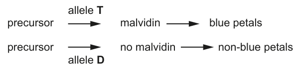
Fig. 3.1
A genetic cross was carried out between two plants heterozygous at both gene loci. The resulting offspring genotypes are shown in a Punnett square in Fig. 3.2.
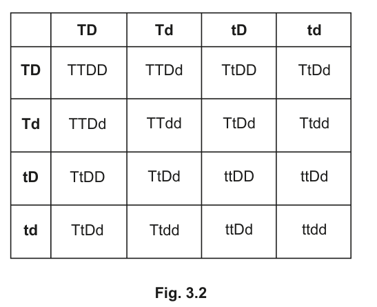
Fig. 3.2
Answer
and
(Each must carry at least one dominant allele to make the malvidin-producing enzyme, AND be homozygous so that no functional inhibitor is present.)
Ttdd and TTdd
Background Concept
A gene codes for a protein; a dominant allele typically produces a functional protein while the homozygous recessive state gives a non-functional protein. In this Primula cross there are two independently assorting autosomal genes, and . codes for a protein that converts a colourless precursor into the blue pigment malvidin, so a plant with at least one has the enzymatic capacity to make malvidin. , however, codes for a protein that suppresses the malvidin pathway. So is dominant and epistatic to : when any is present, malvidin is not made, regardless of the genotype. Blue petals therefore require the simultaneous absence of (i.e. ) and the presence of at least one .
Understanding the Question
The question asks which of the 16 genotypes shown in the printed Punnett square (Fig. 3.2) will produce blue petals. You must apply the rule derived from Fig. 3.1 and the stem: blue = at least one AND homozygous ; everything else is non-blue.
Approach
Read off each of the 16 cells, decide which it is, and select the cells that are simultaneously -containing and . Equivalently, scan the Punnett square for the four cells containing and discard the one that is also .
Step-by-Step Reasoning
- The four cells containing in Fig. 3.2 are: (row 2, column 2), (row 2, column 4), (row 4, column 2) and (row 4, column 4).
- is non-blue because means no functional enzyme is produced, so no malvidin is made.
- The remaining three cells all have at least one and are , so they are blue. They are and the two cells.
- The two genotypes with blue petals are therefore and .
Key Takeaways
- Phenotype depends on the combination of alleles across loci, not on each gene in isolation.
- When the allele of one gene masks the effect of the allele of another, the masking gene is said to be epistatic to the masked gene.
- For blue in this cross the plant must satisfy both conditions: at least one (to make malvidin) and no (so the pathway is not blocked).
Common Mistakes
- Forgetting that is dominant and epistatic: students often include any -containing -containing genotype as 'blue' because it carries .
- Listing as blue — it has no functional product so it cannot make malvidin.
- Writing the genotype without the case of the alleles; the dominant/recessive distinction is the entire point.
Things to Be Careful About
- Use capital / for the dominant alleles and lower-case / for the recessive alleles.
- 'Blue' requires both conditions (at least one AND homozygous ) — do not give partial credit for just one.
- This is a dihybrid cross () so the genotype frequencies are not 1:1; the 16 cells are not all equally common.
Answer
(Non-blue = cells; blue = (1 cell) + (2 cells) = cells.)
13:3
Background Concept
When two heterozygotes are crossed at two unlinked loci (), the standard dihybrid genotypic ratio is the 1:2:1:2:4:2:1:2:1 expansion of the , but the phenotype ratio depends on the gene interaction. Here, because the dominant allele is epistatic to , the 16 offspring collapse into just two phenotype classes (blue and non-blue), giving the classic 13:3 ratio of dominant epistasis.
Understanding the Question
The question asks for the ratio of non-blue to blue petals in the cross of Fig. 3.2. The ratio must be expressed in its simplest whole-number form and in the order requested (non-blue first).
Approach
Reuse the analysis from (a)(i): blue = -containing and = 3 cells. Everything else (16 − 3 = 13 cells) is non-blue. Write the ratio with non-blue first because the question asks for 'non-blue to blue'.
Step-by-Step Reasoning
- Blue cells from (a)(i): (1 cell) + (2 cells) = 3 cells.
- Non-blue cells: every other cell in the Punnett square — all genotypes carrying at least one (which suppresses the pathway), plus the single cell (which has no functional enzyme).
- Total non-blue: cells.
- Simplest ratio: non-blue : blue = 13 : 3.
Key Takeaways
- The same Punnett square yields different phenotype ratios depending on the gene interaction.
- A 13:3 ratio is the diagnostic signature of dominant epistasis.
- For comparison: 9:3:4 indicates recessive epistasis; 9:7 indicates complementary gene action; 9:3:3:1 indicates no interaction.
Common Mistakes
- Inverting the ratio to give 3:13 (the question specifically asks non-blue to blue).
- Counting dihybrid genotype classes (9:3:3:1) and ignoring the gene interaction.
- Forgetting the cell is non-blue.
Things to Be Careful About
- Read the order of the ratio in the question — it is non-blue to blue, not blue to non-blue.
- 13 and 3 share no common factor, so the ratio is already in its simplest form.
Name the type of gene interaction that has caused the offspring ratio you have stated in (a)(ii).
Answer
Epistasis (specifically, dominant epistasis — the dominant allele is epistatic to ).
Epistasis
Background Concept
When the allele of one gene masks the phenotypic expression of the allele of another gene (at a different locus), the interaction is called epistasis. The masking gene is said to be epistatic to the masked gene. The 13:3 phenotype ratio observed in (a)(ii) is the classic signature of dominant epistasis: a single dominant allele at the epistatic locus is enough to mask the effect of the other locus. (By contrast, recessive epistasis gives a 9:3:4 ratio, because only the homozygous recessive state at the epistatic locus masks the other gene.)
Understanding the Question
The candidate must name the type of gene interaction responsible for the 13:3 ratio. The single word 'epistasis' earns the mark; 'dominant epistasis' is the more precise version.
Approach
Recognise the ratio from (a)(ii) as 13:3, recall that this is the diagnostic phenotype ratio for dominant epistasis, and state the term.
Step-by-Step Reasoning
- A 13:3 ratio in a dihybrid cross arises because the dominant allele masks the expression of whenever is present.
- The masking of one gene's alleles by another gene's alleles is called epistasis.
- The gene doing the masking () is epistatic to the gene being masked ().
Key Takeaways
- 13:3 → dominant epistasis
- 9:3:4 → recessive epistasis (e.g. coat colour in mice, where the homozygous recessive at the agouti locus blocks pigment production entirely)
- 9:7 → complementary gene action (both genes needed for the trait to appear)
- 9:3:3:1 → no interaction (two independent dominant/recessive traits)
Common Mistakes
- Writing 'co-dominance' or 'incomplete dominance' — those describe within-locus interactions, not between-locus interactions.
- Writing 'pleiotropy' — that is one gene affecting several phenotypes, not two genes affecting one phenotype.
- Writing 'linked genes' — the two loci in this question assort independently (the standard 16-cell Punnett square is shown).
Things to Be Careful About
- The single word 'epistasis' is the credit-worthy term. Adding 'dominant' is good but not required for the mark.
- Do not confuse 'epistasis' (the interaction) with 'epistatic' (the gene doing the masking).
Gene and gene code for proteins that are involved in the control of the production of malvidin.
Discuss the possible roles of the proteins coded for by gene and gene in the control of the production of malvidin.
Answer
Any five from:
- The dominant allele ( or ) codes for a functional protein; the homozygous recessive ( or ) codes for a non-functional protein.
- could code for an enzyme that catalyses the production of malvidin from the precursor.
- could code for an inhibitor that binds to the enzyme coded for by , so the reaction producing malvidin does not occur.
- Alternatively, the protein coded for by could be a transcription factor that allows the production of the enzyme in the metabolic pathway.
- The protein coded for by could inhibit this transcription factor, switching the pathway off.
See working — five points: dominant allele = functional protein, recessive homozygote = non-functional protein; T could be an enzyme that makes malvidin; D could be an inhibitor of that enzyme; alternatively T could be a transcription factor and D an inhibitor of that transcription factor.
Background Concept
Genes code for proteins, and proteins do the work of the cell. A phenotype is the cumulative result of the proteins operating in a metabolic or developmental pathway. When two genes affect the same pathway they can interact in several molecular ways: one gene's product may be the enzyme that actually performs the conversion; another gene's product may be a regulatory protein (an inhibitor, a repressor, a transcription factor) that switches the first gene's expression or activity on or off. The fact that one allele is dominant usually means its protein is functional; the recessive allele typically produces a non-functional protein (often through a loss-of-function mutation).
Understanding the Question
The question asks the candidate to 'discuss the possible roles' of the proteins coded for by and in the control of malvidin production. The verb 'discuss' signals an open-ended response: at A-Level the strongest answers consider more than one plausible mechanism and explicitly contrast them.
Approach
Establish the general rule that dominant = functional protein, recessive homozygote = non-functional protein. Then offer two coherent mechanistic stories, one of which would suffice for full marks but the combination is more thorough:
- Story A (enzyme / inhibitor): codes for the enzyme that makes malvidin; codes for an inhibitor of that enzyme.
- Story B (transcription factor / inhibitor): codes for a transcription factor needed to switch on the malvidin-producing enzyme; codes for a protein that inhibits that transcription factor.
Both stories predict the same 13:3 ratio and the same blue genotype (-containing and ), and either is acceptable.
Step-by-Step Reasoning
- The dominant allele ( or ) codes for a functional protein; the homozygous recessive ( or ) does not — its protein is non-functional or absent.
- Story A: codes for an enzyme that catalyses the conversion of the colourless precursor to malvidin. Without at least one , the reaction cannot occur and the petals are not blue.
- In Story A, codes for an inhibitor — a regulatory protein that binds to the enzyme produced by and prevents it from working. The reaction does not occur and there is no malvidin.
- Story B: the protein produced by is (or controls) a transcription factor that switches on the gene(s) encoding the malvidin-producing enzyme. Without it, the enzyme is not made.
- In Story B, the protein is a repressor that binds to the transcription factor (or otherwise blocks its action), so the malvidin-producing enzyme is not expressed and no malvidin is made.
- Either story explains why -containing and is the only blue genotype: the enzyme / transcription factor must be functional, and the inhibitor must be absent.
Key Takeaways
- Phenotype = gene × gene × environment; this question exercises the 'gene × gene' part.
- Dominant alleles usually make a working protein; recessive alleles often make a non-working one.
- Regulatory (epistatic) genes typically code for regulators of other genes' expression or activity, not for the metabolic enzymes themselves.
- The 'discuss' command word at A-level often rewards considering more than one mechanism.
Common Mistakes
- Saying 'T makes the colour' or 'D is the colour gene' — without naming what kind of protein the gene codes for, the candidate loses the molecular explanation.
- Confusing the direction of the interaction: suppresses the pathway; it does not itself make the petals non-blue.
- Mixing the two stories incoherently (e.g. 'T is an enzyme and D is a transcription factor' — pick one story at a time, or be explicit that they are alternatives).
- Forgetting to link the molecular mechanism back to the 13:3 ratio or the blue phenotype.
Things to Be Careful About
- The question says 'discuss the possible roles' — it explicitly invites the candidate to consider alternatives. At this level at least one alternative mechanism should appear.
- Avoid vague verbs like 'controls' or 'affects' without naming what the protein actually does (enzyme, transcription factor, inhibitor, repressor).
- Stay genotype-accurate: only the dominant allele ( or ) codes for a functional protein in either story.
Some humans have the inherited condition haemophilia.
Explain the relationship between the gene, factor VIII and the condition haemophilia.
Answer
- The gene is located on the X chromosome — inheritance is sex-linked.
- The allele that causes haemophilia is recessive to the normal allele.
- The recessive allele codes for a non-functioning (or reduced / absent) factor VIII protein.
- Because factor VIII is needed to activate factor X (and ultimately thrombin) in the clotting cascade, blood does not clot quickly enough, and excessive bleeding occurs after injury.
- Because the gene is on the X chromosome, a male with a single copy of the recessive allele () is affected, while a female usually needs two copies () to be affected — hence haemophilia is much more common in males than in females.
F8 is on the X chromosome; the recessive allele codes for a non-functioning (or reduced) factor VIII, so blood does not clot quickly enough and excessive bleeding occurs after injury. As the gene is X-linked, the condition is much more common in males (X to the f, Y) than in females.
Background Concept
The gene codes for coagulation factor VIII, one of the plasma proteins in the blood-clotting cascade. The gene lies on the long arm of the X chromosome (Xq28). A loss-of-function mutation in reduces or abolishes factor VIII activity, which in turn slows the activation of factor X (and ultimately thrombin), so fibrinogen is not converted to fibrin efficiently and a stable blood clot does not form. Because the gene is on the X chromosome, inheritance is sex-linked: a female has two X chromosomes and is usually unaffected unless both carry the mutation (); a male has only one X, so a single copy of the mutant allele () is enough to cause the disease. Different mutations in produce factor VIII of differing activity, so the severity of haemophilia A ranges from mild to severe.
Understanding the Question
The question asks for an explanation of the relationship between three things: the gene, the factor VIII protein, and the clinical condition haemophilia. The candidate must chain genotype to protein to phenotype.
Approach
Lay out the logic in order: gene → protein → physiological role → clinical effect. Be sure to include the sex-linkage aspect (it is haemophilia A) and the recessive nature of the allele.
Step-by-Step Reasoning
- The gene is located on the X chromosome. Inheritance is therefore sex-linked.
- The allele responsible for haemophilia A is recessive to the wild-type allele.
- The recessive allele codes for a factor VIII protein that is non-functional, or is produced in much smaller amounts, or is absent altogether.
- Because factor VIII is required to activate factor X in the clotting cascade, its absence / reduction means the cascade runs too slowly. Thrombin is not activated efficiently, fibrinogen is not converted to fibrin, and the blood clot does not form properly.
- The phenotypic consequence is that bleeding (after injury, surgery, or spontaneously into joints) is excessive and prolonged.
- A male inheriting the recessive allele on his single X () is affected; a female needs two copies () to be affected. Heterozygous females () usually have enough factor VIII from their one functional allele to clot normally — which is why haemophilia A is much more common in males than in females.
Key Takeaways
- Sex-linked recessive inheritance: males are hemizygous and therefore express the recessive phenotype from a single copy.
- The molecular chain is: allele → factor VIII activity → clotting-cascade speed → bleeding phenotype.
- Different mutations give a spectrum of severity, not a single 'all-or-nothing' disease.
- Haemophilia A (factor VIII, ) and haemophilia B (factor IX, , 'Christmas disease') are clinically similar but are caused by mutations in different genes; do not confuse them.
Common Mistakes
- Writing 'haemophilia is caused by a lack of red blood cells / platelets' — it is a clotting-factor deficiency, not a cellular deficiency.
- Calling the gene but not mentioning its location on the X chromosome; the mark scheme requires 'X-linked' or equivalent.
- Saying 'the recessive allele causes the disease' without saying what the recessive allele does at the protein level.
- Writing and instead of / — the allele is on the X chromosome, not on an autosome.
- Mixing up haemophilia A (factor VIII, ) with haemophilia B (factor IX, , 'Christmas disease').
Things to Be Careful About
- Sex-linkage must be explicit: write / to make clear the allele is on the X chromosome.
- Phenotype examples help: normal male, affected male, normal female, carrier female, affected female.
- The 'AVP' mark in the mark scheme is for any valid additional point — e.g. that the severity varies with the mutation, or that the protein is required upstream of thrombin activation.
Genetic engineering is a technique used to modify the genetic material of a specific organism to change a characteristic.
Genetic engineering uses specific enzymes and commonly involves the use of plasmids for the transfer of genes into an organism.
Four enzymes that are used in genetic engineering techniques involving plasmids are:
• restriction endonuclease
• DNA ligase
• DNA polymerase
• reverse transcriptase.
Outline the role of these enzymes in genetic engineering involving plasmids.
restriction endonuclease ______
DNA ligase ______
DNA polymerase ______
reverse transcriptase ______
Answer
- Restriction endonuclease: cuts the DNA / plasmid, producing sticky ends.
- DNA ligase: joins the (desired) DNA to the plasmid, sealing the sugar-phosphate backbones.
- DNA polymerase: makes the complementary (second) DNA strand, forming double-stranded DNA (dsDNA).
- Reverse transcriptase: uses mRNA as a template to synthesise complementary (single-stranded) cDNA.
See working.
Background Concept
Genetic engineering (recombinant DNA technology) is the deliberate modification of an organism's genome. The standard workflow is: obtain a copy of the gene of interest, cut open a vector (a small circular DNA molecule such as a plasmid), insert the gene, seal the plasmid and introduce it into a host cell. Several enzymes are needed because each carries out one specific step.
- Restriction endonucleases recognise short, palindromic DNA sequences and cut the phosphodiester backbone at those sites, leaving either blunt or short single-stranded overhangs ("sticky ends"). The same enzyme must cut both the source DNA and the plasmid so the sticky ends are complementary and can base-pair.
- DNA ligase re-forms the phosphodiester bonds of the sugar-phosphate backbone, joining the gene fragment covalently into the plasmid.
- DNA polymerase synthesises a new strand using an existing strand as a template. In genetic engineering it is most commonly used to make the second strand of cDNA or to copy/amplify DNA, generating fully double-stranded (dsDNA).
- Reverse transcriptase is a viral enzyme that uses an mRNA molecule as a template and synthesises a complementary single-stranded DNA (cDNA). It is needed when the gene of interest must be obtained from mRNA (e.g. because the source is a eukaryotic cell and introns would prevent expression in a bacterial host).
Understanding the Question
The question lists four specific enzymes and asks for an outline (a brief, focused description) of what each does in the context of genetic engineering that uses plasmids. One mark is available for each enzyme, so a single clear sentence per enzyme is enough.
Approach
For each enzyme, state precisely which molecule it acts on and what product it generates. Avoid generic statements like "works on DNA" — the mark scheme wants the action (cut / join / copy / make cDNA).
Step-by-Step Reasoning
- Restriction endonuclease — must be stated as cutting the DNA / plasmid (1 mark). The sticky ends are a useful detail but not the credited point.
- DNA ligase — must be stated as joining DNA to the plasmid (1 mark). "Joins DNA fragments together" is acceptable because it implies the plasmid context.
- DNA polymerase — must be stated as making a complementary strand / forming dsDNA (1 mark).
- Reverse transcriptase — must be stated as using mRNA as a template to make cDNA (1 mark). The scheme explicitly rejects "converts" on its own; the word cDNA (or complementary (single-stranded) DNA) is the required term.
Key Takeaways
Each enzyme performs a single, specific molecular step in a recombinant DNA workflow; the mark scheme rewards action verbs (cuts / joins / synthesises / makes cDNA) and the correct substrate–product pair.
Common Mistakes
- Calling reverse transcriptase "a polymerase that converts mRNA to DNA" without the word cDNA — the mark scheme rejects "converts".
- Saying DNA ligase "glues" DNA — use the formal term "joins".
- Confusing DNA polymerase (used here to make the second strand of cDNA or to copy DNA) with restriction endonuclease (cuts) or with reverse transcriptase (starts from mRNA).
Things to Be Careful About
The mark scheme is strict: one credited phrase per enzyme. Do not over-elaborate at the cost of omitting the key verb or molecule. For reverse transcriptase, the substrate (mRNA) and product (cDNA) must both be named.
Explain why a promoter, as well as the desired gene, is often transferred into an organism.
Answer
Any three of:
- The promoter is the binding site for RNA polymerase, so it must be present for the gene to be transcribed.
- The promoter is the binding site for transcription factors, which then recruit RNA polymerase.
- Without a promoter, the gene would not be expressed (it would not be switched on / activated / transcribed).
- The promoter controls / increases the rate of transcription of the gene of interest (and/or the marker gene).
- A specific (e.g. inducible) promoter ensures transcription occurs only in response to a particular environmental cue, giving controlled expression.
See working.
Background Concept
A promoter is a specific DNA sequence located upstream (5′) of a gene. By itself it is not transcribed. Its role is to act as a landing pad for the transcription factor(s) and for RNA polymerase, which together form the transcription initiation complex. The strength and regulation of a promoter determine when, where and how strongly a downstream gene is expressed.
In recombinant DNA work, the desired gene is often taken from a different species (e.g. the human insulin gene placed in E. coli). The foreign gene will not be recognised by the host's transcription machinery unless a compatible promoter is also transferred with it, so a promoter is always inserted alongside the gene.
Understanding the Question
The command word is explain — the candidate must give reasons, not just a definition. Three marks are available, drawn from a pool of five or six valid points. The question implies the gene is being placed into a new host, so the answer should make clear why the promoter is required, not merely what a promoter is.
Approach
Think of the promoter as the on-switch for transcription. Cover at least three of these linked ideas:
- what binds the promoter (RNA polymerase and/or transcription factors);
- what the consequence of binding is (the gene is transcribed / expressed / switched on);
- what control the promoter offers (rate, tissue-specificity, inducibility by an environmental signal).
Step-by-Step Reasoning
- RNA polymerase binding — without the promoter, RNA polymerase has nowhere to attach and transcription cannot start. This is the most direct reason the gene needs a promoter (1 mark).
- Transcription factor binding — most promoters are recognised only after specific transcription factors have bound; these then recruit RNA polymerase. Transferring a promoter brings this regulatory interaction with the gene (1 mark).
- Gene is expressed / switched on / transcribed — the biological outcome that the candidate must mention explicitly (1 mark). Saying only "RNA polymerase binds" is not the same as saying the gene is expressed.
- Control of transcription — the promoter determines how much transcription occurs and when; transferring a chosen promoter allows the experimenter to control expression level (1 mark).
- Inducible promoter — some promoters respond to a specific environmental change (e.g. lactose, heat shock, IPTG). This is an AVP-style extra mark for an additional valid point.
Key Takeaways
- A promoter is a DNA sequence, not a protein.
- Without a promoter the gene is silent in the host cell — the gene and the promoter must travel together.
- Different promoters give different patterns of expression (constitutive vs tissue-specific vs inducible), which is why the choice of promoter matters in a design.
Common Mistakes
- Describing the promoter as a "binding site for ribosomes" or "for translation" — promoters operate at transcription, not translation.
- Saying the promoter "switches on the protein" or "makes the protein" — the promoter switches on transcription; the protein is the downstream product.
- Treating the promoter as part of the gene itself — it is a separate regulatory sequence placed next to the gene.
Things to Be Careful About
Use the precise terms: transcription factors, RNA polymerase, transcribed / expressed. "Controls expression" alone is too vague — say what is bound or what happens. Remember the question awards up to three marks; the answer must contain at least three distinct creditable points.
The production of insulin by genetic engineering involves the use of plasmids and the bacterium Escherichia coli.
Multiple copies of a gene that codes for an insulin polypeptide are mixed with cut plasmids.
During the process, only some of the plasmids that are taken up by host bacteria will lead to the expression of insulin polypeptides.
Fig. 4.1 shows:
• a cut plasmid and the gene coding for the insulin polypeptide
• three different plasmids that have been formed as part of the genetic engineering process.
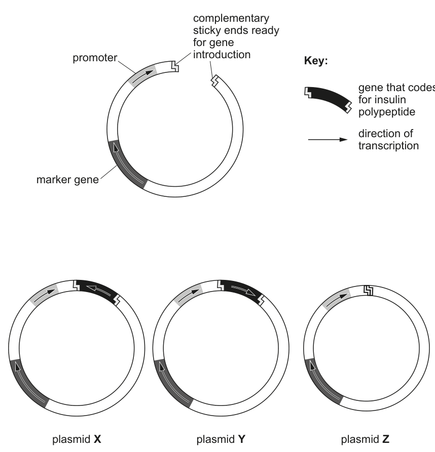
Fig. 4.1
Comment on whether a bacterium will produce the insulin polypeptide if it has either plasmid X or plasmid Y or plasmid Z and explain the reason for your choice.
plasmid X ______
plasmid Y ______
plasmid Z ______
Answer
- Plasmid X — no insulin produced. The insulin gene has been inserted, but in the wrong orientation (back-to-front) relative to the promoter, so RNA polymerase transcribes the antisense strand and a functional mRNA / polypeptide is not made.
- Plasmid Y — insulin produced. The insulin gene is inserted the correct way round, downstream of the promoter and with its direction of transcription matching the promoter's, so the gene is transcribed and translated into the insulin polypeptide.
- Plasmid Z — no insulin produced. The plasmid has re-circularised without the insulin gene (non-recombinant plasmid); the sticky ends have been re-ligated to each other, so there is no gene of interest for the bacterium to express.
See working.
Background Concept
For a recombinant gene to be expressed in a host cell, three conditions must be met:
- The gene must actually be present in the plasmid (i.e. the ligation step must have inserted the fragment, not just re-sealed the empty vector).
- The gene must lie downstream of a promoter that the host's RNA polymerase (with its transcription factors) can recognise.
- The gene must be inserted in the correct orientation — its coding strand must lie on the same strand that the promoter directs RNA polymerase to read, so that a sense mRNA is produced.
If the orientation is reversed, RNA polymerase will transcribe the opposite (template) strand and the resulting mRNA will be the antisense of the gene, which cannot be translated into a functional polypeptide. If the plasmid has simply re-circularised without picking up the gene, the bacterium receives an intact vector but no insulin gene, so again no insulin is made. In real industrial work, only a fraction of the plasmids recovered from a ligation reaction contain the insert in the correct orientation; the rest are either empty, recircularised without insert, or carry the gene in the wrong orientation.
Understanding the Question
The stem tells the student that only some plasmids taken up by host bacteria lead to expression of insulin. The figure shows three possible outcomes of the ligation step:
- Plasmid X — gene present, but inserted in the wrong direction (note the direction-of-transcription arrow on the gene points opposite to that of the promoter).
- Plasmid Y — gene present and in the correct orientation, with the transcription arrow matching that of the promoter.
- Plasmid Z — recircularised plasmid with no gene of interest inside (only the original marker gene and promoter).
The student is asked to comment and explain — so the answer must give the outcome (insulin / no insulin) and the reason (orientation or absence of gene).
Approach
For each plasmid, apply the three conditions above and decide:
- Is the gene present? (look at the figure)
- Is a promoter upstream of it? (look at the figure)
- Is the gene the right way round? (compare the direction of transcription arrow on the gene with that of the promoter)
Step-by-Step Reasoning
- Plasmid X — the gene is present and a promoter is upstream, but the gene's direction-of-transcription arrow points opposite to the promoter's. RNA polymerase will read the wrong strand; no sense mRNA is made, so no insulin polypeptide is produced. The reason to give is therefore gene inserted the wrong way round / backwards.
- Plasmid Y — the gene is present, a promoter is upstream, and the direction-of-transcription arrows on the promoter and the gene point the same way. RNA polymerase will read the coding strand, produce a sense mRNA, and the host's ribosomes will translate it into the insulin polypeptide. The reason is therefore gene inserted the correct way round.
- Plasmid Z — there is no gene of interest inside the plasmid at all; the cut plasmid simply re-ligated to itself (its own sticky ends paired up). Although the marker gene and promoter are present, they have no gene to drive expression of, so no insulin polypeptide is produced. The reason is that the gene was not inserted (the plasmid re-circularised; it is a non-recombinant plasmid).
Key Takeaways
- The presence of a promoter is not enough — the gene must be inserted in the correct orientation.
- A recircularised plasmid (no insert) is a common, useless product of a ligation reaction; this is why the marker gene is also needed for screening.
- This question illustrates why expression efficiency is rarely 100 % in a transformation experiment, even when transformation is successful.
Common Mistakes
- Stating that plasmid Z "does not contain a promoter" — it does contain a promoter; the problem is the absence of the gene of interest.
- Saying plasmid X produces insulin because the gene is present — ignoring the orientation issue.
- Vague wording such as "the gene is not the right one" or "the gene is damaged"; the specific point is the orientation of insertion.
Things to Be Careful About
- The question is a comment with explanation — both elements are needed for full credit.
- Three independent marks: one per plasmid, each requiring a clear reason.
- The figure is the evidence: read the direction of the arrows on promoter and gene carefully; do not rely on what a "correct" plasmid usually looks like.
BRCA2 is a tumour-suppressor gene. Its gene product, BRCA2, is involved in DNA repair. If the DNA cannot be repaired, BRCA2 has a role in causing the cell to die. BRCA2 is found in cells of breast tissue.
When a mutation occurs in BRCA2, damaged DNA may not be repaired and this increases the risk of breast cancer.
When a double-stranded piece of DNA breaks, BRCA2 binds to the damaged DNA directly and interacts with the enzyme RAD51 to repair the damage.
Repairing DNA prevents other mutations and gene rearrangements from occurring which could otherwise lead to breast cancer.
Double-stranded DNA breaks occur naturally during meiosis.
State the event that is initiated as a result of double-stranded breaks during meiosis.
Answer
Crossing over (chiasma formation).
Crossing over (chiasma formation).
Background Concept
During prophase I of meiosis, homologous chromosomes pair up to form bivalents (tetrads). Programmed double-stranded breaks (DSBs) are deliberately introduced into the DNA of these chromosomes by the enzyme SPO11. These breaks are not damage in the harmful sense — they are a normal part of meiosis and initiate the physical exchange of DNA segments between non-sister chromatids of homologous chromosomes. The visible manifestation of this exchange is a chiasma (plural: chiasmata), which is the point where two non-sister chromatids of a homologous pair cross over one another.
Understanding the Question
The stem of the question establishes that BRCA2 is involved in repairing double-stranded DNA breaks, and Part (a) asks specifically what biological event in meiosis is initiated by these double-stranded breaks. The question is essentially asking you to identify the natural, programmed use of DSBs during gamete formation.
Approach
Recognise that DSBs in meiosis are not pathological here — they are a controlled, deliberate part of the mechanism that generates genetic variation. Recall the sequence: SPO11 cuts DNA → strands are resected → single-stranded overhangs invade the homologous chromatid → crossover resolution → chiasma formation. So the event initiated is crossing over.
Step-by-Step Reasoning
- Double-stranded DNA breaks are made at chosen sites in prophase I.
- The 3' single-stranded ends produced by resection invade the homologous (non-sister) chromatid.
- Repair of the breaks using the homologous chromatid as a template produces recombinant chromatids.
- The cytological point at which the exchange is visible is the chiasma.
- The biological process that this represents is crossing over — a key source of genetic variation through recombination.
Key Takeaways
- Programmed DSBs in meiosis initiate crossing over / chiasma formation, not damage.
- This is the basis of genetic recombination and one of the major sources of genetic variation in sexually reproducing populations.
- BRCA2's role in repairing DSBs is essential because unrepaired breaks in somatic cells can lead to mutations and cancer, but in meiosis the same kind of break is exploited to generate diversity.
Common Mistakes
- Writing "replication" — DSBs do not initiate replication; they initiate recombination.
- Writing "mutation" — DSBs in meiosis are not random mutations; they are programmed events leading to legitimate crossing over.
- Writing "separation of homologues" or "independent assortment" — these occur later (anaphase I) and are consequences of alignment, not DSBs.
Things to Be Careful About
The mark scheme accepts "crossing over" or "chiasma formation" as alternatives. Either is a complete answer. Do not give a long description of the whole prophase I — the question asks for the single event initiated by the breaks.
Scientists have identified hundreds of mutations in BRCA2, but not all of these mutations will increase the risk of cancer. One specific mutation in BRCA2, known as 999del5, is found in 0.6% of the general global population.
Iceland is an island country in the North Atlantic Ocean. The ancestors of most of the current population are people who arrived to settle in Iceland in AD 874.
In the Icelandic population, mutation 999del5 is the cause of 7–8% of breast cancer cases in women and 40% of breast cancer cases in men. This is much higher than the percentage of breast cancer cases in the general global population.
Suggest and explain how mutation 999del5 accounts for a very high percentage of breast cancer cases in Iceland compared with the general global population.
Answer
Any three from:
- The mutation occurred a long time ago (in one of the original settlers / a common ancestor) and has been inherited by their descendants.
- Founder effect: the modern Icelandic population descended from a small number of original settlers, so any allele carried by those founders starts at a measurable frequency in the new population.
- Iceland has a small, isolated population, so the gene pool is restricted / genetic diversity is low.
- Much of the Icelandic population is closely related (inbreeding), so the allele has been passed on to many descendants rather than diluted by out-breeding.
- The combination of founder effect and isolation has caused the frequency of the 999del5 / mutant BRCA2 allele to increase in the Icelandic population compared with the global population.
Founder effect in a small, isolated, inbred population has increased the frequency of the 999del5 mutation.
Background Concept
Allele frequencies in a population are not fixed — they change over time under the influence of mutation, selection, migration, genetic drift and non-random mating. Founder effect is a special case of genetic drift: when a new population is established by a very small number of individuals (the founders), the allele frequencies in the new population are simply a sample of those in the original population, biased by which alleles the founders happened to carry. If one of the founders carried a rare allele, that allele can become disproportionately common in the descendant population, especially if the population stays small, isolated, and inbred.
Understanding the Question
The question gives two pieces of data: globally, 999del5 is found in 0.6% of people, but in Iceland it accounts for 7–8% of female and 40% of male breast cancers. The stem also tells you the Icelandic population descends from settlers who arrived in AD 874, so the population is both small (in absolute terms) and isolated (an island in the North Atlantic). You have to explain why this specific mutation is so much more important in Iceland than globally.
Approach
The demographic history of Iceland (small founding population, long-term isolation, inbreeding) maps directly onto the founder effect. The mark scheme is essentially asking you to spell out: (1) the mutation was present in the founders, (2) the population that received it was small, (3) the population is isolated so no new genetic input dilutes the allele, and (4) the result is an inflated frequency of that allele.
Step-by-Step Reasoning
- Origin of the mutation: The 999del5 mutation must have existed in at least one of the original settlers who arrived in AD 874. Such a mutation could have happened once, long ago, in a common ancestor of today's Icelanders.
- Founder effect: Because the modern Icelandic population is descended from that relatively small group of original settlers, any allele carried by them — including 999del5 — starts at a higher frequency in the descendant population than in the global population from which the founders came. This non-representative sample of alleles is the founder effect.
- Small population: A small population means each new generation is a small sample of the previous one. Random fluctuations in allele frequency (genetic drift) are larger in small populations, so a once-rare allele can drift to a much higher frequency.
- Isolated / inbred / small gene pool: Iceland is an island, so migration in and out has been very limited. People marry within the population, so the gene pool is restricted and genetic diversity is low. An allele present in the founders is therefore likely to be passed on to many descendants rather than diluted by outbreeding.
- Result: The frequency of 999del5 has risen (or remained high) in Iceland, so it now accounts for a much higher proportion of breast cancer cases than the global average, even though the mutation is rare worldwide.
Key Takeaways
- Founder effect + small isolated population = high frequency of an allele that was rare in the source population.
- The same principle explains the high frequency of particular disease alleles in many geographically or culturally isolated groups (e.g. the high frequency of certain alleles in Ashkenazi Jewish, Amish, or Finnish populations).
- The question is testing your ability to apply the founder-effect concept, not just name it — you must explicitly link Iceland's history to the present-day allele frequency.
Common Mistakes
- Saying only "founder effect" without describing the small founding population or its consequences. Founder effect on its own is a label, not an explanation.
- Saying "natural selection" increased the frequency. There is no evidence the allele confers any selective advantage; the increase is due to drift / founder effect, not selection.
- Confusing founder effect with bottleneck. A bottleneck is a sharp reduction in an existing population; the Icelandic population is better described by founder effect (although a small effective population size also matters going forward).
- Saying the mutation "arose" in Iceland. It is more accurate to say the mutation existed in the founders — it may have arisen anywhere.
Things to Be Careful About
The mark scheme wants a suggest and explain answer, so simply naming "founder effect" earns one mark only; the other two marks require you to develop the explanation (e.g. small population, isolated, inbreeding, long time ago, increased allele frequency).
One of the largest global genetic screening programmes for breast cancer involves identifying people with mutations in BRCA2.
Outline the advantages of genetic screening for mutations in BRCA2.
Answer
Any three from:
- If the result is negative, the individual is reassured and avoids unnecessary worry.
- If the result is positive, the individual can make lifestyle changes (e.g. diet, exercise, reducing alcohol intake) that may lower their risk.
- A positive result allows early / more frequent monitoring, early treatment or prophylactic surgery (e.g. mastectomy), improving the chance of successful treatment.
- A positive result allows the individual to make an informed decision about having children (e.g. considering IVF with embryo screening, or informing existing children who may also carry the mutation).
- Genetic counselling can be offered alongside screening so individuals understand the implications of their result.
- Identifying carriers allows earlier detection of breast cancer, which lowers the death rate from the disease.
Reassurance if negative; informed lifestyle choices, earlier monitoring / treatment, informed reproductive decisions, access to counselling, and reduced mortality from breast cancer if positive.
Background Concept
Genetic screening tests individuals or populations for the presence of a particular allele, often before any symptoms appear. For late-onset, highly penetrant cancer-predisposition genes such as BRCA1 and BRCA2, a positive result does not mean the person has cancer — it means they have a much higher lifetime risk and can act on that information. The value of screening therefore lies in what people do with the information, both before and after a result is given.
Understanding the Question
Part (b)(ii) asks for the advantages of genetic screening for BRCA2 mutations — that is, what does the individual, and the wider population, gain from being tested? The command word "outline" means you should give a brief but reasoned list of the main benefits, not a single one-line answer. The mark scheme accepts up to six possible points and you need any three.
Approach
Think about the two possible outcomes of a screening test (positive or negative) and what flows from each. Negative results bring reassurance and peace of mind; positive results open up a chain of actions — counselling, lifestyle changes, increased surveillance, prophylactic surgery, and informed reproductive choices. Beyond the individual, screening reduces mortality because cancers are caught earlier when they are more treatable.
Step-by-Step Reasoning
- Negative result: The person is told they do not carry the mutation, so they avoid years of unnecessary worry. This is a real, though sometimes under-emphasised, benefit of screening.
- Positive result — lifestyle: Knowing one carries BRCA2 allows changes that may lower risk (e.g. avoiding hormone-replacement therapy, reducing alcohol, maintaining a healthy weight, breastfeeding).
- Positive result — surveillance / treatment: Carriers can enrol in more frequent screening (e.g. annual MRI / mammography from a younger age than the general population) and consider prophylactic surgery (mastectomy, oophorectomy). Earlier detection dramatically improves survival.
- Positive result — reproduction: A carrier can make informed choices about having children. Options include prenatal testing, pre-implantation genetic diagnosis (PGD) during IVF, and informing relatives who may also carry the allele.
- Counselling: Genetic screening is usually paired with genetic counselling so that individuals understand the meaning, limitations and implications of their result before and after testing.
- Population health: Because earlier detection leads to better outcomes, screening ultimately lowers the death rate from breast cancer in the screened population.
Key Takeaways
- Genetic screening has value for both outcomes: a negative result reassures; a positive result enables prevention, early detection, and informed choices.
- The benefit is only fully realised when screening is coupled with counselling and follow-up clinical action.
- BRCA2 screening is a presymptomatic test, so benefits are about risk management and prevention, not about curing an existing disease.
Common Mistakes
- Giving only the disadvantages or ethical concerns — the question asks for advantages.
- Confusing screening with treatment. Screening tells you your risk; it does not itself treat cancer.
- Suggesting screening "prevents" breast cancer. It does not; it allows earlier detection and risk-reducing actions, which improve outcomes.
- Missing the value of a negative result (reassurance / reduced worry).
Things to Be Careful About
- "Outline" means a short reasoned account — three distinct points is enough for full marks. Do not pad with repeats.
- The mark scheme accepts points either as stand-alone statements (e.g. "reduce worry if negative") or as conditional ones (e.g. "lifestyle changes if positive"). Either is fine, but make sure the logic is clear.
Suggest one advantage to a country of a genetic screening programme for breast cancer that screens for specific mutations in BRCA2 in the population.
Answer
Any one from:
- It is cost-effective, because the screen only tests for the specific mutation(s) known to occur at high frequency in that population rather than screening for hundreds of rare mutations.
- It allows early diagnosis / early / targeted treatment, reducing the cost of treating advanced breast cancer.
- It identifies a mutation that is known to increase the risk of breast cancer, avoiding the cost of testing for neutral variants of unknown significance.
- It could lead to lower death rates from breast cancer in the country.
Screening for specific high-frequency mutations is cost-effective and identifies known high-risk alleles, enabling earlier treatment and lower national mortality.
Background Concept
Hundreds of different BRCA2 mutations have been identified, but their penetrance and population frequency vary enormously. From a public-health perspective, it matters whether the country is screening broadly for all possible mutations (expensive, low yield) or targeting the small number of mutations that are actually common in their population. A targeted approach concentrates resources where they have the greatest impact.
Understanding the Question
Part (b)(iii) shifts the focus from the individual advantages of screening (Part b(ii)) to the national advantage. The phrase "advantage to a country" is the key — you are being asked to think about the programme as a public-health intervention. Only one mark is available, so a single, well-targeted point is enough.
Approach
The mark scheme lists five possible advantages; the strongest country-level arguments are the cost-effectiveness of testing only for common mutations, and the population-level health benefit (lower mortality through earlier / targeted treatment).
Step-by-Step Reasoning
- Cost-effectiveness: A country's health budget is finite. If a particular BRCA2 mutation (e.g. 999del5 in Iceland) accounts for a large share of breast cancer cases, then designing the screen to detect that specific mutation is much cheaper per detected case than screening for hundreds of rare variants. The cost per life-year saved is therefore lower.
- Early / targeted treatment: Knowing the precise mutation present in the population lets clinicians tailor monitoring and treatment. Earlier detection of breast cancer in carriers is cheaper for the health system than treating late-stage disease, and saves lives.
- Avoids neutral variants: Many rare BRCA2 mutations are of unknown clinical significance. Testing only for known high-risk mutations reduces the number of ambiguous results that need expensive follow-up and counselling.
- Population mortality: Aggregated across the country, the screening programme will, over time, lower the death rate from breast cancer.
Key Takeaways
- The same intervention (screening) can be evaluated at the individual level (Part ii) or at the national / health-economics level (Part iii).
- Targeted screening for high-frequency mutations is a recurring real-world strategy: e.g. Tay-Sachs in Ashkenazi Jewish populations, thalassaemia in Mediterranean and South-East Asian populations.
Common Mistakes
- Repeating an individual-level point (e.g. "people can change their lifestyle") — the question specifically asks for an advantage to a country.
- Saying "it is cheaper" without explaining why it is cheaper (e.g. only testing for the specific high-frequency mutation rather than all possible mutations).
- Confusing "cost-effective" with "cheap". Cost-effective means good value for money relative to outcome, not low absolute cost.
Things to Be Careful About
The mark scheme accepts any one of five alternatives. Pick the one you can express most clearly. "Cost-effective because it only screens for high-frequency mutations" is the single most economical answer and the one most directly tied to the stem.
Lipocalin 2 (Lcn2) is a cancer-promoting gene (oncogene). When Lcn2 is expressed, it can result in breast cancer.
Research is being carried out to see if gene editing of Lcn2 could be used to treat breast cancer.
Gene editing was used to treat human cancer cells that had been implanted into mice to form a tumour. The treatment stopped Lcn2 from being expressed in the cancer cells and resulted in a significant reduction in the growth of the tumour. There was no negative effect in normal tissues.
Answer
Any two from:
- The editing tool inserted, deleted or replaced a section of the Lcn2 DNA (or its regulatory sequence).
- This change prevented transcription of the Lcn2 gene, so no mRNA (and therefore no protein) was produced.
- A mutated / faulty section of DNA was replaced with the correct sequence, restoring a functional (or, in the case of an oncogene, removing the harmful) version of the gene.
The editing tool cut, inserted, deleted or replaced DNA in (the regulatory sequence of) Lcn2, preventing its transcription.
Background Concept
Gene editing refers to a family of molecular tools (notably CRISPR-Cas9, but also TALENs and zinc-finger nucleases) that can be programmed to make a precise double-stranded break at a specific DNA sequence. The cell's own repair machinery then repairs the break, but if a DNA template is provided alongside the editing tool, the repair can be hijacked to insert a new sequence or correct a faulty one. Gene editing can therefore (1) disrupt a gene by introducing small insertions or deletions (indels) at the cut site, often producing a non-functional protein, or (2) replace a faulty sequence with a corrected one using homology-directed repair.
Understanding the Question
The stem tells you that gene editing was used to stop Lcn2 being expressed in cancer cells, and that this reduced tumour growth. The task is to suggest how — at the molecular level — the editing produced that effect.
Approach
Work backwards from the observed outcome (no expression of Lcn2). No expression means either no transcription or no functional mRNA. Both follow from changes to the DNA that the editing tool makes. The mark scheme offers three creditworthy points: describing the DNA change, the consequence for transcription, and the idea of replacing faulty DNA with the correct sequence.
Step-by-Step Reasoning
- DNA-level change: A guide-RNA directs the editing nuclease to a specific site in (or near) the Lcn2 gene. The nuclease introduces a double-stranded break; the cell repairs it imperfectly, producing small insertions or deletions that disrupt the gene, or, if a template is supplied, the cell uses homology-directed repair to replace the faulty sequence with a correct one.
- Effect on transcription: Because the coding or regulatory sequence of Lcn2 has been altered, RNA polymerase can no longer transcribe a functional mRNA from the gene. No mRNA → no protein → the oncogene's cancer-promoting activity is lost.
- Specificity of action: The change is made in the DNA itself, so the effect is permanent for that cell and its descendants, not a temporary knockdown as one would get with RNA interference.
Key Takeaways
- Gene editing changes the DNA; gene silencing (e.g. RNAi) changes the mRNA. The mechanism of action is therefore different, and editing is heritable for the cell's lineage.
- Stopping expression of a cancer-promoting gene can be achieved by disrupting the gene, deleting it, or replacing the harmful sequence with a benign one.
- Editing can target either the coding region (no functional protein) or a regulatory region (no transcription at all).
Common Mistakes
- Saying the editing "removed the protein" — editing works on DNA, not on protein directly. The protein stops being made because the gene is no longer expressed.
- Confusing gene editing with gene therapy in general terms. Editing is one tool within gene therapy; the question is specifically about editing.
- Saying the cell "dies" because of the edit. The stem says there was "no negative effect in normal tissues"; the edit is targeted to the cancer cells' DNA, not a general cytotoxic event.
Things to Be Careful About
- Use the precise term "transcription" if you are referring to the synthesis of mRNA. "Expression" is a broader term covering everything from transcription to active protein, but the mark scheme specifically wants you to say transcription is prevented.
- Either "inserted / deleted / replaced" wording is acceptable; pick one and state it clearly.
Answer
The editing tool only cuts DNA at a specific target sequence / site that is unique to Lcn2; other genes, which have different sequences, are not recognised and so are not cut.
The editing tool only targets a specific DNA sequence found in Lcn2 and so does not cut other genes.
Background Concept
Modern gene-editing tools such as CRISPR-Cas9 are programmed to cut at a specific DNA sequence by a short guide RNA whose sequence is complementary to the target site. The nuclease will only cut DNA that closely matches the guide sequence, so a different guide RNA would target a different gene. This sequence-specificity is the basis of the technology's selectivity and is what allows a single gene to be edited without affecting the rest of the genome.
Understanding the Question
The stem tells you that the edit affected Lcn2 and that there was "no negative effect in normal tissues". The question asks you to suggest why only Lcn2 was affected, i.e. why the edit did not knock out other genes at the same time.
Approach
The answer turns on the sequence-specificity of the editing tool. The guide RNA was designed to match a site that is unique to Lcn2; other genes, having different sequences, were not recognised, so the nuclease did not cut them.
Step-by-Step Reasoning
- The guide RNA of the editing tool was designed to be complementary to a short, unique sequence in (or near) the Lcn2 gene.
- The nuclease only cuts DNA where the guide RNA binds; the rest of the genome, which has different sequences, is not a substrate for cutting.
- Therefore, only the Lcn2 gene is disrupted; other genes continue to be transcribed and translated normally, and normal tissues are unaffected.
Key Takeaways
- Sequence-specificity is the defining safety feature of modern gene editing.
- Off-target effects are theoretically possible if a similar sequence exists elsewhere in the genome, but in a well-designed experiment the chosen guide RNA targets a unique site, minimising this risk.
- This is why gene editing is considered a more precise tool than, for example, chemotherapy, which affects any rapidly dividing cell.
Common Mistakes
- Saying "it was injected only into the tumour". That may be true experimentally, but the question is asking about the molecular reason only Lcn2 was affected, not the delivery method.
- Saying "only the cancer cells were edited". This is a delivery / cell-biology point, not a sequence-specificity point. The mark scheme wants the molecular answer.
- Saying "the gene is special" or "the cells are different". Genes are not intrinsically different in a way that makes them more editable; it is the guide sequence that is specific.
Things to Be Careful About
The mark scheme accepts either of two phrasings: "only specific sites/sequences on the DNA are targeted" or "the target site is only found in Lcn2". Both convey the same idea — choose the wording that feels most natural to you and be concise.
In plants, stomata open and close in response to changes in environmental conditions.
Explain why stomata need to open and close according to environmental conditions.
Answer
- Stomata need to open to allow diffusion of carbon dioxide into the leaf for photosynthesis (Calvin cycle).
- Stomata need to close to prevent water loss from the leaf by transpiration.
- Stomata can close at night / in the dark (when there is no photosynthesis) to reduce water loss.
- Stomata must also close during conditions that increase water loss, e.g. high light intensity, high temperature, high wind speed or drought / water stress, to prevent the plant wilting.
Stomata open to obtain CO2 for photosynthesis but close to reduce water loss by transpiration, especially at night or during drought, high temperature, high light intensity or high wind speed.
Background Concept
Stomata are small pores, mainly on the underside of leaves, each flanked by a pair of bean-shaped guard cells. They are the main route by which CO2 enters the leaf for photosynthesis and by which water vapour leaves the leaf (transpiration). Because CO2 is needed for the Calvin cycle but water is constantly lost through the same pore, the plant faces a fundamental trade-off: it must keep stomata open enough to feed photosynthesis, but not so open that it loses more water than its roots can replace.
Guard cells respond to several environmental signals. Blue light (via the phototropin receptors) and low internal CO2 stimulate opening, while darkness, drought (via abscisic acid, ABA) and high CO2 inside the leaf stimulate closure. This is a classic homeostatic compromise: a single opening is regulated by multiple, sometimes opposing, environmental cues.
Understanding the Question
The command word is explain, so for each marking point you need to state WHY the response helps the plant — not just that the response happens. The question gives you 3 marks, so you need any three valid points from the mark scheme: one point about why opening is needed, and two about why and when closing is needed. Always link the stomatal behaviour to a plant process (photosynthesis, transpiration) and to an environmental trigger.
Approach
Start with the photosynthesis side (why open), then the water-balance side (why close), and finally give a specific example of an environmental cue that forces closure. Three short, distinct sentences — each making a clear cause-and-effect link — will score full marks.
Step-by-Step Reasoning
- Photosynthesis requires CO2 — atmospheric CO2 (~0.04%) enters the leaf through the open stomata by diffusion down its concentration gradient. Without an open pore, the Calvin cycle in the chloroplasts would run out of CO2 and photosynthesis would stop.
- Transpiration loses water — water evaporates from the wet cell walls of the mesophyll and diffuses out of the leaf through the stomata. If stomata stayed permanently wide open in hot, dry or windy conditions, the plant would lose water faster than the roots could replace it, leading to wilting and death.
- Darkness removes the need to be open — at night there is no light, so the light-dependent reactions of photosynthesis cannot occur and CO2 fixation is not needed. Closing stomata in the dark conserves water without any cost to photosynthesis.
- Stressful conditions force closure — high light intensity, high temperature, low humidity, high wind speed and drought all increase the rate of transpiration. The guard cells respond (often via abscisic acid in drought) by becoming flaccid and closing the pore, limiting water loss.
Any three of these four points earn the 3 marks.
Key Takeaways
- Stomata must balance two competing demands: CO2 uptake for photosynthesis and water retention.
- Closure is triggered by conditions that increase evaporative loss (heat, wind, drought) and by conditions where photosynthesis cannot occur (darkness).
- The opening/closing response is a homeostatic mechanism that maintains the plant's internal water status.
Common Mistakes
- Saying stomata open "to let air in" — vague; the mark requires the specific gas (carbon dioxide) and its role in photosynthesis / the Calvin cycle.
- Stating that stomata close "to save water" without naming the process (transpiration) — be specific.
- Forgetting to give a trigger — closing in response to high temperature is a different marking point from closing at night.
Things to Be Careful About
- "Explain" demands both the observation and the reason; "describe" only requires the observation.
- The mark scheme accepts "high light intensity / high temperatures / high wind speed / drought" as alternative stressful triggers — give any one to score the point.
Answer
- Blue / light activates a proton pump (H-ATPase) in the guard cell plasma membrane.
- H ions are actively transported out of the guard cells, lowering the H concentration inside and setting up an electrochemical / proton gradient.
- This hyperpolarises the membrane and causes K channels to open, so K ions (and Cl ions) move into the guard cells down their electrochemical gradient.
- The accumulation of K (and Cl) lowers the water potential of the guard cells (it becomes more negative).
- Water enters the guard cells by osmosis, down the water potential gradient.
- The guard cells expand and become (more) turgid.
- Because the inner wall (bordering the pore) is thicker and less elastic than the outer wall, the outer wall stretches more than the inner wall, causing the guard cells to bow outwards and the stoma to open.
Light activates an H-ATPase that pumps H out of the guard cells; K then enters, lowering the water potential so water enters by osmosis; the guard cells become turgid and, because the inner wall is thicker than the outer, they bow apart to open the stoma.
Background Concept
Guard cells are specialised epidermal cells that change shape to open and close the stomatal pore. Their behaviour is driven by turgor — when they are turgid they bow apart and the pore opens; when they lose solutes and become flaccid the pore closes. The opening mechanism is a beautiful example of how plants use active transport to drive a passive osmotic response.
The plasma membrane of a guard cell contains an H-ATPase (proton pump) and voltage-gated K channels. Because the inner wall (next to the pore) is thicker and reinforced with radial cellulose microfibrils, while the outer wall is thinner and more elastic, a uniform increase in volume forces the guard cells to bend away from each other. This asymmetry converts a turgor change into an aperture change.
Understanding the Question
The command word is describe, so you must set out the sequence of events clearly. Six marks = six distinct creditable points (or fewer if you also give some of the AVP points). The mark scheme explicitly warns that if you describe CLOSURE instead of opening you can earn at most 3 marks — so be sure your answer is about opening. The mechanism starts with light (or another trigger) and ends with the pore opening.
Approach
Work through the mechanism in strict order:
light / trigger → H pumped out → K moves in → solute concentration rises → water potential falls → water enters by osmosis → guard cells become turgid → differential wall stretching bends the cells apart → pore opens.
Each arrow is a marking point. Keep the chain tight so the examiner can credit every step.
Step-by-Step Reasoning
- Trigger and pump activation — Blue light (absorbed by the phototropin receptor) activates the H-ATPase in the guard cell plasma membrane. This pump uses ATP to actively transport protons (H) out of the guard cell into the surrounding apoplast. The active transport step is essential; passive movement of H would not produce the required gradient.
- Establishing the gradient — Pumping H out lowers the H concentration inside the cell and hyperpolarises the membrane (the inside becomes more negative). This sets up both a chemical gradient and an electrical gradient that together drive the next step.
- K uptake — The hyperpolarisation opens voltage-gated inward-rectifying K channels in the plasma membrane. K ions flow into the guard cells down their electrochemical gradient. Chloride ions (Cl) and malate often accompany the K to balance charge.
- Solute accumulation lowers water potential — The high internal concentration of K (and accompanying anions) makes the cytoplasm and vacuole hypertonic to the surrounding apoplast. The water potential () of the guard cell therefore decreases (becomes more negative).
- Osmotic water entry — Water moves into the guard cell by osmosis, down the water potential gradient. Aquaporins in the plasma membrane and tonoplast facilitate rapid water flow.
- Turgor increases — As water enters, the central vacuole swells and the protoplast pushes against the cell wall. The guard cell becomes more turgid.
- Differential wall stretching opens the pore — The inner wall (next to the stomatal pore) is thicker, more rigid and reinforced by radially arranged cellulose microfibrils, so it resists stretching. The outer wall is thinner and more elastic, so it stretches much more. The combined effect is that the guard cells bow outwards (like a pair of curved sausages), pulling the inner walls apart and opening the pore between them.
Any six of these points earn the marks; the seventh (asymmetric walls) is often the one students forget and is well worth including.
Key Takeaways
- Stomatal opening is driven by an active proton pump, but the bulk water movement is passive (osmosis).
- Solute accumulation (K and accompanying anions) lowers water potential, drawing water in.
- The asymmetric wall structure converts isotropic turgor into a directional opening of the pore.
- The sequence is: active H efflux → K influx → osmotic water uptake → turgor → shape change → open pore.
Common Mistakes
- Describing closure (loss of K, ABA signalling, Ca channels) instead of opening — capped at 3 marks.
- Saying water "enters by active transport" — water only moves by osmosis.
- Saying K is actively transported in — K moves down its electrochemical gradient through channels; it is the H pump that uses ATP.
- Omitting the wall asymmetry point and so losing a free mark — the bowed shape of the guard cells is the mechanical reason the stoma actually opens.
- Confusing the location of the thicker wall (it is the INNER wall, bordering the pore, that is thicker and less elastic — students often reverse this).
Things to Be Careful About
- Mark scheme accepts either "outer wall thinner, allows more stretching" OR "inner wall thicker, allows less stretching" — either phrasing is fine.
- "AVP" points (mark point 8) include: light stimulating ATP production, Cl co-transport, and aquaporin-mediated water entry — include any of these for extra credit if you have covered the core points.
- The mechanism is initiated by blue light, not by photosynthesis directly; photosynthesis lowers internal CO2 which also contributes, but the standard CIE mechanism starts with H-ATPase activation by light.
Experiments were carried out to determine the effect of light intensity on the rate of photosynthesis of a species of the unicellular protoctist, Chlorella. A cell suspension of Chlorella was used.
Carbon dioxide uptake was used as a measure of the rate of photosynthesis.
• The suspension of Chlorella was illuminated at a light intensity of 3 lux for 20 seconds.
• The carbon dioxide uptake by Chlorella was measured at the end of the 20 second period of illumination.
• The experiment was repeated at 6 lux, 9 lux, 12 lux and in a dark room.
• The suspension was maintained at a temperature of .
Table 7.1 shows the results of the experiments.
Table 7.1
| light intensity / lux | total uptake after 20 seconds / | rate of photosynthesis / |
|---|---|---|
| 0 | 0 | 0.0 |
| 3 | 20 | 1.0 |
| 6 | 44 | ............ |
| 9 | 72 | 3.6 |
| 12 | 80 | 4.0 |
Use Table 7.1 to calculate the rate of photosynthesis at a light intensity of 6 lux.
Complete Table 7.1 by writing your calculated value in the space provided.
Working
Answer
2.2 µmol s⁻¹
Background Concept
The rate of a process is defined as the quantity of product (here, taken up) accumulated per unit time. When the total amount taken up over a fixed interval is known, the mean rate over that interval is simply that total divided by the duration. The units of rate are therefore the units of the quantity divided by the unit of time (e.g. ).
Understanding the Question
Table 7.1 records both the total uptake (in ) at the end of 20 s and the corresponding rate (in ). The cell at 6 lux is empty: the total uptake is and the time is . We must insert the rate that the experimenter would have calculated, and it must agree with the units used elsewhere in the table.
Approach
Apply the definition of rate:
Use the same units the table is using (total in , time in s) so the answer is in , matching the other filled cells in the rate column.
Step-by-Step Reasoning
- From the table, at the total taken up in is .
- Divide this by the time interval: .
- Quote the answer with the units used in the rest of the rate column: .
This is consistent with the surrounding points: at 3 lux the rate is and at 9 lux it is , so a value of at 6 lux sits sensibly on the rising portion of the curve.
Key Takeaways
- Rate = total quantity ÷ time, with units of quantity per unit time.
- Always quote the answer to the same precision and in the same units as the rest of the column.
Common Mistakes
- Forgetting to divide by 20 and writing 44.
- Quoting the answer as instead of (units flipped).
- Using the rate column's existing value at 3 lux (1.0) as a reference and giving an answer not consistent with the 44 µmol total.
Things to Be Careful About
- Keep the time units consistent: 20 s, not 0.20 min or 1/3 min.
- Match the precision of the question's table (one decimal place is appropriate here).
Plot a graph of the data in Table 7.1 on the grid in Fig. 7.1 to show the effect of light intensity on the rate of photosynthesis.
Draw a curve and extend your curve to show what would happen to the rate of photosynthesis if the experiment is carried out at 18 lux.
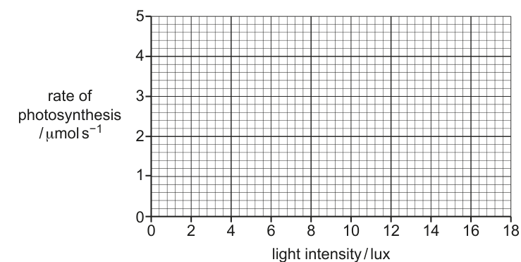
Fig. 7.1
Answer
Points to plot on Fig. 7.1:
| light intensity / lux | rate of photosynthesis / |
|---|---|
| 0 | 0.0 |
| 3 | 1.0 |
| 6 | 2.2 |
| 9 | 3.6 |
| 12 | 4.0 |
- Plot each point clearly with a small cross or dot, exactly at the intersection of the grid lines.
- Draw a smooth best-fit curve through the five points.
- Extend the curve to 18 lux. The line should level off (the gradient becomes much smaller) so that at 18 lux the rate is approximately the same as at 12 lux (≈ , or only very slightly higher).
Five points plotted correctly, a smooth best-fit curve drawn through them, and the curve levelled off between 12 and 18 lux at a rate of about 4.0 µmol s⁻¹.
Background Concept
The rate of photosynthesis depends on several environmental factors. At low light intensities, light is the factor that limits how fast the light-dependent reactions can run, so increasing the light intensity speeds up the overall rate. As light intensity continues to rise, another factor (commonly concentration or temperature) becomes the bottleneck; once that happens, further increases in light no longer increase the rate, and the graph plateaus. This produces the classic saturating curve.
Understanding the Question
The student is given a pre-printed grid (Fig. 7.1) with:
- y-axis: rate of photosynthesis / , running 0 to 5 in steps of 1;
- x-axis: light intensity / lux, running 0 to 18 in steps of 2.
The five data points (including the new one calculated in (a)(i)) must be plotted accurately, joined with a smooth best-fit curve, and the curve extended from 12 lux to 18 lux in a way that is biologically realistic.
Approach
- Choose a sensible scale on the y-axis: the data range from 0.0 to 4.0 ; the printed axis already goes to 5, which is appropriate.
- Use a cross or a clear dot in a circle for each point, exactly at the intersection of the grid lines corresponding to its (x, y) values.
- Draw a smooth freehand curve through the points — the points describe a curve that rises steeply at first and then flattens, so a single smooth curve, not a series of straight segments, is needed.
- Extrapolate to 18 lux. Biologically, light is no longer limiting, so the curve should level off rather than continuing to rise steeply. The mark scheme accepts either a horizontal continuation or a continuation with a much smaller gradient that still ends essentially flat at 18 lux.
Step-by-Step Reasoning
- At (0, 0.0) the curve must pass through the origin.
- At 3 lux the rate is 1.0; at 6 lux it is 2.2; at 9 lux it is 3.6; at 12 lux it is 4.0. The increments between consecutive points are 1.0, 1.2, 1.4, 0.4 — the rise is steepest in the middle and is already decelerating by 12 lux.
- A best-fit smooth curve through these five points therefore bends over clearly as it approaches 12 lux.
- Extending this trend to 18 lux, the curve continues to flatten. The rate at 18 lux is unlikely to be much above 4.0; a small further rise (e.g. to about 4.1 or 4.2) is acceptable, but the curve must clearly level off (a continued steep rise would be biologically wrong).
Key Takeaways
- Always check that each plotted point lies on the correct grid intersection.
- A 'best-fit' curve is smooth — never a zig-zag of straight line segments between points.
- Extrapolation must be consistent with the trend in the data, not just continued in the same direction.
- Recognise the saturating shape of a light-intensity vs photosynthesis-rate graph.
Common Mistakes
- Plotting (6, 2.2) incorrectly (e.g. as 2.0 or 2.5 because of a misread of the table).
- Joining the points with straight lines, or drawing a curve that wiggles above and below individual points instead of passing through them in a smooth way.
- Extending the curve to 18 lux with the same steep gradient, so that the rate keeps climbing well above 5 — this is biologically unrealistic because light is no longer the limiting factor.
- Forgetting the (0, 0) point.
Things to Be Careful About
- Read the question's table to one decimal place — the calculated value of 2.2 must be plotted as 2.2, not 2.0.
- Use a sharp pencil; small crosses are easier for the examiner to verify than large blobs.
- The mark scheme credits three separate points: five points plotted correctly, a best-fit curve, and a levelling-off shape between 12 and 18 lux. Each must be visibly present on the graph.
Answer
- Light intensity is no longer the limiting factor above 12 lux.
- Temperature / concentration is now limiting, so increasing the light intensity further does not increase the rate.
Light is no longer limiting; another factor (temperature or CO2 concentration) is now limiting the rate.
Background Concept
The rate of photosynthesis at any moment is set by whichever environmental factor is in shortest supply relative to the cell's needs. This is the limiting-factor principle. At low light intensities, raising the light speeds up the light-dependent reactions and therefore the whole process. Once light is plentiful, the next bottleneck takes over: usually the supply of to the Calvin cycle or the temperature (which controls the kinetic energy of enzyme-catalysed steps in the Calvin cycle, especially the rubisco-catalysed carboxylation).
Understanding the Question
Part (a)(iii) follows the graph in (a)(ii). Between 12 and 18 lux the curve levels off rather than continuing to rise. The student must explain why further light does not produce a faster rate of photosynthesis.
Approach
State the limiting-factor principle explicitly: identify that light has ceased to be the bottleneck and name a factor that has become limiting. Use the information given in the question stem — the temperature was held at and the supply was set by the cell suspension — to decide which factor is most plausibly limiting in this particular set-up.
Step-by-Step Reasoning
- As light intensity rises, the rate of the light-dependent reactions increases. This pushes more reduced NADP and ATP into the Calvin cycle.
- The rate at which the Calvin cycle can use these products is set by enzyme activity (mostly temperature-dependent) and by the availability of .
- Once light is in excess, the light-dependent reactions can produce ATP and reduced NADP faster than the Calvin cycle can consume them. ATP and reduced NADP accumulate, and the rate of the overall process is capped by the slower, light-independent stage.
- Therefore, between 12 and 18 lux the rate stops increasing because light intensity is no longer limiting, and the limiting factor is now the temperature or the concentration.
Key Takeaways
- A plateau on a rate vs light-intensity graph is the classic signature that a different factor has become limiting.
- The two factors that most commonly take over from light are concentration and temperature.
- The limiting-factor principle can be stated as: at any given time, the rate of photosynthesis is limited by the factor in shortest supply.
Common Mistakes
- Saying 'the chlorophyll is saturated' without explaining what this means in terms of a downstream bottleneck.
- Naming a single specific factor (e.g. 'temperature') without acknowledging that is also a plausible candidate.
- Failing to state that light is no longer the limiting factor — saying only that the rate is 'constant' or that 'the plant is photosynthesising as fast as it can' does not earn the mark.
Things to Be Careful About
- The question asks for an explanation, not just a description. A bare statement of the shape ('it levels off') earns nothing; the reasons for the shape must be given.
- Use the term 'limiting factor' — examiners reward this precise wording.
In photophosphorylation, photoactivation of chlorophyll results in the synthesis of ATP.
Describe how photoactivation of chlorophyll results in the synthesis of ATP in photophosphorylation.
Answer
- Light is absorbed by chlorophyll and electrons in the chlorophyll are excited / emitted (photoactivation).
- The excited electrons travel along the electron transport chain, releasing energy.
- This energy is used to pump (protons) from the stroma into the thylakoid space / lumen.
- This increases the concentration in the thylakoid space, creating a proton gradient across the thylakoid membrane.
- / protons diffuse back from the thylakoid space into the stroma through ATP synthase.
- The flow of through ATP synthase drives the synthesis of ATP from ADP and (chemiosmosis).
Excited electrons pass along the electron transport chain; the energy released pumps H⁺ into the thylakoid space, creating a proton gradient. H⁺ then diffuses back into the stroma through ATP synthase, driving ATP synthesis (chemiosmosis).
Background Concept
The light-dependent reactions of photosynthesis take place on the thylakoid membranes inside the chloroplast. The thylakoid membrane is the site of two key pieces of molecular machinery: the photosystems (which capture light energy) and the electron transport chain with ATP synthase (which convert that energy into ATP). ATP is made indirectly, by building up a difference in concentration across the thylakoid membrane and then letting those protons flow back down their gradient through ATP synthase. The synthesis of ATP in this way, driven by a proton gradient, is called chemiosmosis — the same principle that operates in mitochondria during oxidative phosphorylation.
Understanding the Question
Part (b) asks the candidate to describe, in a clear sequence, how the absorption of a photon by chlorophyll leads to the synthesis of ATP. The mark scheme explicitly offers seven creditable points (any four of which are needed for full marks), and the candidate should arrange them in the correct causal order so that the mechanism reads as a continuous story rather than a list of disconnected facts.
Approach
Walk through the pathway in the order the events actually happen on the membrane:
- Light absorption → photoactivation (an electron in chlorophyll is raised to a higher energy level).
- The high-energy electron leaves chlorophyll and is passed along a series of electron carriers (the electron transport chain), losing energy at each step.
- Some of that released energy is used by the carriers to actively transport across the membrane, from the stroma into the thylakoid lumen.
- The accumulation of inside the lumen sets up a proton gradient (and an electrochemical gradient) across the thylakoid membrane.
- flows back down its gradient through the channel part of ATP synthase, into the stroma.
- The energy of this proton flow drives the catalytic part of ATP synthase to combine ADP and into ATP — this is chemiosmosis.
A complete answer should make all of these points, but the mark scheme only requires four of the seven listed.
Step-by-Step Reasoning
- Photoactivation. A photon of light is absorbed by a chlorophyll molecule in a photosystem. An electron in the chlorophyll is promoted to a higher energy level; this is photoactivation. The excited electron can leave the chlorophyll and enter the electron transport chain.
- Electron transport. The electron is passed from one carrier to the next along the chain of electron carriers embedded in the thylakoid membrane. At each transfer, a small amount of energy is released.
- Proton pumping. Some of the carriers use the released energy to actively pump from the stroma, across the membrane, into the thylakoid space (lumen). This raises the concentration inside the thylakoid space relative to the stroma.
- Proton gradient. The difference in concentration across the thylakoid membrane, together with the membrane potential that builds up, constitutes a proton motive force (an electrochemical gradient).
- ATP synthase. ions flow back from the thylakoid space into the stroma through a channel in the enzyme ATP synthase. The enzyme is shaped so that this flow rotates part of its structure, and the rotation drives the catalytic synthesis of ATP from ADP and inorganic phosphate () in the stroma.
- Chemiosmosis. The entire process — building a gradient by active transport and then harvesting it through ATP synthase — is called chemiosmosis, the same mechanism that operates in respiration.
Key Takeaways
- Photoactivation of chlorophyll is the start of the process; the energy of the excited electron is not used directly to make ATP, but is converted into a proton gradient.
- The proton gradient is set up across the thylakoid membrane, with being high in the thylakoid space and low in the stroma.
- ATP synthase is the only route for protons to flow back; the energy released as they flow drives the synthesis of ATP from ADP and .
- The whole process is called chemiosmosis and is essentially the same idea as oxidative phosphorylation in mitochondria.
Common Mistakes
- Saying that ATP is made 'in the thylakoid space' — ATP is actually made in the stroma, where ADP and are available to ATP synthase.
- Confusing the direction of proton flow: protons are pumped into the thylakoid space, but they flow out of the thylakoid space (back into the stroma) through ATP synthase.
- Omitting the electron transport chain and jumping straight from 'excited electron' to 'ATP is made'.
- Forgetting to say where the energy for proton pumping comes from (it comes from the electron transport chain).
- Calling the thylakoid space the 'thylakoid' (it is the fluid-filled space inside the thylakoid, often called the lumen).
Things to Be Careful About
- Use the term 'chemiosmosis' at least once — it is a specific mark-scheme point.
- The mark scheme requires the candidate to state both that the protons are pumped AND that this creates a gradient. Writing only one of the two is a common half-credit mistake.
- The direction of proton flow through ATP synthase (from thylakoid space to stroma) must be stated; examiners penalise the reverse direction.
Fig. 8.1 is a diagram of a mitochondrion.
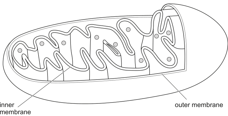
Fig. 8.1
Outline the roles played by the mitochondrial membranes in respiration.
Answer
Any four of:
- Outer and inner membranes allow entry of oxygen (for oxidative phosphorylation) and pyruvate (for the link reaction).
- The membranes allow exit of carbon dioxide produced by the Krebs cycle / link reaction.
- The double membrane provides compartmentalisation (separating the matrix from the intermembrane space and cytosol).
- The inner membrane is the location of the electron transport chain, which releases energy used to pump H+ into the intermembrane space.
- The inner membrane is the location of ATP synthase for the production of ATP.
- The cristae (infoldings of the inner membrane) increase the surface area available for many electron transport chains and ATP synthase complexes.
- The inner membrane is impermeable to H+ ions, maintaining the proton / H+ gradient (and a high H+ concentration in the intermembrane space).
- The inner membrane is the site of oxidative phosphorylation / chemiosmosis.
See answer
Background Concept
A mitochondrion is bounded by a smooth outer membrane and a highly folded inner membrane (the folds are called cristae). The two membranes enclose two compartments: the intermembrane space (between the membranes) and the matrix (inside the inner membrane). This double-membrane system is essential to aerobic respiration because the inner membrane houses the protein complexes of the electron transport chain (ETC) and ATP synthase. As electrons pass along the ETC, protons (H+) are pumped from the matrix into the intermembrane space, generating an electrochemical gradient. Protons flow back into the matrix through ATP synthase, driving the phosphorylation of ADP to ATP — this is chemiosmosis. For this gradient to be harnessed for ATP synthesis, the inner membrane must remain largely impermeable to H+, and the surface area available for ETCs and ATP synthases must be large — which is exactly what the cristae provide.
Understanding the Question
Part (a) carries a Fig. 8.1 of a mitochondrion with the outer membrane and the folded inner membrane labelled. The command word is "outline", which means a brief description of the main features. The question asks specifically about the roles of the membranes (not the matrix, not the enzymes) in respiration, so the answer should be membrane-centred. Four marks are available, and the mark scheme supplies eight possible points; the candidate only needs any four of them.
Approach
Think about what each membrane physically does: it is a barrier and a surface. As a barrier it (i) controls the movement of respiratory substrates and products (O2, pyruvate in; CO2 out), (ii) maintains the H+ gradient by being selectively impermeable, and (iii) creates compartments (matrix vs intermembrane space) so that the different stages of respiration are separated. As a surface it provides the location for the ETC and ATP synthase, and its folding (cristae) enlarges that surface.
Step-by-Step Reasoning
- Substrate and product exchange: small, non-polar molecules (O2, CO2) and pyruvate must cross the membranes to reach their sites of use. The outer membrane has large non-specific porins; the inner membrane is more selective, but specific carriers admit pyruvate. CO2 produced by the link reaction and Krebs cycle must diffuse out.
- Compartmentalisation: the matrix contains the enzymes of the Krebs cycle, the link reaction and (in prokaryotic-like relics) mitochondrial DNA and ribosomes. The intermembrane space is the destination of the H+ pumped by the ETC. Keeping these regions separate is essential to the chemiosmotic mechanism.
- Site of oxidative phosphorylation: the ETC complexes I–IV and ATP synthase are integral membrane proteins of the inner membrane. Pumping H+ across the membrane as electrons travel along the chain stores energy as a proton-motive force.
- Surface area: the inner membrane is thrown into cristae, which can occupy a large proportion of the volume of a mitochondrion in active cells (e.g. cardiac muscle). This means many ETCs and ATP synthases can be packed in, increasing the rate of ATP production.
- Impermeability to H+: the inner membrane does not allow protons to leak back into the matrix except through ATP synthase, so the proton gradient built up by the ETC is preserved and used for ATP synthesis rather than being dissipated.
Key Takeaways
- The mitochondrion's two membranes do three jobs: control what enters and leaves, separate the matrix from the intermembrane space, and provide the surface for oxidative phosphorylation.
- Cristae = increased surface area for ETCs and ATP synthases.
- An intact, H+-impermeable inner membrane is a prerequisite for chemiosmosis.
Common Mistakes
- Saying the Krebs cycle occurs on the inner membrane — it occurs in the matrix (the mark scheme explicitly ignores this).
- Describing the outer membrane as the site of oxidative phosphorylation — this is wrong; it is the inner membrane.
- Saying the membrane "produces ATP" without mentioning ATP synthase or chemiosmosis — too vague for a mark.
- Confusing the intermembrane space with the matrix when describing where protons accumulate.
Things to Be Careful About
- The command word "outline" does not require extensive detail — short, distinct points, one mark each, are sufficient.
- Link function explicitly to the membrane (e.g. "inner membrane holds ATP synthase", not just "ATP synthase is present").
- Remember that the cristae are infoldings of the inner membrane, not separate structures.
The shapes and numbers of mitochondria are continually changing due to fission. Fission
occurs when one mitochondrion splits to form two mitochondria.
Answer
Any two of:
- To increase the number of mitochondria to match a higher metabolic rate / energy demand of the cell.
- In cells about to undergo mitosis / cell division, so that there are enough mitochondria to be distributed between the two daughter cells.
- (AVP) e.g. to replace damaged mitochondria; to allow redistribution of mitochondria to regions of the cell with high energy demand.
See answer
Background Concept
Mitochondria are not static organelles. They constantly change shape and number through fission (one mitochondrion splitting into two) and fusion (two joining). Together, these processes — known as mitochondrial dynamics — allow the cell to control the number, size and position of its mitochondria in response to demand, and to remove damaged ones.
Understanding the Question
This is a "suggest" question worth 2 marks. The question gives the fact that mitochondria undergo fission (one splitting into two) and asks why a cell would benefit from this. The candidate must give biological reasons, not just describe fission.
Approach
Think of fission as a way of increasing the number of mitochondria. What circumstances make a cell need more mitochondria?
Step-by-Step Reasoning
- Energy demand: cells with high metabolic activity (e.g. muscle, liver) need many mitochondria. When energy demand rises, fission increases mitochondrial number, providing more capacity for oxidative phosphorylation.
- Cell division: before mitosis, the cell doubles its organelles. Fission produces the additional mitochondria so that each daughter cell inherits an adequate complement.
- Quality control: damaged mitochondria can be isolated and removed; fission is a step in the segregation and removal of dysfunctional mitochondria (mitophagy).
- Spatial control: in long cells such as neurons, fission can redistribute mitochondria to regions with high energy demand (e.g. synapses, the axon terminal).
Key Takeaways
- Mitochondrial number is dynamic and responds to the cell's energy demand and the cell cycle.
- Fission complements fusion in mitochondrial quality control and distribution.
Common Mistakes
- Describing how fission occurs (e.g. mentioning dynamin-related proteins) — the question asks why, not how.
- Saying "to make more energy" without linking to the cell's metabolic rate or the need to provide mitochondria to daughter cells.
- Treating fission as a form of respiration — it is a structural division, not a chemical reaction.
Things to Be Careful About
- "Suggest" questions welcome reasonable biological ideas; the candidate does not have to limit themselves to textbook points but should keep suggestions biologically plausible.
- Stay focused on benefits to the cell, not on the mechanism.
Guanosine triphosphate (GTP) is a molecule that is used as a source of energy in some reactions, instead of ATP. Guanosine is composed of a purine, similar to adenine, and ribose.
Suggest why GTP can be a suitable source of energy in some reactions.
Answer
GTP can be hydrolysed (the high-energy phosphate bond is broken) to release energy, in the same way as ATP. / Some G proteins bind to GTP rather than ATP, so GTP can act as an energy source for those reactions.
GTP can be hydrolysed to release energy, in a similar way to ATP.
Background Concept
Adenosine triphosphate (ATP) is the universal energy currency of the cell, but it is not the only one. Guanosine triphosphate (GTP) has the same triphosphate structure and a similar high-energy phosphoanhydride bond. Hydrolysis of the terminal phosphate (GTP → GDP + Pi) releases about the same amount of free energy as ATP hydrolysis. GTP is used by specific enzymes and signalling proteins — for example, GTP-binding proteins (G proteins) in signal transduction bind GTP and use its hydrolysis to GDP to drive conformational changes, and GTP is the energy source in some biosynthetic reactions such as protein synthesis (where it is used in translation and signal-recognition-particle targeting).
Understanding the Question
The question is worth 1 mark and asks the candidate to suggest why GTP can act as a source of energy in some reactions. The mark scheme accepts either a general point about hydrolysis releasing energy or the specific point that certain G proteins use GTP rather than ATP.
Approach
Recognise that GTP and ATP are structurally very similar nucleoside triphosphates and that the energy is stored in the same kind of high-energy phosphate bond.
Step-by-Step Reasoning
- ATP = adenine + ribose + three phosphate groups. GTP = guanine + ribose + three phosphate groups. The purine base differs (adenine → guanine), but the rest of the molecule is the same.
- The energy of ATP and GTP is released by hydrolysis of the terminal phosphate bond, producing a nucleoside diphosphate and inorganic phosphate.
- Therefore GTP can fulfil the same energy-carrying role in any reaction whose enzyme specifically accepts GTP (e.g. the GTPases of the G-protein family and some steps of translation).
Key Takeaways
- GTP and ATP are interchangeable in chemistry: both release energy on hydrolysis of a high-energy phosphate bond.
- Different nucleotides are used in different signalling contexts; GTP-binding proteins (G proteins) are the classic example.
Common Mistakes
- Saying GTP "is the same as ATP" — they are not identical; the purine base differs. Saying "GTP has the same high-energy bonds as ATP" is more accurate.
- Implying that GTP can completely replace ATP — GTP has a more limited, specific role.
Things to Be Careful About
- Keep the answer short: a single mark is awarded for a single clear point.
- The mark scheme's two alternatives are: (1) hydrolysis releases energy, or (2) G proteins bind GTP. Either is acceptable; either one is sufficient.
Old or damaged mitochondria reduce the ability of a cell to carry out aerobic respiration and produce the ATP needed for the metabolic processes of the cell.
Suggest what occurs to these mitochondria to allow the cell to maintain the same overall rate of respiration and ATP production.
Answer
- The old/damaged mitochondria are degraded / broken down by (hydrolytic) enzymes in lysosomes.
- The products / components of this breakdown are reused to produce new mitochondria, so the total number (and overall rate of respiration) is maintained.
Old mitochondria are broken down by lysosomal hydrolytic enzymes, and the components are recycled to make new mitochondria.
Background Concept
Mitochondria have a limited lifespan and are continually turned over. The average mitochondrion survives for only days to weeks depending on cell type. Old or damaged mitochondria are typically removed by mitophagy — they are engulfed by autophagosomes and fused with lysosomes, whose hydrolytic enzymes (proteases, lipases, nucleases) digest the organelle. The amino acids, fatty acids, sugars and nucleotides released are recycled by the cell and used, together with new mitochondrial proteins encoded by nuclear DNA, to assemble new mitochondria. This turnover maintains a healthy population of functional mitochondria and prevents a fall in the cell's capacity for oxidative phosphorylation.
Understanding the Question
This "suggest" question is worth 2 marks. The stem tells the candidate that old or damaged mitochondria reduce the cell's ability to respire and asks what happens to them so that the cell's overall rate of respiration and ATP production is kept constant. The candidate must therefore describe the removal and recycling of dysfunctional mitochondria, not their repair.
Approach
Think about (i) how the cell destroys an organelle, and (ii) what happens to the breakdown products. The two marks correspond to these two ideas: degradation by lysosomes, and recycling of the components to make new mitochondria.
Step-by-Step Reasoning
- Degradation: damaged mitochondria are tagged (e.g. by PINK1/Parkin in mammals), engulfed and fused with lysosomes, and the proteins, lipids and DNA of the mitochondrion are hydrolysed.
- Recycling: the resulting amino acids, fatty acids, sugars, nitrogenous bases and phosphate are re-used in cellular metabolism. Together with fresh proteins translated from nuclear mRNA, they are assembled into new mitochondria. Because new functional mitochondria replace the old non-functional ones, the total mitochondrial mass and the cell's overall capacity for oxidative phosphorylation are maintained, even though individual mitochondria are short-lived.
Key Takeaways
- Mitochondria are continually turned over by lysosomal degradation (mitophagy).
- The breakdown products are recycled to build new mitochondria.
- This turnover is essential for sustained cellular ATP production and for quality control of the mitochondrial population.
Common Mistakes
- Saying the mitochondria are "repaired" — the question explicitly states they are old or damaged, and the mark scheme expects degradation.
- Saying only that lysosomes digest them, without mentioning the recycling of components (this only earns one of the two marks).
- Confusing lysosomes with proteasomes — proteasomes degrade individual cytosolic proteins, not whole organelles.
Things to Be Careful About
- The question asks how the cell maintains the same overall rate of respiration — the answer must therefore show that new mitochondria are produced to compensate for the lost ones; the word "overall" is the key.
- Use precise language: "hydrolytic enzymes of lysosomes" or "lysosomal digestion" is the correct phrasing, not just "broken down".
Fig. 9.1 shows how the mean global atmospheric carbon dioxide concentration has changed over the 800 000 () years leading up to the year 2020.
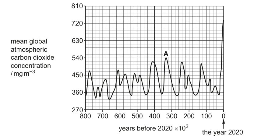
Fig. 9.1
Calculate the percentage increase in carbon dioxide concentration between point A and the year 2020.
Show your working. Write your answer to one decimal place.
percentage increase = ______
Working
Read the two values from Fig. 9.1:
- Concentration at point A (≈ 330 000 years ago)
- Concentration in the year 2020
Answer
percentage increase
36.7 %
Background Concept
Atmospheric carbon dioxide concentration fluctuates naturally on geological timescales, driven by orbital (Milankovitch) cycles that change the balance between volcanic outgassing, weathering, photosynthesis and the burial of organic carbon. Over the last ~800 000 years these natural cycles have kept CO₂ oscillating roughly between 180 and 300 ppm (≈ 360 to 540 mg m⁻³ in the units used here, since 1 ppm ≈ 2 mg m⁻³ at sea level). In the last few centuries the burning of fossil fuels and deforestation have driven atmospheric CO₂ sharply upwards — well outside the natural envelope.
Percentage change is a single-figure way to compare two quantities with different absolute sizes:
The denominator is always the initial (old) value, so the percentage describes how much bigger or smaller the new value is relative to the starting point.
Understanding the Question
The graph in Fig. 9.1 plots mean global atmospheric CO₂ concentration (in mg m⁻³) against time (in thousands of years before 2020). Point A is a peak about 330 000 years ago, and the curve ends at the year 2020. The question asks for the percentage increase in CO₂ from A to 2020, to one decimal place.
The command word is calculate — the answer is numerical and you must show working. Two marks are available: 1 for the working (substituting numbers into the formula) and 1 for the correct final numerical answer.
Approach
- Take the CO₂ value at A from the graph.
- Take the CO₂ value at 2020 from the graph.
- Apply the percentage-change formula using A as the initial value.
- Round to one decimal place.
The graph's grid lines are spaced at 90 mg m⁻³ (270, 360, 450, 540, 630, 720, 810), so careful reading is needed near the 540 line and near the 2020 endpoint.
Step-by-Step Reasoning
- Reading the graph: Point A is a peak that sits exactly on the 540 mg m⁻³ gridline, so its value is 540 mg m⁻³. The 2020 endpoint sits about two-thirds of the way between 720 and 810, i.e. ≈ 738 mg m⁻³ (this is the value that gives the mark-scheme answer of 36.7%).
- Subtracting to find the absolute change: 738 − 540 = 198 mg m⁻³.
- Dividing by the initial value: 198 ÷ 540 = 0.3666…
- Multiplying by 100: 36.666… %
- Rounding: to one decimal place this is 36.7 %.
The mark scheme also accepts 36.66666667 with the substitution shown, awarding one mark for the working and a second for the final rounded answer.
Key Takeaways
- Always divide by the initial value when calculating percentage change.
- Read graph values as precisely as the gridlines allow; here the minor grid is at 10 mg m⁻³ intervals (since 90 is split into nine squares).
- Show the substitution explicitly: even if your final number is wrong, the correct formula earns a mark.
- One decimal place means 36.7, not 37 or 36.66.
Common Mistakes
- Dividing by the wrong value: using 738 (the final value) as the denominator gives 26.8 % — wrong denominator.
- Rounding the intermediate steps too early: 198/540 should be carried to several decimals before multiplying by 100.
- Reading the graph too coarsely: the 2020 point is not at 720 or 810; both readings matter.
- Forgetting the % sign — the answer is a percentage, not a raw number.
Things to Be Careful About
- Do not write
37— the question demands one decimal place. - Do not omit the working — the formula earns a method mark even if the final answer is miscalculated.
- A common error is to compute
(738 − 540)/738 × 100; this would be the percentage decrease relative to 2020, which is the wrong reference point for an increase.
Suggest how the changes in carbon dioxide concentration between A and the year 2020 may have affected the environment and biodiversity.
Answer
Any four of:
- (Increase in) global warming / rise in mean global temperature ;
- Sea levels rise (leading to flooding / loss of coastal habitats) ;
- Habitat change / loss ; e.g. more forest fires, ocean acidification, coral bleaching, desertification ;
- Food webs / food chains / ecosystems disrupted ;
- Change in biodiversity ;
- (Increased) extinction (of species) ;
- AVP ; e.g. migration of species to cooler regions, spread of tropical diseases, loss of pollinator–plant partnerships.
Any four creditable effects from the mark scheme.
Background Concept
Carbon dioxide is a greenhouse gas. Rising atmospheric CO₂ enhances the greenhouse effect: short-wave solar radiation still reaches the surface, but more long-wave (infrared) radiation emitted by the Earth is absorbed and re-emitted by CO₂ (and CH₄, N₂O, water vapour) in the lower atmosphere. The extra trapped energy raises mean global temperatures — global warming.
Warming has cascading consequences:
- Thermal expansion of seawater and melting of land ice raise sea levels.
- Climate belts shift, changing where particular temperatures and rainfall patterns occur.
- Oceans absorb about 25 % of anthropogenic CO₂, which lowers ocean pH — ocean acidification.
- Species have evolved for specific thermal and precipitation envelopes; rapid change forces migration, local extinction, or death.
- Warmer, drier conditions promote forest fires, drought, and coral bleaching.
Understanding the Question
Part (a)(ii) follows directly from part (a)(i). The candidate has just calculated that atmospheric CO₂ rose by ~37 % in a few thousandths of the time span shown by the rest of the curve. The question asks the candidate to suggest — i.e. propose plausible consequences — for the environment and biodiversity.
Because the command word is suggest, the marking scheme is generous: any biologically reasonable effect of elevated CO₂ that touches the environment or biodiversity earns credit, up to four marks. There is no requirement to be exhaustive.
Approach
- Start with the direct, physical consequence: greenhouse effect → warming.
- Move to second-order physical consequences: ice melt and thermal expansion → sea-level rise.
- Move to ecological consequences: habitat change (fire, ocean acidification, bleaching, desertification) and disruption of food webs.
- Move to biodiversity consequences: species migration, local extirpation, and extinction.
Aim for four distinct points. Candidates who list only consequences at the physical level (warming + sea-level rise) lose marks because they have not addressed biodiversity; candidates who list only vague statements about "loss of biodiversity" without mechanism also lose marks.
Step-by-Step Reasoning
- Point 1 — Global warming. The most direct consequence. Atmospheric CO₂ traps infrared radiation, raising mean global temperature. Worth one mark.
- Point 2 — Sea-level rise. Warmer water expands; ice sheets and glaciers melt; low-lying land is flooded. Worth one mark.
- Point 3 — Habitat change / loss. Many ecosystems are sensitive to small changes in temperature or precipitation. Examples accepted by the mark scheme include forest fires, ocean acidification, coral bleaching, and desertification. Worth one mark for the general point plus possibly more for the example.
- Point 4 — Disrupted food webs / ecosystems. Climate-driven species migrations and phenological mismatches (e.g. flowers opening before their pollinators emerge) break established predator–prey and mutualistic interactions. Worth one mark.
- Point 5 — Change in biodiversity / extinction. Direct statement of the consequence. Some species will thrive in the new conditions; others will not survive. Worth one mark; "extinction" is a stronger, separate credit.
A good four-mark answer combines one physical consequence, one habitat-level consequence, and one biodiversity-level consequence, plus one further development (e.g. specific example).
Key Takeaways
- CO₂ is a greenhouse gas; its rise drives warming, which is the root cause of the cascade.
- Environmental and biodiversity effects are interlinked — physical climate change reshapes habitats, which reshapes communities.
- "Suggest" questions reward breadth and plausible mechanisms, not exhaustive detail.
Common Mistakes
- Only mentioning warming and stopping — this earns one mark at most.
- Repeating the same idea in different words (e.g. "temperature rises" and "the climate gets hotter") — counts as one point, not two.
- Ignoring biodiversity — the question explicitly asks for effects on biodiversity as well as the environment.
- Naming a single effect as the entire answer — four marks require four distinct points.
Things to Be Careful About
- The mark scheme accepts forest fires and ocean acidification as concrete examples of habitat change — both are common, biologically meaningful and worth including if space allows.
- "Extinction" is credited separately from "change in biodiversity", so candidates should not substitute one for the other.
- Avoid causal leaps that are not biologically supported (e.g. "CO₂ directly causes cancer in animals").
Answer
Any four of:
- Plants may have (future) use(s) that we do not yet know about ;
- Aesthetic / ethical / cultural reasons ;
- Medical uses / example ; e.g. plants are a source of drugs such as aspirin, quinine, morphine, vincristine, digitalis ;
- Ecotourism ;
- Maintain stability of ecosystems / food chains / food webs ; (e.g. keystone species, pollination services, nutrient cycling)
- Resource material ; e.g. wood for building, fibres for clothing, food for humans, agriculture ;
- Maintain / increase the gene pool / genetic diversity ; (raw material for breeding programmes)
- Maintain soil stability / prevent desertification.
Any four creditable reasons from the mark scheme.
Background Concept
Biodiversity — the variety of living organisms — has value to humans at several levels:
- Utilitarian value (instrumental): direct uses such as food, timber, fibres, medicines, and the breeding stock for agriculture.
- Ecosystem-service value: functioning ecosystems provide pollination, soil formation, nutrient cycling, climate regulation, flood control and water purification.
- Genetic-resource value: wild relatives of crops carry alleles for disease resistance, drought tolerance and other traits that breeders may need in the future.
- Intrinsic value: aesthetic, ethical, cultural and spiritual reasons — the argument that species have a right to exist independent of human utility.
- Option value: the future, unknown uses of species we have not yet discovered (e.g. a wild plant whose secondary metabolites may one day treat a new disease).
Cambridge syllabus explicitly expects students to be able to outline these categories and give at least one example for each.
Understanding the Question
This is a "outline" question worth four marks. The candidate must give four distinct reasons for maintaining plant biodiversity. The mark scheme accepts any four from a list of eight, and rewards a single supporting example where it is offered. There is no requirement to structure the answer in any particular way, but clearly separated points (bullet list, numbered list) help the examiner to award the marks.
The question is specifically about plant biodiversity, so examples and reasons should be drawn from the plant kingdom (e.g. timber, fibre, medicines derived from plants, wild relatives of crops).
Approach
- Decide on four distinct categories. Avoid repeating the same idea in two different wordings.
- For each category, give a short justification. A concrete example strengthens the point.
- Order the points for readability (e.g. from utilitarian to intrinsic, or vice versa).
A strong four-point answer picks one each from: a direct economic use, a medical/option use, an ecosystem-service use, and an aesthetic/ethical/cultural reason. Candidates can also include genetic-resource value and soil stability if preferred.
Step-by-Step Reasoning
- Reason 1 — Future / option value. We do not yet know all the ways plants might be useful; some plants may contain as-yet-undiscovered medicines or industrial chemicals. Mark-scheme line 1.
- Reason 2 — Aesthetic / ethical / cultural. Plants have beauty (wildflowers, forests), and many people believe species have an intrinsic right to exist. Cultural value includes sacred groves, national emblems, and traditions tied to particular plants. Mark-scheme line 2.
- Reason 3 — Medical. Plants are a major source of pharmaceutical drugs: aspirin (from willow bark, Salix), quinine (from Cinchona), morphine (from opium poppy, Papaver somniferum), vincristine (from Catharanthus roseus), digitalis (from Digitalis). Mark-scheme line 3.
- Reason 4 — Ecological stability. Plant communities underpin food webs and provide ecosystem services (pollination, soil formation, water regulation, climate buffering). Loss of plant diversity destabilises these systems. Mark-scheme line 5.
- Reason 5 — Economic resources. Plants supply wood for building, fibres (cotton, linen, jute) for clothing, food directly (cereals, fruit, vegetables), and feedstock for agriculture. Mark-scheme line 6.
- Reason 6 — Genetic resource. Wild relatives of crops carry alleles that breeders can use to improve yield, disease resistance or environmental tolerance. Maintaining genetic diversity preserves this raw material. Mark-scheme line 7.
- Reason 7 — Soil stability. Plant roots bind soil; loss of vegetation (e.g. through deforestation or overgrazing) accelerates erosion. Mark-scheme line 8.
- Reason 8 — Ecotourism. Wild plant communities attract tourists, supporting local economies and giving those communities a financial incentive to conserve rather than exploit. Mark-scheme line 4.
Any four of these earns full marks.
Key Takeaways
- Biodiversity has multiple categories of value — utilitarian, ecological, genetic, aesthetic/ethical, option.
- Concrete examples are always rewarded; vague platitudes ("plants are important") are not.
- "Outline" questions reward breadth over depth — four crisp points beat one long paragraph.
Common Mistakes
- Repeating the same idea twice (e.g. "food" and "agriculture" as two separate points) — only one mark.
- Omitting an example when one would clearly earn extra credit.
- Confusing reasons for plant biodiversity with reasons for animal biodiversity — the question is specifically about plants, so animal-only examples (e.g. "pandas in zoos") are off-target.
- Forgetting the "plant" focus and listing general biodiversity reasons without plant-specific grounding.
Things to Be Careful About
- The mark scheme gives any four from eight, so the order in which reasons are presented does not matter.
- "Maintain stability of ecosystems" is a stronger answer than "keeps nature balanced" — the examiner rewards mechanistic understanding.
- "Gene pool / genetic diversity" should be tied to a use (e.g. crop breeding), otherwise it is just a phrase.
Fig. 10.1 is a diagram of part of a neurone membrane while the resting potential is maintained.
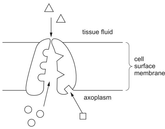
Fig. 10.1
On Fig. 10.1, use label lines and letters to label:
K – potassium ions
A – ATP.
Answer
On Fig. 10.1, add two label lines to the axoplasm (lower) side of the membrane:
- K – label line drawn to a circle in the axoplasm (a potassium ion, )
- A – label line drawn to the square in the axoplasm (the ATP molecule supplying energy to the pump)
Label K on a circle (K⁺) and label A on the square (ATP) in the axoplasm.
Background Concept
The resting potential of a neurone (about −70 mV inside relative to outside) is maintained by the sodium–potassium (Na⁺/K⁺) pump, an active-transport protein embedded in the cell surface membrane. For every one cycle it uses one molecule of ATP to pump 3 Na⁺ out of the axon and 2 K⁺ in, against their respective concentration gradients. This unequal pumping makes the inside of the axon negative relative to the outside and keeps the Na⁺ and K⁺ gradients steep — gradients that are then exploited during the action potential.
In a typical textbook diagram of the pump:
- Triangles (△) are used to represent sodium ions (Na⁺).
- Circles (○) are used to represent potassium ions (K⁺).
- The pump protein is the bulky structure spanning the membrane.
- The square (□) symbol represents the ATP molecule that binds to the pump on its cytoplasmic (axoplasm) side to provide energy for active transport.
The pump operates only on the axoplasm side of the membrane, because that is where ATP is generated (by mitochondria) and where the relevant ion-binding sites face.
Understanding the Question
Part (a) gives Fig. 10.1, a simplified diagram of the Na⁺/K⁺ pump. The candidate must add two label lines and the letters K and A to the correct features. The question provides the key:
- K – potassium ions
- A – ATP
The mark scheme awards one mark for each correct placement, so both must be correct to score 2.
Approach
Read the question first: it tells you exactly what each label refers to. Then look at the symbols in the figure. The trick is to notice that both potassium ions and the ATP symbol are located in the axoplasm (lower side), so both labels are added below the membrane.
Step-by-Step Reasoning
- Look at the symbols. Triangles are in the tissue fluid (top); from the question and the standard convention these are Na⁺. Circles are in the axoplasm (bottom) — these are the K⁺ ions. The square is the ATP molecule that drives the pump.
- Draw a label line from the letter K to any circle in the axoplasm. (1 mark)
- Draw a label line from the letter A to the square in the axoplasm. (1 mark)
- Common trap: do not point K to a triangle (that would be Na⁺), and do not point A to a circle (that would be a K⁺ ion). Both labels belong in the axoplasm because that is the side of the membrane where K⁺ is concentrated by the pump and where ATP binds to the pump protein.
Key Takeaways
- In standard CIE diagrams, triangles = Na⁺ and circles = K⁺.
- The Na⁺/K⁺ pump is an active-transport protein — it needs ATP, and the ATP-binding site is on the axoplasm (cytoplasmic) side.
- Always read the key supplied with the question before labelling, since symbol conventions can vary between papers.
Common Mistakes
- Pointing K to a triangle (Na⁺): the candidate has confused the ion shapes.
- Pointing A to a circle or triangle: ATP is the square symbol, not an ion.
- Drawing both labels above the membrane: both K⁺ and ATP are on the axoplasm side of the membrane, not in the tissue fluid.
Things to Be Careful About
- Use label lines (straight lines ending in a small touch, not arrows pointing into structures) and place the letter at the end of the line, as required by CIE drawing conventions.
- Do not write the answer in prose in the margin — the marks are for the labels on the figure itself.
Answer
- Sodium ion channels open and Na⁺ enters the axon by facilitated diffusion, down its electrochemical gradient.
- This causes the membrane to become depolarised (the inside becomes less negative / more positive).
- Once the threshold potential is reached, more voltage-gated sodium channels open (positive feedback), producing the rapid depolarisation of the action potential.
- Potassium ion channels open and K⁺ moves out of the axon, down its electrochemical gradient.
- This causes repolarisation of the membrane (the inside becomes negative again).
- The membrane briefly becomes more negative than the resting potential – hyperpolarisation.
- The Na⁺/K⁺ pump (and ion channels) restore the original ion distributions and the membrane returns to its resting potential.
Na⁺ channels open → Na⁺ enters → depolarisation past threshold → K⁺ channels open → K⁺ exits → repolarisation → hyperpolarisation → return to resting potential.
Background Concept
An action potential is a rapid, transient reversal of the membrane potential of an excitable cell, propagating along the axon without losing amplitude. It is generated by the coordinated opening and closing of voltage-gated sodium (Na⁺) channels and voltage-gated potassium (K⁺) channels in the axon membrane. The Na⁺/K⁺ pump has not finished its work — it has already established the steep ion gradients (high Na⁺ outside, high K⁺ inside) during the resting potential; the action potential is the controlled, brief collapse and restoration of those gradients.
The key terms to use precisely are:
- Depolarisation – the inside of the membrane becoming less negative (more positive) than the resting potential.
- Repolarisation – the inside returning towards the negative resting potential.
- Hyperpolarisation – the inside becoming more negative than the resting potential (an 'undershoot').
- Threshold – the critical membrane voltage (about −55 mV) at which enough Na⁺ channels are open that the depolarisation becomes self-propagating.
Understanding the Question
This is a 'describe the sequence' question worth 4 marks. The mark scheme credits any 4 points from a list of 7 specific statements, plus an AVP. The candidate must give a clear ordered account using the correct technical vocabulary — marks are tied to specific terms such as 'depolarised', 'threshold', 'repolarised' and 'hyperpolarised'.
Approach
Mentally run through the action potential as it would appear on a voltage-versus-time trace: a flat baseline (resting), an upward stroke (depolarisation), a downward stroke (repolarisation), a dip below the baseline (hyperpolarisation), then a return to baseline. Match each phase to the ion channel responsible and to the precise vocabulary the mark scheme requires.
Step-by-Step Reasoning
- Na⁺ channels open (voltage-gated). Because Na⁺ is more concentrated outside the axon and the inside is negative, Na⁺ rushes into the axon down its electrochemical gradient. (MP1)
- The influx of positive charge makes the inside less negative — the membrane depolarises. (MP2)
- If depolarisation reaches the threshold potential, an all-or-nothing action potential fires. Below threshold, the signal simply decays. (MP3)
- At the peak of the action potential, Na⁺ channels inactivate and voltage-gated K⁺ channels open. K⁺ diffuses out of the axon, taking positive charge with it. (MP4)
- Loss of positive charge from the axoplasm repolarises the membrane — the inside becomes negative again. (MP5)
- K⁺ channels are slow to close, so K⁺ continues to leave briefly, driving the potential below resting level — this is hyperpolarisation. (MP6)
- The Na⁺/K⁺ pump and leak channels restore the original ion distribution, and the membrane returns to resting potential. (MP7)
- (AVP) A strong candidate may add that the entry of Na⁺ makes the local membrane potential more positive, which in turn opens more voltage-gated Na⁺ channels — the positive-feedback loop that produces the rapid upstroke of the action potential.
Any four of these marking points (1–7) score full marks; the AVP is an alternative route to the fourth mark.
Key Takeaways
- The action potential is a sequence of voltage-gated channel events; describe it in order.
- Memorise the exact vocabulary: depolarised, repolarised, hyperpolarised, threshold, resting potential. Vague terms such as 'the charge changes' or 'ions move' will not earn marks.
- The Na⁺/K⁺ pump does not generate the action potential — it sets up the gradients that make it possible. The action potential itself is generated by facilitated diffusion of Na⁺ in and K⁺ out through voltage-gated channels.
Common Mistakes
- Writing 'sodium ions move in and potassium ions move out' without naming the channels or the resulting change in membrane potential.
- Saying the membrane 'reverses polarity' instead of using depolarised / repolarised.
- Forgetting to mention threshold, or describing the action potential as graded rather than all-or-nothing.
- Omitting hyperpolarisation — it is a separate, named phase worth a mark.
- Confusing the role of the Na⁺/K⁺ pump (which restores gradients afterwards) with the channels that generate the action potential.
Things to Be Careful About
- 'Describe the sequence' demands an ordered account; marks can be lost for jumping backwards and forwards.
- The mark scheme credits 'Na⁺ enters' or 'sodium channels open' (and similarly for K⁺); the candidate may give either but it is safer to name the channel and the direction of ion movement.
- The word 'refractory period' is closely linked to this topic but is not required to answer this specific question — do not pad the answer with it unless it is the AVP you choose to use.
Table 10.1 shows the axon diameter, myelination and transmission speed of impulses of motor neurones for three animals: squid, cockroach and cat.
Table 10.1
| animal | axon diameter / mm | myelination | transmission speed / |
|---|---|---|---|
| squid | 1.5 | no | 30 |
| cockroach | 0.05 | no | 10 |
| cat | 0.02 | yes | 100 |
Describe and suggest explanations for the results shown in Table 10.1.
Answer
Description
- Comparing cat (myelinated, 100 m s⁻¹) with squid or cockroach (unmyelinated): myelination produces a faster transmission speed than an unmyelinated axon of similar function.
- Comparing squid (1.5 mm, 30 m s⁻¹) with cockroach (0.05 mm, 10 m s⁻¹), both unmyelinated: the larger the axon diameter, the faster the transmission speed (and vice versa).
- Comparing the cat axon (0.02 mm, myelinated, 100 m s⁻¹) with the squid axon (1.5 mm, unmyelinated, 30 m s⁻¹): myelination has a greater effect on transmission speed than axon diameter — a thin myelinated axon still conducts faster than a much thicker unmyelinated axon.
Explanation
- In a myelinated axon the action potential 'jumps' from one node of Ranvier to the next (saltatory conduction), so depolarisation only has to occur at the nodes, giving a much higher conduction velocity.
- In an unmyelinated axon, a larger diameter means a smaller internal resistance to the longitudinal flow of ions, and proportionally less ion leakage across the membrane relative to the volume of axoplasm, so the action potential is conducted faster.
Myelination increases speed (saltatory conduction between nodes of Ranvier); in unmyelinated axons, larger diameter gives faster conduction (lower resistance / less leakage); myelination has a greater effect than diameter.
Background Concept
The speed at which an action potential travels along an axon depends on two main structural features of the axon:
-
Myelination. Schwann cells (PNS) or oligodendrocytes (CNS) wrap the axon in a myelin sheath made largely of lipid. The myelin insulates the axon so that the membrane is only electrically 'exposed' at gaps called nodes of Ranvier. Voltage-gated Na⁺ channels are concentrated at the nodes, so the action potential effectively 'jumps' from node to node — this is saltatory conduction (from Latin saltare, to leap). Myelinated axons therefore conduct much faster than unmyelinated axons of similar diameter.
-
Axon diameter. In an unmyelinated axon, the action potential is regenerated continuously along the entire membrane. A larger diameter:
- gives a larger cross-sectional area of axoplasm, so internal longitudinal resistance to ion flow is lower (resistance is inversely proportional to cross-sectional area, just as for a wire);
- provides a smaller surface-area-to-volume ratio, so proportionally fewer ions leak across the membrane per unit volume of axoplasm — the signal decays less and is conducted further before needing to be regenerated.
So in the data we would expect: myelinated > unmyelinated, and within unmyelinated, larger diameter > smaller diameter.
Understanding the Question
Table 10.1 gives three rows of data. The command word is 'describe and suggest explanations', which means the candidate must (a) state the patterns shown in the data and (b) give a biological reason for each pattern. There are 4 marks, awarded for any 4 of 5 listed points (3 descriptive + 2 explanatory). The two invertebrates (squid, cockroach) have unmyelinated axons of different diameters; the mammal (cat) has a thin but myelinated axon. This pairing lets the question separate the effects of diameter and myelination.
Approach
Read the table once for the trend, then once for the explanation. Identify three comparisons:
- Cat vs squid: same function (motor), very different diameter, but cat is myelinated. → effect of myelination.
- Squid vs cockroach: both unmyelinated, very different diameters. → effect of diameter alone.
- Cat vs cockroach: similar small diameters but cat is myelinated. → effect of myelination controlling for diameter.
The 'greatest effect' comparison (point 3) is the most demanding: it requires a cross-row comparison.
Step-by-Step Reasoning
- Myelination increases speed. Cat (0.02 mm, myelinated) conducts at 100 m s⁻¹, vastly faster than either unmyelinated axon. The explanation is saltatory conduction: depolarisation only needs to occur at the nodes of Ranvier, so the impulse effectively 'leaps' between nodes, and the conduction velocity is much higher than in an unmyelinated axon where depolarisation has to be regenerated at every point along the membrane. (MP1 + MP4)
- Larger diameter → faster speed in unmyelinated axons. Squid (1.5 mm, 30 m s⁻¹) is much faster than cockroach (0.05 mm, 10 m s⁻¹) — a 30-fold increase in diameter, a 3-fold increase in speed. (MP2)
- The effect of myelination exceeds the effect of diameter. The cat axon is 75× thinner than the squid axon (0.02 vs 1.5 mm) but conducts >3× faster (100 vs 30 m s⁻¹). The only structural difference that can explain a thin myelinated axon out-performing a giant unmyelinated one is myelination. (MP3)
- Mechanism for the diameter effect. A wider axon has a larger cross-sectional area, so internal (axoplasmic) resistance to the flow of local currents is lower. It also has a smaller surface-area-to-volume ratio, so proportionally less ion leakage occurs per unit volume of axoplasm, and the local current decays more slowly with distance. (MP5)
The mark scheme awards any 4 of these 5 points. A complete answer that scores all 4 marks would give MP1, MP2, MP4 and MP5, or alternatively MP1, MP3, MP4 and MP5.
Key Takeaways
- A 4-mark 'describe and explain' data question typically wants both an observation and a reason for each pattern. A pure description will earn only the descriptive marks; a pure mechanism will earn only the explanatory marks.
- Two structural features control conduction velocity: myelination (saltatory conduction) and axon diameter (resistance / leakage). Examiners love pairing them, so be ready to discuss them together.
- Always quantify your comparison: 'cat (myelinated, 0.02 mm) conducts at 100 m s⁻¹ — 3× faster than the squid (unmyelinated, 1.5 mm) at 30 m s⁻¹, despite being 75× thinner.' Numbers win marks.
Common Mistakes
- Writing only a description with no mechanism, or only a mechanism with no description.
- Saying the larger axon has 'more ions' or 'more space' — the mark-scheme answer is less resistance to ion flow or lower surface-area-to-volume ratio.
- Confusing the direction of the effect — claiming myelination slows conduction, or that larger axons conduct more slowly (a common misconception derived from thinking of plumbing resistance).
- Stating the cause of saltatory conduction incorrectly as 'insulation' alone, without the linked idea that depolarisation only occurs at the nodes.
- ORA-style errors: stating 'smaller diameter gives faster conduction in unmyelinated axons' — the data show the opposite.
Things to Be Careful About
- The mark scheme states 'a larger diameter (for unmyelinated)' — the diameter effect is most clearly seen in unmyelinated axons, where the action potential is regenerated continuously. Do not extend the diameter argument to myelinated axons without qualification.
- The cockroach's 0.05 mm axon is described as unmyelinated — insects have unmyelinated axons, which is why they compensate with very thin axons for fine motor control or with giant axons for escape responses.
- The command word is 'describe and suggest explanations' — the two halves of the answer are scored separately, so make sure both appear.