Biology 9700/41 — May/June 2023
Cambridge A-Level · A Level Structured Questions · worked solutions for every part, with the mark scheme
Topics Energy and Respiration · Selection and Evolution · Control and Coordination · Inheritance · Photosynthesis · Classification, Biodiversity and Conservation · +2 more
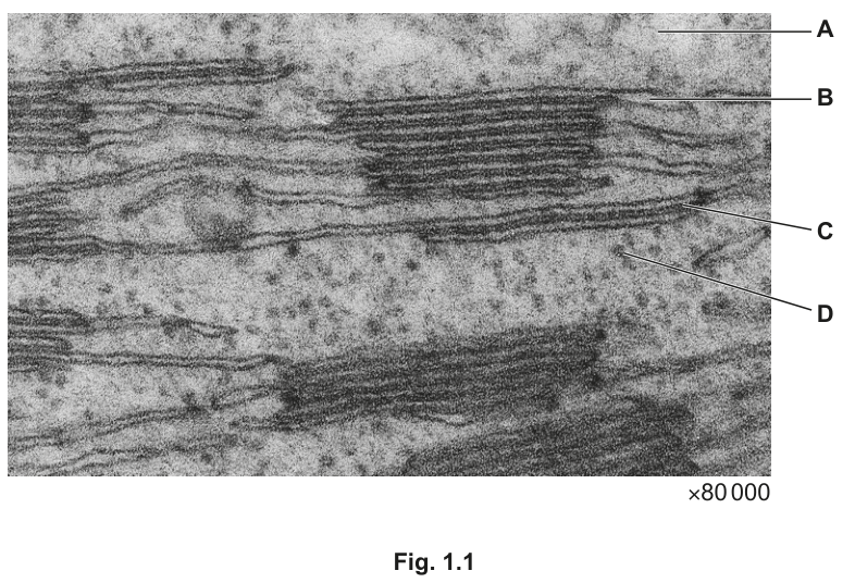
Fig. 1.1 shows a transmission electron micrograph of part of a chloroplast.
Table 1.1 describes some functions that occur in different parts of a chloroplast.
Complete Table 1.1 by identifying the letter on Fig. 1.1 that is a location matching the description. Each letter may be used once, more than once, or not at all.
Table 1.1
| description | letter |
|---|---|
| accumulates (builds up) a high concentration of protons | |
| makes triose phosphate | |
| makes some chloroplast proteins | |
| pumps protons |
Answer
| description | letter |
|---|---|
| accumulates (builds up) a high concentration of protons | B |
| makes triose phosphate | A |
| makes some chloroplast proteins | D |
| pumps protons | C |
B, A, D, C
Background Concept
A chloroplast is a double-membraned organelle enclosing a fluid stroma, in which the thylakoid membrane system is suspended. The thylakoid membrane is folded into stacks called grana (singular: granum), and the fluid interior of the thylakoid is the thylakoid (lumen) space. The thylakoid membrane carries the electron transport chain, ATP synthase, photosystems I and II, and the pigment–protein complexes that absorb light. The stroma contains the enzymes of the Calvin cycle, chloroplast DNA, chloroplast ribosomes, and starch grains.
The light-dependent reaction takes place on the thylakoid membrane:
- Photolysis of water at photosystem II releases into the thylakoid space and electrons into the electron transport chain.
- The electron transport chain pumps additional from the stroma into the thylakoid space, creating a high proton concentration (low pH) inside the thylakoid.
- Protons flow back out through ATP synthase, driving the phosphorylation of ADP to ATP (chemiosmosis / photophosphorylation).
The light-independent reaction (Calvin cycle) takes place in the stroma, where is fixed by rubisco onto RuBP, producing GP, which is reduced (using ATP and reduced NADP from the light-dependent reaction) to triose phosphate (TP).
Some chloroplast proteins are encoded by chloroplast DNA and translated on chloroplast ribosomes, which are 70S ribosomes located in the stroma — distinct from the cytoplasmic 80S ribosomes.
Understanding the Question
The candidate is shown a TEM of a chloroplast at with four labels:
- A = stroma (the lighter, granular matrix between thylakoids)
- B = granum (a stack of thylakoids)
- C = thylakoid membrane (a single dark line)
- D = a small dense body in the stroma — a ribosome (this is the only interpretation consistent with the mark scheme entry 'makes some chloroplast proteins')
The task is to match each description of a process to the letter of the location where it occurs.
Approach
Each description is a clue to a specific process; identify the location of that process in the chloroplast. The four clues are proton accumulation (light-dependent), TP synthesis (Calvin cycle), chloroplast protein synthesis (chloroplast ribosomes), and proton pumping (electron transport chain).
Step-by-Step Reasoning
- Accumulates a high concentration of protons → B (granum). Photolysis and the electron transport chain together pump into the thylakoid space. Because the thylakoid membrane is relatively impermeable to , protons build up inside the thylakoid (and therefore inside the granum), creating the proton gradient used by ATP synthase.
- Makes triose phosphate → A (stroma). TP is the first sugar made in the Calvin cycle. All Calvin-cycle enzymes (including rubisco) are dissolved in the stroma, so the stroma is where is fixed and reduced to TP.
- Makes some chloroplast proteins → D (ribosome). Chloroplasts contain their own DNA and 70S ribosomes. These ribosomes translate mRNA from chloroplast genes to make a small subset of chloroplast proteins (most chloroplast proteins are encoded by nuclear DNA and imported from the cytoplasm). The ribosomes appear as small dense granules in the stroma on a TEM.
- Pumps protons → C (thylakoid membrane). The electron transport chain components (plastoquinone, the cytochrome b₆f complex) are embedded in the thylakoid membrane and actively pump from the stroma into the thylakoid lumen as electrons pass along the chain.
Key Takeaways
- The light-dependent reaction occurs on the thylakoid membrane; its products are ATP, reduced NADP, and , and it generates a proton gradient across the thylakoid membrane.
- The light-independent reaction (Calvin cycle) occurs in the stroma and produces triose phosphate.
- Chloroplast ribosomes, located in the stroma, translate a small number of proteins encoded by chloroplast DNA; the rest are encoded in the nucleus and imported.
- Chemiosmotic ATP synthesis depends on the proton gradient built up inside the thylakoid space.
Common Mistakes
- Confusing the thylakoid space (where accumulates) with the stroma (where the Calvin cycle occurs).
- Confusing the thylakoid membrane (where the ETC pumps ) with the granum (where accumulates).
- Identifying D as a starch grain rather than a ribosome — starch is a product, not a site of protein synthesis; the dense spot at D in the micrograph is a ribosome, the only structure that fits the 'makes proteins' description.
Things to Be Careful About
- The granum (B) is a stack of thylakoids, not the membrane itself. The membrane is C. The thylakoid space enclosed by the granum is where protons accumulate.
- 'Pumps protons' (C) is a different process from 'accumulates protons' (B) — the membrane is the active site, the lumen is the reservoir.
Membranes of the type labelled C in Fig. 1.1 were made into a liquid extract. Chromatography was then used to separate and identify the coloured components (pigments) in this extract. The resulting chromatogram showed that these membranes contain a yellow pigment, an orange pigment, a green-brown pigment and two different green pigments.
Describe how you would carry out chromatography to separate and identify the coloured pigments in the liquid extract of C.
Answer
Any four of:
- Draw a pencil line near the bottom of a strip of chromatography paper and place a spot of the liquid extract of C on the line (allow it to dry; repeat / concentrate the spot to make it more intense).
- Suspend the paper in a suitable solvent (e.g. propanone / petroleum ether / named solvent) with the solvent level below the pencil line, in a closed container.
- Leave until the solvent has moved a suitable distance up the paper, then remove the paper and immediately mark the solvent front in pencil.
- Measure the distance moved by each pigment spot from the origin and the distance moved by the solvent front.
- Calculate the value for each pigment:
- Compare each value with the values of known pigments (chlorophyll a, chlorophyll b, carotene, xanthophyll) run under the same conditions to identify each pigment.
See working
Background Concept
Paper chromatography separates a mixture of solutes by partitioning them between a stationary phase (the paper, with traces of water bound to the cellulose) and a mobile phase (a solvent that moves up the paper by capillary action). Each solute has a characteristic partition coefficient between these two phases, so each travels a characteristic distance relative to the solvent front. This ratio is the value:
For a given solvent and paper type, is reproducible and can be used to identify a solute by comparison with a known standard run under identical conditions. Pigments from the thylakoid membrane (chlorophyll a, chlorophyll b, carotene and xanthophyll) can be separated and identified this way.
Understanding the Question
The question asks the candidate to describe the practical procedure that would be used to separate and identify the pigments in a liquid extract of the thylakoid membrane (C). This is the standard Paper 3 / Paper 4 / Paper 5 chromatography description, and the mark scheme accepts any four of seven possible points. The candidate is being tested on practical procedure and on the use of values for identification.
Approach
Write out the procedure in the order in which it is carried out: preparation of the paper, application of the sample, running the chromatogram, marking the solvent front, measuring distances, calculating , and identifying pigments by comparison with known standards.
Step-by-Step Reasoning
- Preparation of the paper. A pencil line is drawn near the bottom of the chromatography paper. (Pencil — not pen — is used because the graphite will not dissolve in the solvent.) The extract is spotted onto this baseline using a fine capillary tube; the spot is allowed to dry and the spotting is repeated several times to concentrate the pigment and produce an intense band.
- Choice and use of solvent. The strip of paper is suspended in a beaker of solvent (commonly a hydrocarbon solvent such as petroleum ether, or propanone) with the solvent level below the pencil line so the spot does not wash off into the bulk solvent. The beaker is covered (e.g. with a watch glass or lid) to saturate the atmosphere with solvent vapour and prevent the solvent front from advancing unevenly.
- Running and stopping the chromatogram. The paper is left until the solvent has risen most of the way up the strip but has not reached the top. The paper is then removed and the solvent front is marked immediately in pencil (it disappears as the solvent evaporates).
- Measuring distances. The distance from the origin to the centre of each pigment spot and the distance from the origin to the solvent front are measured in mm.
- Calculating . Each is calculated as the ratio of the two distances. is dimensionless and always lies between 0 (pigment does not move) and 1 (pigment moves with the solvent front).
- Identification. Each is compared with the values of known pure pigments (chlorophyll a, chlorophyll b, carotene, xanthophyll) run on the same paper in the same solvent. A match identifies the pigment. (Note: 'yellow', 'orange', 'green-brown' and 'green' bands on a real chromatogram correspond to xanthophyll, carotene, chlorophyll a, and chlorophyll b respectively, but the question does not require the candidate to name them from their colour.)
- Detail of method. Other valid practical details include using a fine capillary / pipette, keeping the atmosphere solvent-saturated, ensuring the paper hangs vertically, and using a small volume of solvent.
Key Takeaways
- values are reproducible under fixed conditions (paper, solvent, temperature) and are used to identify solutes by comparison with known standards.
- The pencil line / origin must be above the solvent level, otherwise the sample dissolves into the bulk solvent.
- The solvent front must be marked immediately because it evaporates quickly.
- Each of chlorophyll a, chlorophyll b, carotene and xanthophyll can be separated and identified by paper chromatography.
Common Mistakes
- Drawing the origin line in ink rather than pencil (the ink runs with the solvent and contaminates the chromatogram).
- Placing the solvent level above the origin so the sample washes into the bulk solvent.
- Failing to mark the solvent front before the paper dries.
- Forgetting to measure the distance the solvent has moved (not the distance between the origin and the top of the paper).
- Calculating as the distance from the top of the paper to the spot, instead of origin to spot divided by origin to solvent front.
- Not comparing with known values — a calculated is only useful if it is matched to a standard.
Things to Be Careful About
- The mark scheme does not require the candidate to name the pigments or to mention a specific solvent; a general description is enough. Four of the seven points are sufficient for full marks.
- 'Concentrate the spot' (point 2 of the mark scheme) is a legitimate way to improve the visibility of faint bands and is worth a mark on its own.
- Always state the formula for explicitly — 'distance moved by spot divided by distance moved by solvent' is the credit-worthy wording.
Explain why membrane C has many different coloured pigments to function efficiently.
Answer
Any three of:
- Different pigments absorb / capture / harvest light (energy of different wavelengths) so more wavelengths of light can be absorbed.
- This increases the range / number of wavelengths / colours of light absorbed, so more of the incident light is used.
- This increases the efficiency / rate of the light-dependent reaction (and therefore of photosynthesis overall) or gives a broader action spectrum.
- e.g. chlorophyll a, chlorophyll b, carotene and xanthophyll — each absorbs a different set of wavelengths.
Different pigments absorb different wavelengths; together they widen the range of light absorbed, increasing the efficiency of photosynthesis.
Background Concept
Chloroplast pigments are molecules that absorb visible light. Each pigment has a characteristic absorption spectrum — a plot of absorbance against wavelength — with peaks in the parts of the spectrum it absorbs strongly. The four main pigments of the thylakoid membrane are:
- Chlorophyll a — blue-violet (~430 nm) and red (~660 nm); the principal pigment, present in both photosystems.
- Chlorophyll b — blue (~450 nm) and red-orange (~640 nm); an accessory pigment that broadens absorption.
- Carotene — blue-violet (~450 nm); an orange carotenoid accessory pigment.
- Xanthophyll — blue-violet; a yellow carotenoid accessory pigment.
The overall action spectrum of photosynthesis (the rate of photosynthesis at each wavelength) is broader than the absorption spectrum of any single pigment, because the accessory pigments pass the energy they absorb to chlorophyll a in the reaction centres via resonance transfer.
Understanding the Question
The question stem states that the chromatogram of membrane C reveals five pigments: a yellow, an orange, a green-brown and two greens. The candidate is asked to explain why the thylakoid membrane has so many different coloured pigments — i.e. what the biological advantage is of carrying this pigment diversity.
Approach
Identify what each pigment does (absorbs light), observe that different pigments absorb different wavelengths, and link that to the consequence for photosynthesis (more wavelengths can be used → higher rate).
Step-by-Step Reasoning
- Each pigment absorbs light. Pigments function by absorbing photons; the energy of the absorbed photon is then used in the light-dependent reaction (directly in chlorophyll a at the reaction centre, or via resonance transfer from accessory pigments).
- Each pigment absorbs a different set of wavelengths. The four / five pigments have different absorption spectra, so together they cover a wider range of wavelengths than any one pigment could.
- More wavelengths absorbed → more light energy captured → more photosynthesis. White sunlight contains a wide spread of wavelengths; if only one pigment were present, only a narrow band of wavelengths would be useful and the rest would pass through unused. By having several pigments, the thylakoid membrane harvests more of the incident light, increasing the rate of the light-dependent reaction and the rate of photosynthesis overall. This is reflected in a broader action spectrum (the rate of photosynthesis plotted against wavelength) that more closely matches the white-light spectrum.
- Named pigments. Chlorophyll a, chlorophyll b, carotene and xanthophyll are the four named pigments expected. Naming any two of them is sufficient for the mark.
Key Takeaways
- Pigments harvest light energy for the light-dependent reaction.
- Different pigments absorb at different wavelengths, so a combination of pigments widens the range of usable wavelengths.
- This raises the rate of photosynthesis and gives a broader action spectrum.
- Chlorophyll a is the only pigment that can directly drive the photochemical reactions; accessory pigments (chlorophyll b, carotene, xanthophyll) pass the energy to chlorophyll a by resonance transfer.
Common Mistakes
- Saying only that 'pigments give leaves their colour' — this is a description, not a function in photosynthesis, and does not earn the mark.
- Saying 'the pigments reflect different colours' — pigments absorb the colour they do not appear; the colour seen is the wavelength that is reflected / transmitted, but the function is absorption.
- Naming the pigments without saying why having several is beneficial — the mark is for the explanation, not the list.
- Confusing the absorption spectrum (absorbance of a pigment against wavelength) with the action spectrum (rate of photosynthesis against wavelength). They are similar but not identical.
Things to Be Careful About
- The mark scheme asks for three of the four points; any three is sufficient. Make sure each point is a distinct idea rather than three ways of saying the same thing.
- 'Increase the efficiency of photosynthesis' or 'broader action spectrum' is a single point — choose one or the other (the mark scheme offers them as alternatives).
- When naming pigments, two are enough. CIE examiners generally accept any reasonable name (e.g. β-carotene for carotene, lutein for xanthophyll).
The rest of this paper
9 more questions- Q2Classification, Biodiversity and Conservation14M
- Q3Energy and Respiration · Selection and Evolution · Control and Coordination12M
- Q4Genetic Technology · Selection and Evolution11M
- Q5Homeostasis · Control and Coordination12M
- Q6Inheritance13M
- Q7Energy and Respiration · Control and Coordination10M
- Q8Inheritance4M
- Q9Selection and Evolution · Energy and Respiration7M
- Q10Inheritance6M