Biology 9700/43 — May/June 2021
Cambridge A-Level · A Level Structured Questions · worked solutions for every part, with the mark scheme
Topics Homeostasis · Control and Coordination · Energy and Respiration · Classification, Biodiversity and Conservation · Genetic Technology · Photosynthesis · +2 more
Fig. 1.1 is a diagram of a part of a sarcomere in striated muscle.
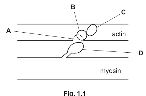
With reference to Fig. 1.1, name A, B, C and D.
A ______
B ______
C ______
D ______
Answer
- A – binding site
- B – tropomyosin
- C – troponin
- D – myosin head (accept: ATPase)
A: binding site; B: tropomyosin; C: troponin; D: myosin head (ATPase)
Background Concept
A sarcomere is the basic contractile unit of striated (skeletal and cardiac) muscle, bounded at each end by a Z-line. Two types of filament slide past one another to shorten the sarcomere:
- Thin filaments are made of the globular protein actin, twisted into a double helix. Wound around the actin helix is the long, rod-like protein tropomyosin, and attached at intervals to tropomyosin is the three-subunit globular complex troponin. On each actin monomer there is a binding site for the myosin head — this site is normally blocked by tropomyosin in a relaxed muscle.
- Thick filaments are bundles of the motor protein myosin. Each myosin molecule has a tail and two globular myosin heads; the heads possess both actin-binding and ATPase (ATP-hydrolysing) activity.
The interplay of these four components (A, B, C, D) is what the question is testing.
Understanding the Question
The diagram (Fig. 1.1) shows part of a sarcomere with the thick (myosin) and thin (actin) filaments, and four structures labelled A–D. The candidate must name each one. The marking scheme accepts the structural roles rather than insisting on a single phrasing.
Approach
Match the visible shape and position of each labelled structure to the sarcomere component it represents:
- A small circle sitting on the actin filament = the actin binding site for myosin.
- A long strand running along the actin = tropomyosin.
- A globular complex attached to the strand = troponin.
- A projection from the thick filament = myosin head.
Step-by-Step Reasoning
- A points to a small circle on the actin filament. This is the site at which the myosin head binds during cross-bridge formation, so it is the binding site.
- B is the long strand that wraps around the actin filament, covering the binding sites in a relaxed muscle — this is tropomyosin.
- C is the globular complex sitting on tropomyosin; it is the Ca²⁺-sensing component that moves tropomyosin out of the way — this is troponin.
- D projects from the thick filament and contains both the actin-binding and ATPase activities — this is the myosin head (the marking scheme also accepts ATPase, because that is the enzymatic function of the head).
Key Takeaways
- Four players to keep distinct on a sarcomere diagram: actin binding site, tropomyosin, troponin and the myosin head.
- Tropomyosin is the long inhibitory strand; troponin is the Ca²⁺-binding globular complex attached to it.
- The myosin head is both a binding site and an ATPase — the "engine" of contraction.
Common Mistakes
- Confusing troponin and tropomyosin — the names are too similar. Remember: troponin is the complex (globular), tropomyosin is the strand (rod-like).
- Naming the actin filament rather than the binding site on it for label A.
- Calling D the "myosin filament" or just "myosin" — the structure shown is specifically the head, and that precision is what is credited.
Things to Be Careful About
- The marking scheme accepts either "myosin head" or "ATPase" for D; either is fine.
- Spelling: troponin and tropomyosin are both single words, easily misspelled.
When a muscle cell is stimulated, calcium ions are released from the sarcoplasmic reticulum.
With reference to Fig. 1.1, describe the role of calcium ions in the contraction of the sarcomere.
Answer
Any four from:
- Calcium ions are released from the sarcoplasmic reticulum (SR).
- Ca²⁺ binds to troponin (C).
- This causes tropomyosin (B) to move / change shape.
- The binding site (A) on actin is exposed.
- The myosin head (D) binds to actin, forming a cross-bridge.
Ca²⁺ released from SR → binds to troponin → tropomyosin moves → binding site exposed on actin → myosin head binds actin / cross-bridge forms.
Background Concept
In a relaxed striated muscle, the binding site on actin is hidden because tropomyosin is lying across it. Troponin holds tropomyosin in this blocking position. Contraction cannot occur until this blockage is removed, and the trigger is the calcium-ion concentration in the sarcoplasm rising.
The sarcoplasmic reticulum (SR) is a modified endoplasmic reticulum that wraps around each myofibril. It actively pumps Ca²⁺ into its lumen (using Ca²⁺-ATPases) to keep sarcoplasmic [Ca²⁺] very low in a resting cell. When an action potential travels down the T-tubules, it triggers voltage-sensitive receptors that open ryanodine receptors on the SR, releasing Ca²⁺ into the sarcoplasm.
Understanding the Question
Part (b) asks for a description of what Ca²⁺ does once it has been released. The question explicitly refers to Fig. 1.1, so the answer should name the labelled structures A–D as it goes. The command word "describe" requires the sequence of events, not just a single statement.
Approach
Follow the calcium signal through the molecular cascade in the order the marking scheme lists it: release → binding to troponin → tropomyosin moves → binding site exposed → myosin head binds. Pick any four of these five well-articulated points to reach the four marks.
Step-by-Step Reasoning
- Release from the SR — the action potential causes the SR to open Ca²⁺ channels and release calcium ions into the sarcoplasm around the myofibril.
- Binding to troponin (C) — Ca²⁺ binds to specific sites on the troponin complex. This binding changes troponin's shape.
- Tropomyosin (B) moves — the troponin shape change pulls tropomyosin deeper into the groove of the actin helix, away from the myosin-binding site.
- Binding site (A) exposed — the actin site is now uncovered and available.
- Myosin head (D) binds — the energised myosin head attaches to the exposed binding site on actin, forming a cross-bridge, after which the head performs its power stroke.
Because the marking scheme accepts any four of the five, a candidate who covers the full logical chain of release → troponin binding → tropomyosin movement → site exposure → myosin binding will earn all four marks.
Key Takeaways
- The Ca²⁺ signal is the molecular switch that turns contraction on; removing Ca²⁺ (by re-pumping into the SR) turns it off.
- Troponin is the Ca²⁺ sensor; tropomyosin is the inhibitor it controls.
- Cross-bridge formation is the outcome: myosin head binding to the now-exposed actin site.
Common Mistakes
- Saying "calcium binds to actin" or "calcium binds to tropomyosin" — it specifically binds to troponin.
- Skipping the step where the binding site is exposed and going straight to "myosin binds to actin" — the exposure is a separate, creditable point.
- Writing about ATP, the power stroke or detachment — these are later events in the cross-bridge cycle, not the role of Ca²⁺ in initiating contraction.
- Failing to refer to the structures A–D in Fig. 1.1 when the question explicitly says "with reference to Fig. 1.1".
Things to Be Careful About
- The "describe" command requires a sequence; one-sentence answers rarely earn all four marks.
- "Changes shape" is accepted in place of "moves" for tropomyosin, so do not worry if your wording differs from the mark scheme as long as the meaning is the same.
- This is the role of Ca²⁺ only — the subsequent power stroke, ATP-driven detachment and re-cocking of the myosin head are not part of the answer.
When a mammal dies, aerobic respiration stops. The striated muscles contract and remain contracted for a few hours after death.
Suggest why the muscles remain contracted for a few hours after death.
Answer
- No / little ATP is produced because aerobic respiration has stopped.
- Therefore myosin heads cannot be released from actin / cross-bridges cannot be broken, so the muscle remains contracted.
No ATP available (respiration stopped) → myosin heads cannot detach from actin / cross-bridges persist → muscle stays contracted (rigor mortis).
Background Concept
A complete cross-bridge cycle has four steps: (1) the energised myosin head binds to actin, (2) the power stroke slides the actin filament as the head pivots, (3) a new ATP binds to the myosin head causing it to detach from actin, and (4) hydrolysis of that ATP re-cocks the head. Step 3 — detachment — is strictly ATP-dependent. Without ATP, the myosin head stays bound.
When a mammal dies, circulation stops, so oxygen delivery ceases. Cells quickly become anaerobic and ATP production falls towards zero; existing ATP reserves are used up within minutes by ion pumps, leak channels and the continuing cross-bridge cycle.
Understanding the Question
Part (c) uses the command word "suggest", which means apply biological knowledge to a novel scenario and give a reasoned explanation. The information given is that aerobic respiration has stopped, yet the muscles are contracted and remain so for hours. The candidate must explain what is preventing the muscles from relaxing.
Approach
Recall what is required for a striated muscle to relax: myosin heads must release from actin, and that step needs ATP. Then ask: is ATP available after death? If not, what state will the muscle be locked in?
Step-by-Step Reasoning
- After death, no oxygen reaches the cells, so aerobic respiration stops and the small ATP made anaerobically is rapidly exhausted. No / little ATP is available.
- Detachment of the myosin head from the actin binding site requires a new molecule of ATP to bind to the myosin head. With no ATP, cross-bridges cannot be broken and the myosin heads remain bound to actin.
- Because cross-bridges persist, the sarcomeres (and therefore the whole muscle) stay in the contracted state. This phenomenon is called rigor mortis. It only ends many hours later, when endogenous proteolytic enzymes break down the muscle proteins and the muscle finally softens.
Key Takeaways
- Relaxation, not contraction, is the ATP-requiring step in the cross-bridge cycle.
- Without ATP, myosin heads remain locked onto actin → contracted muscle.
- Rigor mortis is the textbook example of this principle in action.
Common Mistakes
- Saying "the muscles contract because there is no ATP" — it is the failure to relax / release that keeps them contracted, not the act of contracting.
- Stating "no energy" instead of "no / little ATP" — ATP is the precise currency the mark scheme requires.
- Bringing in calcium ions, which would actually be irrelevant here — the calcium has long since leaked out of the SR into the extracellular space after death, and troponin is no longer activated; the cause of the persistent contraction is downstream of Ca²⁺.
Things to Be Careful About
- "Suggest" allows some biological reasoning rather than demanding pure recall — the link between ATP and myosin detachment is the key insight.
- The muscle does not stay contracted forever; the mark scheme only needs the reason it stays contracted, not what eventually reverses it.
The rest of this paper
9 more questions- Q2Energy and Respiration · Homeostasis12M
- Q3Classification, Biodiversity and Conservation12M
- Q4Genetic Technology · Classification, Biodiversity and Conservation11M
- Q5Photosynthesis · Energy and Respiration11M
- Q6Homeostasis11M
- Q7Inheritance9M
- Q8Selection and Evolution9M
- Q9Genetic Technology15M
- Q10Homeostasis · Control and Coordination15M