Biology 9700/42 — May/June 2019
Cambridge A-Level · A Level Structured Questions · worked solutions for every part, with the mark scheme
Topics Selection and Evolution · Control and Coordination · Genetic Technology · Inheritance · Classification, Biodiversity and Conservation · Homeostasis · +3 more
Fig. 1.1 is an electron micrograph showing a cross-section of a myelinated neurone.
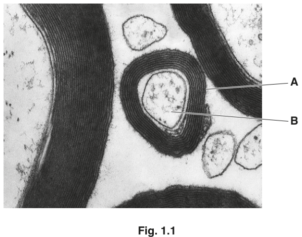
Name A and B.
A ______
B ______
Answer
A – myelin sheath (Schwann cell) ;
B – axon (axoplasm / cytoplasm) ;
A: myelin sheath / Schwann cell; B: axon / axoplasm / cytoplasm
Background Concept
A myelinated motor or sensory neurone in the peripheral nervous system has its axon wrapped along most of its length by Schwann cells. Each Schwann cell wraps itself many times around a short section of axon, and the concentric layers of phospholipid-rich plasma membrane form the myelin sheath. Between adjacent Schwann cells are short, unmyelinated gaps called nodes of Ranvier. The axon itself is the long projection that carries the action potential, and its contents (axoplasm) is continuous with the cytoplasm of the cell body.
In an electron micrograph the myelin sheath appears as a thick, dark, multi-layered ring because the heavy-metal stain (osmium tetroxide) binds to the lipid-rich membrane layers. The axon inside appears as a lighter, more granular, central core because it is cytoplasm with cytoskeletal elements and organelles, not a stack of membranes.
Understanding the Question
You are shown a cross-section of a myelinated neurone in Fig. 1.1. Label A points to the dark, layered outer ring, and label B points to the lighter material inside that ring. The question simply asks you to name these two structures.
Approach
Look at where the labels point. The dark, multi-layered outer ring is the myelin sheath (formed by a Schwann cell); the lighter central region is the axon. Either of the alternative names is acceptable, so you can write whichever you are more confident about.
Step-by-Step Reasoning
- The dark, concentric, multi-layered band is characteristic of the myelin sheath — many wrappings of Schwann-cell membrane, each stained darkly by the osmium. The mark scheme also accepts Schwann cell because the sheath is made by that cell.
- The lighter, granular, central region is the axon. Inside the axon is the axoplasm (the cytoplasm of the axon) — both terms are accepted.
Key Takeaways
- The myelin sheath appears as a dark, layered ring on an EM because of its stacked lipid-rich membranes.
- The axon is the lighter central core, containing the axoplasm.
Common Mistakes
- Calling A simply "Schwann cell membrane" or "lipid" — the marking point is the whole structure (myelin sheath / Schwann cell), not a single membrane.
- Calling B "nerve fibre" or "dendrite" — a cross-section of a myelinated fibre in the PNS shows a single axon, not a dendrite, and the term nerve fibre is not accepted here.
Things to Be Careful About
- Both A and B in the image are on the same axon (the cross-section in the centre); nearby cross-sections of other fibres are distractors.
- Use the terms that the mark scheme credits — myelin sheath or Schwann cell for A, and axon, axoplasm or cytoplasm for B.
Explain what is meant by saltatory conduction and describe its effect on the transmission of a nerve impulse.
Answer
- The action potential / impulse 'jumps' from one node of Ranvier to the next ;
- Local circuits / local currents are set up between nodes (longer, due to insulation by the myelin sheath) ;
- This makes the transmission of the impulse faster / increases the speed of conduction ;
Saltatory conduction is where the action potential 'jumps' between nodes of Ranvier via long local circuits, increasing the speed of impulse transmission.
Background Concept
In a myelinated axon, the axonal membrane is exposed to the extracellular fluid only at the nodes of Ranvier — short, regularly-spaced gaps between adjacent Schwann cells. Everywhere else, the myelin sheath acts as an electrical insulator: it prevents ion movement across the membrane in the myelinated regions, and it greatly increases the resistance and decreases the capacitance of the membrane there.
Because the membrane is insulated between the nodes, the local circuit currents that flow when an action potential is regenerated at one node spread passively through the axoplasm all the way to the next node before they decay. This makes the local circuits longer than they would be in a non-myelinated axon of the same diameter.
Understanding the Question
This part has two parts to address: explain what saltatory conduction is (i.e. the mechanism) and describe its effect on transmission (i.e. what the mechanism achieves). The mark scheme splits these into 3 points — 2 for the mechanism and 1 for the speed effect.
Approach
First describe the movement of the action potential: it does not travel continuously along the membrane but jumps from node to node. Then explain why — the myelin sheath insulates the membrane between nodes, so local currents flow passively between nodes. Finally, state the consequence — this greatly speeds up transmission compared with a non-myelinated axon of similar diameter.
Step-by-Step Reasoning
- Jumping: the action potential is regenerated only at the nodes of Ranvier, so the impulse effectively 'jumps' (Latin saltare = to jump) from node to node. [Mark 1]
- Mechanism: the myelin sheath insulates the axonal membrane between nodes, so the local circuit / local current set up by an action potential at one node spreads passively through the axoplasm to the next node, depolarising it to threshold. These local circuits are longer than in a non-myelinated axon. [Mark 2]
- Effect: because the impulse is regenerated only at widely-spaced nodes rather than at every micrometre of membrane, conduction is much faster than in a non-myelinated axon. [Mark 3]
Key Takeaways
- Saltatory conduction = the action potential jumps between nodes of Ranvier.
- Myelin makes local circuits longer by insulating the membrane between nodes.
- The result is a faster nerve impulse (and also less energy use, because fewer Na⁺/K⁺-ATPase cycles are needed per unit length).
Common Mistakes
- Saying only "the impulse travels faster" with no mechanism — the question requires both an explanation and a description of the effect.
- Saying "the action potential jumps over the myelin sheath" — it is regenerated at the nodes; the myelin is the insulating material between them.
- Confusing saltatory conduction with continuous conduction (the non-myelinated type).
Things to Be Careful About
- The word longer (qualifying local circuits) is what the mark scheme expects — it explains why the impulse can leap such a distance without decaying.
- Do not bring in unrelated detail about refractory periods, action potential mechanism or the sodium-potassium pump — they are not credited here.
A type of sea snail, Conus purpurascens, produces a toxin that blocks calcium ion channels in the presynaptic knob of a cholinergic synapse. The presence of this toxin results in no action potentials in the postsynaptic neurone.
Explain why the presence of this toxin results in no action potentials in the postsynaptic neurone.
Answer
- No calcium ions enter the presynaptic knob (calcium ion channels blocked) ;
- (So) vesicles do not move towards / fuse with the presynaptic membrane ;
- No exocytosis / release of acetylcholine (ACh) ;
- ACh does not diffuse across the synaptic cleft ;
- No binding with receptor (proteins) on the postsynaptic membrane ;
- Sodium ion channels do not open ;
- Sodium ions do not enter the postsynaptic neurone ;
- No depolarisation (of the postsynaptic membrane), so no action potential is generated ;
Blocking Ca²⁺ entry stops vesicle exocytosis, so no ACh is released into the cleft, no Na⁺ channels open postsynaptically, and the postsynaptic membrane cannot depolarise to threshold.
Background Concept
A cholinergic synapse is a synapse that uses acetylcholine (ACh) as its neurotransmitter. Transmission across it follows a precise sequence:
- An action potential arrives at the presynaptic knob and depolarises its membrane.
- This opens voltage-gated calcium (Ca²⁺) ion channels; Ca²⁺ flows down its electrochemical gradient into the knob.
- The Ca²⁺ causes synaptic vesicles containing ACh to move to and fuse with the presynaptic membrane.
- ACh is released by exocytosis into the synaptic cleft.
- ACh diffuses across the cleft and binds to specific receptor proteins on the postsynaptic membrane, opening ligand-gated Na⁺ channels.
- Na⁺ flows into the postsynaptic neurone, depolarising it; if threshold is reached, a new action potential is generated.
Each step depends on the one before it, so blocking a single step — such as Ca²⁺ entry — collapses the entire cascade.
Understanding the Question
You are told that the toxin from Conus purpurascens blocks calcium ion channels in the presynaptic knob of a cholinergic synapse, and the observation is that no action potentials occur in the postsynaptic neurone. You must explain why the toxin has this effect by walking through the steps that depend on Ca²⁺ entry. The mark scheme credits up to 5 of the 8 logical points.
Approach
Apply the synaptic transmission sequence above. Identify which step is blocked first (Ca²⁺ entry) and then trace every subsequent step that consequently fails. Each failed step that is correctly identified and linked back to the previous one earns a mark.
Step-by-Step Reasoning
- Step blocked: The toxin blocks voltage-gated Ca²⁺ channels, so no Ca²⁺ enters the presynaptic knob. [Mark 1]
- Vesicle step: Without the Ca²⁺ trigger, synaptic vesicles do not move to or fuse with the presynaptic membrane. [Mark 2]
- Release step: No vesicle fusion means no exocytosis of ACh into the synaptic cleft. [Mark 3]
- Diffusion step: With no ACh released into the cleft, none can diffuse across to the postsynaptic membrane. [Mark 4]
- Receptor binding: There is therefore no ACh to bind to receptor proteins on the postsynaptic membrane. [Mark 5]
- Na⁺ channel opening: Without ACh-receptor binding, the ligand-gated Na⁺ channels on the postsynaptic membrane do not open. [Mark 6]
- Na⁺ entry: So no Na⁺ flows into the postsynaptic neurone. [Mark 7]
- Depolarisation: Without Na⁺ influx there is no depolarisation of the postsynaptic membrane, and threshold is never reached — so no new action potential can be generated. [Mark 8]
Any five of these eight points earn the five marks; in practice a strong answer gives the early, defining marks and finishes with the depolarisation point, because that explicitly answers the question's no action potentials wording.
Key Takeaways
- Ca²⁺ entry into the presynaptic knob is the indispensable trigger for neurotransmitter release.
- Synaptic transmission is a strict cascade; blocking the first step stops every later step.
- A cholinergic synapse specifically uses ACh and ligand-gated Na⁺ channels on the postsynaptic side.
Common Mistakes
- Vague wording such as "the toxin stops the nerve signal" — you must name the specific ions, structures and processes that fail.
- Saying the toxin "blocks the synapse" or "prevents messages being passed" — the mark scheme rejects this; you must identify the Ca²⁺ channel as the blocked structure.
- Confusing the role of Ca²⁺ at the synapse with its role in muscle contraction or in the action potential itself.
- Stating that ACh release is prevented by the toxin binding to ACh — the toxin blocks the channel, not the transmitter.
- Saying "no exocytosis of vesicles" — the mark scheme accepts no exocytosis of ACh but specifically rejects exocytosis of vesicles because vesicles are the structure, not the released product.
Things to Be Careful About
- Always link each point to the one before it: the cascade is what makes the explanation logical.
- Use precise terms: presynaptic knob, synaptic cleft, postsynaptic membrane, depolarisation, threshold.
- Notice that the question says no action potentials (plural) in the postsynaptic neurone — finishing with "no depolarisation, so threshold is not reached" directly addresses the observation in the stem.
The rest of this paper
9 more questions- Q2Selection and Evolution · Genetic Technology · Inheritance16M
- Q3Genetic Technology9M
- Q4Inheritance · Selection and Evolution12M
- Q5Homeostasis13M
- Q6Selection and Evolution10M
- Q7Classification, Biodiversity and Conservation6M
- Q8Energy and Respiration9M
- Q9Photosynthesis · Classification, Biodiversity and Conservation15M
- Q10(outdated) Aspects of Human Reproduction · Control and Coordination · Selection and Evolution15M