Biology 9700/43 — May/June 2017
Cambridge A-Level · A Level Structured Questions · worked solutions for every part, with the mark scheme
Topics Inheritance · Homeostasis · Energy and Respiration · Classification, Biodiversity and Conservation · Control and Coordination · Photosynthesis · +2 more
The mammalian kidney is an organ involved in homeostasis.
Explain what is meant by the term homeostasis.
Answer
Homeostasis is the maintenance of a constant internal environment (within narrow limits) inside the body.
Homeostasis is the maintenance of a constant internal environment within narrow limits inside the body.
Background Concept
Homeostasis is the process by which the body maintains a stable internal environment despite changes in the external environment. The 'internal environment' refers to the conditions of the tissue fluid that bathes the cells – things like temperature, pH, water potential (), and the concentrations of glucose, ions (especially Na⁺, K⁺, Ca²⁺) and waste products (such as urea). The concept was first articulated by the French physiologist Claude Bernard in the 19th century as the 'milieu intérieur'.
Homeostasis is not a single event but a continuous, dynamic process of detection, correction and re-establishment of the optimum. The body detects deviations from the set-point and brings the variable back into range. In mammals the main homeostatic systems deal with temperature, blood glucose, blood water potential and blood carbon dioxide.
Understanding the Question
This is a 1-mark 'explain what is meant by' question. The mark scheme wants three elements for the single mark: an action verb (maintain/keep/restore), a description of the state (constant/stable/within narrow limits), and what is being kept stable (internal environment/in the body). Missing any of the three weakens the answer.
Approach
Write one or two sentences that cover all three elements. Do not add examples (e.g. temperature regulation) – the mark is for the definition, not for illustration, and an example can never replace a missing element.
Step-by-Step Reasoning
The mark scheme credits three components for this mark:
- An action verb: 'maintain', 'keep' or 'restore'. Avoid 'control' (too vague) and 'balance' (implies equilibrium between two opposing forces rather than maintenance of a steady state).
- A description of the state achieved: 'constant', 'stable', 'at a set-point' or 'within narrow limits'.
- The target: 'internal environment' or 'in the body'. 'Blood' alone is not enough – the internal environment is broader than blood and includes tissue fluid.
A complete answer might be: 'The maintenance of a constant internal environment (within narrow limits) inside the body.' This is the cleanest single-line form of the answer that captures all three required elements.
Key Takeaways
- Homeostasis is the maintenance of a stable internal environment, not the prevention of all change.
- The 'internal environment' is the tissue fluid surrounding the cells; blood is the means of communicating changes between cells and the homeostatic control centres.
- A complete definition needs three parts: action, quality and target.
Common Mistakes
- Saying 'keeping a balance' or 'maintaining balance' – balance is a different concept (equilibrium between two things); homeostasis is about a steady state.
- Saying 'the body keeps things the same' without specifying what is being kept the same (the internal environment).
- Defining homeostasis as 'the regulation of body temperature' or 'the control of blood glucose' – these are examples, not the definition.
- Saying 'keeping a constant environment' without saying what is constant and where (internal environment of the body).
Things to Be Careful About
- The mark is awarded for the definition, not for examples. Do not waste the mark by listing examples of variables that are homeostatically controlled.
- 'Internal environment' is more precise than 'inside the body' – both are accepted, but the former is the CIE preferred phrasing.
- 'Narrow limits' is the standard qualification; the body does allow some fluctuation around the set-point.
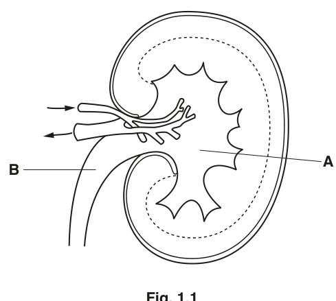
Fig. 1.1 shows a section through a kidney.
With reference to Fig. 1.1, name structures A and B.
A ______
B ______
Answer
A – renal pelvis
B – ureter
A – renal pelvis; B – ureter
Background Concept
A mammalian kidney has a recognisable gross anatomy. The outer layer is the renal cortex, beneath which is the renal medulla (made up of cone-shaped pyramids). The inner cavity of the kidney is the renal pelvis, a funnel-shaped space that collects urine from the minor and major calyces as it drains from the papillae of the medullary pyramids. From the pelvis, urine passes into the ureter – a muscular tube lined with transitional epithelium that conveys urine to the bladder by peristalsis. The renal artery and renal vein enter and leave the kidney at the hilum, alongside the ureter.
Understanding the Question
Fig. 1.1 shows a longitudinal section of a kidney. Label A points to a branching, star-shaped structure in the centre of the kidney (the renal sinus). Label B points to a tube exiting the kidney at the hilum. Both are 1-mark identifications, worth 2 marks in total.
Approach
Use the position and shape of each label to deduce the structure. The renal pelvis is a central, branching cavity; the ureter is the single tube that exits the hilum and is the route by which urine leaves the kidney.
Step-by-Step Reasoning
- Structure A: the label line points to a fan-like or star-shaped region in the centre of the kidney. This is the renal pelvis, the funnel that receives urine from the calyces and channels it into the ureter.
- Structure B: the label line points to the tube that exits the kidney at the hilum and travels down towards the bladder. This is the ureter.
The mark scheme explicitly notes an ecf in part (b)(ii) if A is mislabelled as the medulla – so confusing the central pelvis with the surrounding medulla is a common error, but here A is unambiguously the pelvis.
Key Takeaways
- The renal pelvis is the central cavity; the medulla is the tissue around it.
- The ureter is the only tube that exits the kidney to the bladder; the renal artery and renal vein also leave at the hilum but they carry blood, not urine.
Common Mistakes
- Calling A the medulla – the medulla is the inner tissue (containing the pyramids), not the central cavity.
- Calling B the urethra – the urethra connects the bladder to the outside, not the kidney to the bladder.
- Confusing the renal pelvis with a calyx (a calyx is a smaller cup that drains a single pyramid; the pelvis is the larger funnel that collects from all the calyces).
Things to be Careful About
- A is a central, branching structure, not a region of tissue. The renal pelvis is a space, not a solid body.
- The ureter and the renal vein both leave the kidney at the hilum; only the ureter carries urine, so the position alone is not enough – you must recognise the tube as the one leading to the bladder.
On Fig. 1.1, use label lines and letters to label where:
U – ultrafiltration occurs
L – the loop of Henle is found
C – blood urea concentration is low.
Answer
Add three new label lines and letters to Fig. 1.1:
- U – label line pointing to the renal cortex (the outer layer where Bowman's capsules and glomeruli are located).
- L – label line pointing to the medulla (the inner region containing the loops of Henle).
- C – label line pointing to the renal vein (the vessel that leaves the kidney carrying blood with the lowest urea concentration).
U – cortex; L – medulla; C – renal vein
Background Concept
The kidney performs its work in a clearly compartmentalised way, and each process has a specific location:
- Ultrafiltration takes place in the renal corpuscles (glomerulus + Bowman's capsule), which are found only in the cortex. Blood is forced under high hydrostatic pressure from the glomerular capillaries into the Bowman's capsule, producing a filtrate that is essentially plasma without the cells and large proteins.
- The loops of Henle descend from the cortex into the medulla. The longer the loop, the deeper into the medulla it goes. This is essential for the counter-current multiplier system that concentrates the urine.
- Blood urea is removed by filtration at the glomerulus and is not reabsorbed in any significant amount. Therefore, the concentration of urea falls as blood passes through the kidney. Blood entering the kidney via the renal artery has the highest urea concentration, and blood leaving via the renal vein has the lowest.
Understanding the Question
You are asked to add three labels to the printed figure: U (where ultrafiltration occurs), L (where the loop of Henle is found) and C (where blood urea is lowest). Each label is worth one mark.
Approach
Match each process to its anatomical location based on kidney structure. Use the labels U, L and C with label lines, as instructed.
Step-by-Step Reasoning
- U (ultrafiltration) → cortex. The renal corpuscles lie in the cortex; this is the only site of ultrafiltration in the nephron.
- L (loop of Henle) → medulla. The loops of Henle are the long hairpin-shaped tubules that run from the cortex down into the medulla and back up. Most of the loop sits in the medulla.
- C (low blood urea) → renal vein. As blood passes through the kidney, urea is removed by ultrafiltration and is not reabsorbed. The blood that leaves the kidney in the renal vein therefore has the lowest urea concentration.
The mark scheme also allows an error carried forward (ecf) if the candidate mislabelled A as the medulla in part (i). In that case L pointing to the pelvis (formerly mislabelled as medulla) and U pointing to the medulla (provided 'cortex' is written by U) still earn credit.
Key Takeaways
- Ultrafiltration is exclusively a cortical process because all glomeruli are in the cortex.
- The medulla houses the loops of Henle and the vasa recta; this is where the urine is concentrated.
- The renal vein is the only vessel that carries blood with reduced urea because urea is removed but not put back.
Common Mistakes
- Pointing U to the medulla because 'filtration' sounds like 'deeper' work. Ultrafiltration is a cortical event.
- Pointing L to the cortex – the start of the loop is in the cortex, but the loop as a whole extends into the medulla; the mark scheme credits the medulla.
- Pointing C to the renal artery. The renal artery carries blood with the highest urea, not the lowest.
Things to be Careful About
- The label letters U, L and C must be on label lines (not just written next to the structure) – the figure must be unambiguously annotated.
- The renal vein is the vein that leaves the kidney and joins the inferior vena cava. Do not confuse it with the renal artery or with the ureter.
Describe the roles of the hypothalamus and the posterior pituitary in osmoregulation.
Answer
- The hypothalamus contains osmoreceptors that detect the water potential of the blood.
- ADH is produced by neurosecretory cells in the hypothalamus.
- ADH is transported down the axons of these neurosecretory cells to the posterior pituitary, where it is stored.
- When blood water potential is low, ADH is released from the posterior pituitary into the blood.
- ADH increases the permeability of the distal convoluted tubule and collecting duct to water (by stimulating the insertion of aquaporins into the cell-surface membrane).
- More water is reabsorbed, producing a smaller volume of more concentrated urine.
Hypothalamus detects low blood water potential; ADH is made in the hypothalamus and transported to the posterior pituitary; ADH is released when water potential is low; ADH increases water reabsorption (via aquaporins) in the collecting duct, producing more concentrated urine.
Background Concept
The body controls its water balance by adjusting how much water is reabsorbed from the kidney filtrate. The key hormone is antidiuretic hormone (ADH, also called vasopressin). 'Antidiuretic' literally means 'against passing urine' – more ADH means less urine, and a more concentrated one.
ADH is unusual among mammalian hormones in that it is made by neurones rather than by glandular cells. It is produced in the cell bodies of neurosecretory cells whose cell bodies sit in the hypothalamus and whose axons run down into the posterior pituitary. The posterior pituitary does not synthesise ADH; it stores it and releases it into the blood. This is why the hypothalamus and the posterior pituitary are discussed as a single functional unit.
ADH acts on the distal convoluted tubule (DCT) and especially the collecting duct. Without ADH, the collecting duct is relatively impermeable to water and the body loses water in dilute urine. With ADH, water-channel proteins (aquaporins) are inserted into the luminal membrane of the collecting duct cells, water flows out down the osmotic gradient into the hypertonic medulla, and the urine becomes more concentrated.
Understanding the Question
The question is 5 marks and uses the command word 'describe'. It specifically asks for the roles of BOTH the hypothalamus AND the posterior pituitary, so any answer that does not address both organs is unlikely to score full marks. The mark scheme lists 8 possible points, of which any 5 earn full marks. You should aim to give a connected, logical description that follows the path of the signal from detection to effect.
Approach
Structure the answer as a short narrative: detection (hypothalamus) → synthesis and transport (hypothalamus to posterior pituitary) → release (posterior pituitary) → action (kidney). Cover at least one point for each organ and at least one point for the action on the kidney.
Step-by-Step Reasoning
A good five-point answer would cover:
- The hypothalamus detects the water potential of the blood via osmoreceptors (1 mark).
- ADH is produced by the cell bodies of neurosecretory cells in the hypothalamus (1 mark).
- ADH is transported down the axons of these neurosecretory cells to the posterior pituitary, where it is stored (1 mark for transport, 1 mark for storage in posterior pituitary if you want to separate them).
- When blood water potential is low, ADH is released from the posterior pituitary into the blood (1 mark).
- ADH increases the permeability of the collecting duct (and DCT) to water, by stimulating the insertion of aquaporins into the membrane (1 mark for either permeability or aquaporins).
- This results in more water being reabsorbed, producing a smaller volume of more concentrated urine (1 mark for the effect on urine).
The mark scheme offers these as alternative paths:
- 'osmoreceptors shrink when blood water potential is low' is an alternative to (or extension of) the detection point.
- 'ADH causes more water reabsorption / smaller volume / more concentrated urine' is the physiological outcome.
- 'aquaporins' is the molecular mechanism.
For full marks, make sure you say WHERE ADH is made (hypothalamus), WHERE it is stored/released (posterior pituitary) and WHERE it acts (collecting duct / DCT). Do not simply say 'ADH is released' without naming the gland.
Key Takeaways
- ADH is produced in the hypothalamus and stored in the posterior pituitary – not the other way around.
- The collecting duct is the main target of ADH; the DCT is a secondary target.
- ADH works through aquaporins (water-channel proteins) inserted into the luminal membrane of collecting-duct cells.
- Low blood water potential triggers the release of ADH; high water potential suppresses it.
- Neurosecretory cells are modified neurones that release hormones into the blood – they bridge the nervous and endocrine systems.
Common Mistakes
- Saying ADH is produced in the posterior pituitary. The posterior pituitary is a storage and release site, not a production site.
- Saying ADH is released when blood water potential is high. The opposite is true – it is released when water potential is low (the blood is too concentrated).
- Saying ADH increases water reabsorption by 'making the collecting duct work harder'. Be specific: ADH increases permeability by inserting aquaporins.
- Confusing ADH with aldosterone (which controls Na⁺ reabsorption, not water directly).
- Saying the kidney 'produces' ADH or 'detects' the change. The kidney responds to ADH; detection happens in the hypothalamus.
Things to be Careful About
- The posterior pituitary is neural tissue, not a glandular tissue. It is essentially the end of the hypothalamus, connected by the hypothalamo-hypophyseal tract.
- 'Osmoreceptors shrink when the water potential of the blood decreases' is the precise statement. Saying 'osmoreceptors detect dehydration' is too vague.
- 'Negative feedback': when the reabsorbed water returns the blood water potential to normal, ADH release is reduced. Mentioning the loop closes the description, though it is not required for the 5 marks.
- The mark scheme explicitly rejects the idea that ADH is made in the posterior pituitary – this is a common misconception in textbooks and a guaranteed mark-loser.
The rest of this paper
9 more questions- Q2Photosynthesis9M
- Q3Genetic Technology · Inheritance14M
- Q4Energy and Respiration7M
- Q5Selection and Evolution · Inheritance · Classification, Biodiversity and Conservation14M
- Q6Control and Coordination · Energy and Respiration10M
- Q7Inheritance9M
- Q8Classification, Biodiversity and Conservation11M
- Q9Homeostasis15M
- Q10Control and Coordination15M