Biology 9700/52 — May/June 2014
Cambridge A-Level · Planning, Analysis and Evaluation · worked solutions for every part, with the mark scheme
Topics Planning · Analysis, Conclusions and Evaluation
A group of students was given the task of planning a method to compare the effect of two different restriction enzymes on some DNA extracted from peas.
The students used the following procedure to extract and digest DNA from the peas.
• 3 g of sodium chloride was dissolved in of distilled water in a beaker.
• of liquid detergent was added to the salt solution and stirred.
• 50 g of peas were ground up using a glass rod and then added to the salty detergent solution.
• The beaker containing the mixture of peas and salty detergent solution was left in a water bath at for exactly 15 minutes.
• The mixture was then cooled in an ice-water bath for 5 minutes, stirring frequently.
• The mixture was filtered into another beaker.
• of filtrate was transferred into a boiling tube and 2–3 drops of a protease solution were added. This was left for a few minutes.
• Ice-cold ethanol was then poured down the side of the boiling tube to form a layer on top of the filtrate. This was left undisturbed for a few minutes to form a layer of precipitated DNA.
• The precipitated DNA was separated from the cold ethanol layer by twisting the DNA onto a glass hook.
• The DNA was mixed with EDTA buffer solution.
• samples of the EDTA buffer solution containing the DNA extract were placed into three separate test-tubes, 1, 2 and 3.
• Two different restriction enzymes, Eco RI and Hin dIII, were added to the samples as shown in Table 1.1.
Table 1.1
| sample | 1 | 2 | 3 |
|---|---|---|---|
| restriction enzymes added | Eco RI | Hin dIII | Eco RI and Hin dIII |
• These were left for three hours to complete digestion of the DNA into fragments of different sizes.
Suggest a reason for each of the following steps in the procedure for extracting DNA.
using detergent
Answer
Detergent disrupts the (phospholipid) cell membranes, releasing the DNA from the cells.
Detergent disrupts the (phospholipid) cell membranes, releasing the DNA from the cells.
Background Concept
Every plant cell is bounded by a cell-surface membrane and contains membrane-bound organelles, including the nucleus where the DNA is stored. The basic structure of each of these membranes is a phospholipid bilayer — phospholipid molecules with hydrophilic phosphate heads facing outwards and hydrophobic fatty acid tails facing inwards. To extract DNA, every one of these membrane barriers must be broken so that the DNA can escape into solution. Detergents are amphipathic molecules: they have a hydrophilic head and a hydrophobic tail, so they can insert into the bilayer and disrupt it in the same way as the phospholipids themselves.
Understanding the Question
The students' procedure lists several steps to extract DNA from peas. The very first chemical added (along with salt solution) is liquid detergent. The question asks why detergent is used. The detergent is added before the peas are ground up, so it is present to act on the cellular structures of the pea tissue as it is disrupted.
Approach
Think about what detergent molecules do to phospholipid membranes at the molecular level, and which cellular structures must be opened to liberate DNA. The mark scheme requires a specific answer naming the membrane (or phospholipid) — not a vague statement about "cells".
Step-by-Step Reasoning
- The peas are made of cells, each surrounded by a phospholipid cell-surface membrane and containing a nuclear membrane (also a phospholipid bilayer) around the DNA.
- Detergent is amphipathic; its hydrophobic tail interacts with the hydrophobic interior of the phospholipid bilayer, while its hydrophilic head interacts with water.
- This interaction breaks up the bilayer, disrupting the membrane structure and forming mixed detergent–lipid micelles.
- The membrane is therefore no longer a continuous barrier, so the cellular contents (including the nucleus and the DNA) are released into the surrounding salty solution.
- The mark scheme explicitly wants the answer framed in terms of disrupting the (phospholipid) membranes — "makes the membrane permeable", "destroys the membrane" and vague "lyses the cell" are weaker answers that may not score the same mark.
Key Takeaways
- Detergents disrupt phospholipid membranes because they are themselves amphipathic.
- Releasing DNA from a cell requires breaking both the cell-surface membrane and the nuclear membrane.
- This is a general principle used whenever biological membranes must be solubilised (e.g. in lysis buffers, in COVID-19 spike protein work, etc.).
Common Mistakes
- Saying the detergent "kills the cell" or "lyses the cell" without naming the membrane: the mark scheme specifically wants membranes / phospholipids.
- Saying the detergent breaks down the cell wall: cellulose cell walls are not disrupted by detergent — mechanical grinding and/or enzymes are needed for that.
- Mentioning pH or alkalinity of the detergent: not the key point.
- Saying the detergent denatures DNA: incorrect and not credited.
Things to Be Careful About
- Specify "membranes" or "phospholipids" (or both) — "cells" on its own is too vague.
- Do not credit references to enzymes in detergent disrupting membranes: that is a confused statement.
leaving at
Answer
The 60 °C temperature denatures / inactivates enzymes (such as DNases) that would otherwise break down the DNA.
The 60 °C temperature denatures / inactivates enzymes (such as DNases) that would otherwise break down the DNA.
Background Concept
Enzymes are proteins, and most enzymes are denatured by temperatures well below the boiling point of water (typically 40–60 °C). Once denatured, an enzyme's tertiary structure is irreversibly disrupted, its active site is lost, and it can no longer catalyse its reaction. Importantly, DNA is much more heat-stable than proteins — its two strands only separate at around 90 °C — so a temperature that destroys enzymes does not damage the DNA itself. Plant cells also contain nucleases (especially DNases) that would rapidly digest any DNA released from the nucleus if they remained active.
Understanding the Question
The procedure places the beaker of peas, salt and detergent in a water bath at 60 °C for exactly 15 minutes. The question asks why this heating step is included. The word "exactly" is a clue: 60 °C is a deliberately chosen temperature — high enough to denature enzymes but low enough not to damage the DNA.
Approach
Identify what biological component would be destroyed at 60 °C. The candidates are proteins (enzymes) and DNA itself. The mark scheme rewards "denaturing / inhibiting enzymes" and explicitly rejects answers about denaturing or melting the DNA.
Step-by-Step Reasoning
- The peas contain enzymes, including nucleases (DNases), that can digest DNA.
- 60 °C is hot enough to denature these enzymes (their active sites are lost) without denaturing the DNA.
- With the DNases inactive, the DNA released from the nuclei is not broken down and is preserved for the later steps of the extraction.
- The time of "exactly 15 minutes" ensures denaturation is complete without unnecessary exposure.
- The mark scheme explicitly excludes: denaturing DNA, melting DNA, strand separation of DNA, killing microbes, and "constant temperature". None of these is the credited reason.
Key Takeaways
- A temperature that destroys enzymes (≈60 °C) leaves DNA intact.
- Inactivating DNases is essential if extracted DNA is to be recovered in a useful form.
- The word "exactly" in the procedure is a hint that the temperature must be carefully controlled.
Common Mistakes
- Saying the 60 °C denatures the DNA: the mark scheme rejects this — the strands only separate at ~90 °C.
- Saying the 60 °C "melts" or "separates the strands of" the DNA: explicitly rejected.
- Saying the temperature "kills microbes": explicitly rejected.
- Saying it provides a constant temperature: rejected; the credit is for a specific effect on the contents.
Things to Be Careful About
- Be specific that the enzymes being denatured are nucleases / DNases (or simply "enzymes") — the mark scheme rejects references to denaturing DNA or other materials.
- Note that 60 °C is chosen to be just above the denaturation temperature of most enzymes but well below the denaturation temperature of DNA.
filtering the mixture
Answer
Filtering removes / separates the (cell) debris (and other solids) from the liquid containing the DNA, so that only the filtrate (which contains the DNA) is carried forward.
Filtering removes / separates the (cell) debris (and other solids) from the liquid containing the DNA, so that only the filtrate (which contains the DNA) is carried forward.
Background Concept
Filtration separates an insoluble solid from a liquid by passing the mixture through a porous barrier (filter paper or muslin). The pores are small enough to retain solid particles but large enough to let the liquid and dissolved solutes through. In a cell extract, the desired product (DNA) is in solution, while the unwanted material is the insoluble structural debris of broken cells: cell-wall fragments, unbroken tissue, starch grains, and other large particulates.
Understanding the Question
After the peas have been ground, mixed with detergent and salt, heated, and cooled, the mixture is filtered into another beaker. The question asks why this step is carried out. The filtration step produces a filtrate (the liquid that passes through) and a residue (the material left behind).
Approach
Think about what the filter paper holds back and what passes through, and which of these contains the DNA. The DNA is dissolved in the liquid; the solid debris is what should be discarded.
Step-by-Step Reasoning
- After grinding, the mixture contains both liquid (water, detergent, salt, dissolved DNA) and solid material (cell-wall fragments, starch, tissue debris).
- The filter paper / muslin has pores that let the liquid and dissolved molecules (including DNA) through but retain the larger solid particles.
- The filtrate therefore contains the DNA in solution, while the debris is left on the filter.
- Carrying the filtrate forward keeps the DNA-containing liquid and discards the unwanted solid material.
- The mark scheme accepts "removing / trapping / separating cellular debris" and explicit examples such as "cell walls" or "solids", but rejects vague terms such as "impurities", "particles" unqualified, and "detergent" or "salt".
Key Takeaways
- Filtration is a simple physical separation based on particle size.
- In DNA extraction, the DNA is in solution, so it ends up in the filtrate.
- Removing debris at this stage simplifies all later steps.
Common Mistakes
- Saying filtration "removes impurities": the mark scheme explicitly rejects "impurities".
- Saying it "removes detergent" or "removes salt": both rejected; these pass through the filter with the DNA.
- Vague "removes particles" without specifying cell debris / solids.
- Saying it "purifies the DNA": too vague — the mechanism is removing solid debris.
Things to Be Careful About
- The mark scheme rejects "peas" or "components" unqualified. Specify cell debris, cell walls, solids, or similar.
adding protease
Answer
The protease breaks down the proteins / histones associated with the DNA (in the chromosomes), leaving the DNA in a purer form.
The protease breaks down the proteins / histones associated with the DNA (in the chromosomes), leaving the DNA in a purer form.
Background Concept
In the nucleus, DNA is not free in solution. It is wound around histone proteins to form nucleosomes, which are then packaged into chromatin and ultimately into chromosomes. When DNA is extracted, these proteins come out with it. If the proteins are not removed they will interfere with later steps: the DNA will be cloudy, the restriction enzymes used to cut it may not work well, and the gel electrophoresis will be hard to read. Proteases are enzymes that hydrolyse the peptide bonds of proteins, breaking them into smaller peptides or amino acids.
Understanding the Question
After the filtrate has been collected, the procedure adds 2–3 drops of protease solution to a 10 cm³ sample and leaves it for a few minutes. The question asks why the protease is added. The key is what protease actually does and what proteins are present in the extract.
Approach
Identify which proteins are present in the filtrate that would interfere with the DNA work, and explain how protease removes them. The mark scheme requires the answer to be specific to proteins associated with the DNA itself, not a general statement about proteases.
Step-by-Step Reasoning
- The filtrate contains DNA together with the histone proteins that were wrapped around it in chromatin.
- These proteins would otherwise contaminate the DNA and may inhibit the restriction enzymes in the next step.
- Protease hydrolyses the peptide bonds of these proteins, breaking them into smaller peptides / amino acids that no longer co-purify with the DNA.
- The result is a DNA sample that is much purer and more amenable to enzymatic digestion and gel electrophoresis.
- The mark scheme rejects a generic "function of proteases" answer: the credit is specifically for breaking down proteins / histones associated with DNA / chromosomes.
Key Takeaways
- Histones and other DNA-binding proteins are common contaminants of crude DNA preps.
- Protease is included to digest these specific proteins and free the DNA.
- This is the same principle used in commercial DNA-extraction kits (which include proteinase K).
Common Mistakes
- Giving a generic statement about the function of proteases (e.g. "to break down proteins"): the mark scheme explicitly ignores this; the proteins must be specified as those associated with the DNA / chromosomes / histones.
- Saying the protease "removes RNA": incorrect — RNase is required for that.
- Saying the protease "kills the cell": irrelevant by this stage of the procedure.
Things to Be Careful About
- Specify "proteins / histones associated with the DNA (or chromosomes)" — not just "proteins" in general.
The students used gel electrophoresis to separate the DNA fragments produced by the digestion with the restriction enzymes.
Describe the main stages used in gel electrophoresis that the students could use to separate and locate the DNA fragments.
Answer
- Make an agarose gel, with wells formed (e.g. using a comb) for loading the DNA samples.
- Place the DNA samples into the wells at the cathode (negative electrode) end of the gel — DNA is negatively charged and will migrate towards the anode (positive electrode).
- Cover the gel with (electrophoresis) buffer so that a circuit is completed and current can flow through the gel.
- Connect a power supply and apply a potential difference / voltage across the gel for a set time so that the DNA fragments separate by size (smaller fragments move further).
- Stain the DNA (e.g. with ethidium bromide) and view the bands under UV light. Safety: the stain is toxic / the UV light is harmful, so wear gloves (and goggles) to protect skin and eyes.
Prepare an agarose gel with wells; load DNA samples at the cathode; cover with buffer; apply a voltage; stain the DNA and view under UV light, with appropriate safety precautions.
Background Concept
Gel electrophoresis separates charged molecules by size. The molecules are pulled through a gel (a porous jelly-like matrix) by an applied electric field; smaller molecules pass through the pores more easily and travel further in a given time, while larger ones are held back. DNA has a uniform negative charge per unit length (because of the phosphate groups in its sugar–phosphate backbone), so the rate of migration depends mainly on fragment size, not sequence. To make DNA visible after the run, it must be stained — common stains include ethidium bromide (viewed under UV light), methylene blue, crystal violet, and SYBR Green.
Understanding the Question
The students have digested pea DNA with restriction enzymes. They now want to separate the resulting fragments by size and visualise them. The question asks for the main stages of gel electrophoresis they could use. The command word is "describe", which means the answer should be a clear, ordered set of procedural points (a list works well). The mark scheme rewards any five of eight possible points, so a complete description must cover preparation of the gel, the loading and running steps, and visualisation, plus a hazard-and-precaution pair.
Approach
Plan the answer in the order the procedure is actually carried out: (1) make the gel; (2) load the samples; (3) connect the electric field; (4) run the gel; (5) visualise the bands. Add a safety point to cover the hazard credit. Pick the wording that matches the mark scheme's accepted terms — "agarose gel", "wells", "cathode / negative electrode", "buffer", "potential difference / voltage", "stain and observe" — to make sure each line scores.
Step-by-Step Reasoning
- Gel preparation. Agarose powder is dissolved in buffer, poured into a mould and allowed to set with a comb in place. When the comb is removed it leaves wells / pits / chambers in the set gel, into which samples are loaded. Agarose is the standard gel for DNA; agar or acrylamide would also be accepted, but starch is rejected.
- Sample loading. Each DNA sample is mixed with a dense loading dye (e.g. glycerol plus a coloured marker) so that it sinks into the well, and is pipetted into a separate well using a micropipette. The samples must be placed at the cathode (negative end) because DNA is negatively charged and will migrate towards the anode.
- Buffer. The gel is submerged in electrophoresis buffer (e.g. TAE or TBE). The buffer carries the current and keeps the gel from drying out; it is what completes the circuit between the electrodes through the gel.
- Applying the field. A power pack is connected to the electrodes and a potential difference / voltage is applied. The negatively charged DNA fragments migrate towards the anode; smaller fragments move faster and travel further.
- Staining and visualisation. The gel is removed and stained (e.g. with ethidium bromide, methylene blue, crystal violet, SYBR Green, or acridine orange) so that the DNA bands become visible. Bands are then viewed either under UV light (for fluorescent stains) or against a light box. Pre-stained gels are also acceptable.
- Safety. Many of the stains (e.g. ethidium bromide) are toxic / mutagenic and the UV light is harmful to skin and eyes. The matching precaution is to wear gloves when handling the stain and to wear UV-blocking goggles / a face shield when viewing the gel under UV. (Either hazard–precaution pair is acceptable, as long as both are present.)
Key Takeaways
- Gel electrophoresis separates DNA fragments by size because the charge:mass ratio of DNA is constant.
- The procedural order is: prepare gel → load samples at cathode → cover with buffer → apply voltage → stain and view.
- Safety must be paired explicitly: name the hazard and the matching precaution.
Common Mistakes
- Saying the samples are placed at the anode: DNA is negatively charged, so it must be placed at the cathode; "anode" gets no credit.
- Saying "electricity" or "current" without specifying "potential difference" or "voltage": "electricity" unqualified is ignored.
- Naming a stain that does not bind DNA (e.g. iodine) or stating the gel is visualised under a light microscope: not credited.
- Omitting the safety precaution, or giving a hazard without a precaution (or vice versa): only paired hazard + precaution scores.
- Mentioning Southern blotting, radioactive probes or VNTRs: the mark scheme explicitly ignores these for this stage of the procedure.
Things to Be Careful About
- The mark scheme wants agarose, not starch; agarose is spelt with a final "-ose".
- A hazard and its precaution must be a matched pair — e.g. UV light and goggles, or stain toxicity and gloves.
- The well-loading step accepts any specific detail (e.g. using a micropipette, adding loading dye, using a different tip for each sample) but not a specified volume.
- "Starch gel" is explicitly rejected; do not name starch.
Identify a dependent variable that the student could measure.
Answer
Distance moved by (the) DNA fragments / number of DNA fragments (or the position of the fragments, or the length / size of the fragments measured against a size standard / ladder).
Distance moved by the DNA fragments (or number of fragments, or fragment size measured against a size standard).
Background Concept
In any controlled comparison, the dependent variable is the quantity that is measured to give a result — it is what changes in response to the independent variable. In a comparison of restriction-enzyme digests run on a gel, the independent variable is the enzyme used (none, Eco RI, Hin dIII, or both), and the dependent variable is the property of the separated DNA that is actually observed and recorded.
Understanding the Question
The students have three lanes of DNA digested with different enzymes (Table 1.1: lane 1 Eco RI only, lane 2 Hin dIII only, lane 3 both). They then run the digests on a gel. The question asks what the students could measure as the dependent variable. The mark scheme accepts any of: (a) the distance moved by the fragments; (b) the number of fragments; (c) the position of the fragments on the gel; or (d) the size of the fragments by comparison with a size standard / ladder. References to "bands", "lines" or "stripes" on their own are ignored.
Approach
Think about what can actually be read off a stained gel. The gel shows bands at different positions, each band representing many copies of a DNA fragment of a particular size. The directly measurable quantities are: how far each band has travelled, how many bands are present, and the position of each band relative to the wells or to a known size standard.
Step-by-Step Reasoning
- The gel image shows bands at different positions along each lane.
- The distance a band has moved from the well is inversely related to the size of the DNA fragment in that band (smaller fragments travel further in a given time under a given voltage).
- The number of distinct bands in a lane corresponds to the number of distinct fragment sizes produced by that digest.
- The position of each band can be compared to a DNA size standard (a "ladder" of fragments of known size) run in a parallel lane, allowing the size of each fragment to be read off directly.
- Any of these three quantities is an acceptable dependent variable. The mark scheme does not require all of them.
Key Takeaways
- In a restriction-digest comparison, the dependent variable is what is actually measured on the gel.
- The two most natural choices are the distance moved by each fragment and the number of distinct fragments per lane.
- A size standard / ladder converts distance into absolute fragment size.
Common Mistakes
- Saying "the bands": the mark scheme ignores references to bands, lines or stripes alone, because the question is about what the students measure, not what they see.
- Saying "the colour of the bands": bands in a restriction digest are visualised with a single stain; their colour does not vary between lanes in a meaningful way (this is not a sequencing gel).
- Confusing the independent and dependent variable — e.g. saying the enzyme used is the dependent variable.
Things to Be Careful About
- The dependent variable must be something that can be quantified from the gel image (a number or a distance), not just a visual description.
State two variables that the students should standardise to ensure that the results from the different samples shown in Table 1.1 can be compared.
-
______
-
______
Answer
Any two of:
- volume / amount of DNA sample loaded into each well;
- time (and therefore distance) the gel is run for;
- type / pH of the buffer (e.g. EDTA, Tris) and volume of buffer;
- voltage / potential difference / current applied to the gel;
- concentration / thickness / pore size of the gel;
- temperature of the gel during the run;
- (type of) stain and time of staining.
Any two of: volume of DNA sample; time of run; type/pH/volume of buffer; voltage applied; gel concentration/thickness; temperature; type and time of staining.
Background Concept
A controlled variable (or standardised variable) is anything that could affect the dependent variable and which the experimenter therefore keeps the same across every treatment. If such a variable is allowed to differ between treatments, the comparison is no longer fair — any difference in the dependent variable could be due to that variable rather than to the treatment. In gel electrophoresis, many procedural parameters can affect how far a given fragment travels or how intense its band appears: voltage, run time, buffer composition, gel concentration, sample volume, and staining procedure all matter.
Understanding the Question
The students are comparing the fragment patterns produced by Eco RI, Hin dIII and the two enzymes together (Table 1.1). For the comparison to be valid, every condition except the enzyme must be the same in all three tubes. The question asks for two variables that should be standardised.
Approach
Work through the procedure step by step and ask: "If I changed this, would the band pattern change even if the DNA were identical?" If the answer is yes, that variable must be kept the same in all three tubes. The mark scheme accepts any two of a long list, so pick the two most obviously relevant.
Step-by-Step Reasoning
- Volume / amount of DNA sample loaded. If different volumes of DNA are loaded in the three wells, the bands in one lane will be brighter or fainter than those in another lane, simply because more or less DNA is present. This confounds comparison. Keep the volume constant.
- Time / distance the gel is run. If the three lanes are run for different times (or different voltages) the fragments will travel different distances even if they are the same size. Keep the run time and voltage the same.
- Buffer. The pH, ionic strength and volume of buffer affect the migration rate. The mark scheme accepts named buffers (EDTA, Tris) or "same buffer / same pH". The volume of buffer must be enough to cover the gel — the same in all runs.
- Voltage / current. Higher voltage speeds up migration. Apply the same voltage / current to all three lanes (or run all three lanes in the same gel at the same time).
- Gel. The concentration, thickness and pore size of the gel determine the separation range. Use the same gel for all three lanes.
- Temperature. Migration rate is temperature-dependent. Run all lanes at the same temperature.
- Staining. The type of stain and the staining time both affect band intensity. Use the same stain and staining time for all lanes.
Any two of these is enough for the two marks.
Key Takeaways
- A controlled variable is one that, if changed, would change the dependent variable but is not the focus of the experiment.
- In gel electrophoresis the key controlled variables are: loading volume, run time, voltage, buffer, gel and staining.
- The mark scheme explicitly excludes "amount" / "quantity" / "mass" unqualified, "size of wells", "distance between anode and cathode" and "time" alone — be specific.
Common Mistakes
- Saying "the same amount of DNA": the mark scheme ignores "amount" / "quantity" / "mass" / a stated figure — say "volume of DNA sample".
- Saying "time" alone without specifying what time (run time, staining time, etc.).
- Saying "the same gel" or "the same wells": not credit-worthy; specify concentration / thickness / pore size.
- Mentioning the size of the wells: ignored by the mark scheme.
- Naming the independent variable (the enzyme) as something to standardise: the enzyme is what is being changed, not what is held constant.
Things to Be Careful About
- Be specific: "the voltage applied" rather than "electricity"; "the type of buffer" rather than "the chemicals".
- The volume of the DNA sample loaded is a different variable from the concentration of DNA — the mark scheme is specific about "volume of sample added to the wells".
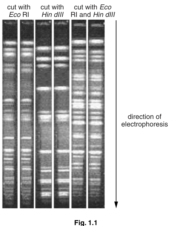
Fig. 1.1 shows the results of the electrophoresis.
The students concluded that the DNA of peas had more restriction sites for Eco RI than for Hin dIII and that some of the Hin dIII sites were within fragments produced by Eco RI.
State one piece of evidence in Fig. 1.1 to support each of these conclusions:
there are more restriction sites for Eco RI than for Hin dIII
there are some Eco RI sites within fragments produced by Hin dIII.
Answer
More Eco RI sites than Hin dIII sites:
The Eco RI lane (lane 1) has more fragments (bands) than the Hin dIII lane (lane 2) — i.e. more, and shorter, fragments from Eco RI than from Hin dIII.
Eco RI sites within fragments produced by Hin dIII:
Some of the bands visible in the single-enzyme lanes (1 and 2) are absent from the double-digest lane (3), and the double-digest lane has additional (shorter) fragments / many more fragments than either single-enzyme digest.
More Eco RI sites: more / shorter fragments in lane 1 than in lane 2. Some Eco RI sites within Hin dIII fragments: some single-enzyme bands are absent in the double-digest lane and the double digest has more / shorter fragments.
Background Concept
Restriction enzymes cut DNA at specific short sequences (recognition sites). The number of fragments produced by a digest is determined by the number of times the enzyme cuts: if the enzyme makes n cuts, the DNA is broken into n + 1 fragments (for circular DNA it is exactly n fragments, but pea chromosomal DNA is essentially linear here). Therefore, more cuts = more fragments = generally smaller fragments. If two enzymes are used together, the result is the intersection of the two single-enzyme digests: any site cut by either enzyme will be cut in the double digest, and any fragment that contains a site for the second enzyme will be cut further.
Understanding the Question
The students have concluded two things from Fig. 1.1:
- The pea DNA has more restriction sites for Eco RI than for Hin dIII.
- Some of the Hin dIII sites lie within fragments produced by Eco RI (i.e. some Eco RI fragments contain Hin dIII sites that further cut them in the double digest).
The question asks for one piece of evidence in Fig. 1.1 to support each conclusion. The mark scheme gives any one of two possibilities for each, so any one of the points below will score.
Approach
Read off the three lanes of the gel carefully. The arrow on the right indicates the direction of electrophoresis — bands further from the wells (closer to the bottom) are smaller fragments. Compare the band patterns of the three lanes and translate the visual differences into statements about enzyme sites.
Step-by-Step Reasoning
For "more Eco RI sites than Hin dIII sites":
- A restriction enzyme produces a separate band for each distinct fragment size.
- Lane 1 (Eco RI) shows more bands than lane 2 (Hin dIII), and several of the Eco RI bands are smaller (further down the gel) than any of the Hin dIII bands.
- Because more bands = more cuts = more restriction sites, this shows that Eco RI has cut the DNA at more sites than Hin dIII has.
- The mark scheme accepts either "the total number of fragments from Eco RI is greater" or "there are more shorter fragments for Eco RI".
For "some Eco RI sites within fragments produced by Hin dIII":
- In the double digest (lane 3), Eco RI cuts at all of its sites, and Hin dIII also cuts at all of its sites. Therefore any Hin dIII site that lies inside an Eco RI fragment will further cut that fragment into smaller pieces.
- Lane 3 shows bands that are not present in lane 1, and bands that are not present in lane 2 — these are new fragments produced only when both enzymes are used together.
- It also shows that some bands present in lane 1 alone, or in lane 2 alone, are absent in lane 3 — those fragments have been further cut by the other enzyme.
- The combination of "missing bands in lane 3" and "new (shorter) bands in lane 3" is evidence that the enzymes cut inside each other's fragments.
- The mark scheme accepts either "some fragments in the single-enzyme digests are not present in the double digest" or "the double digest has many more (shorter) fragments".
Key Takeaways
- Number of cuts = number of fragments − 1 (for a linear DNA molecule).
- A double digest produces all the cuts of both single digests, so new bands appear and some single-digest bands disappear.
- A band that disappears in the double digest is evidence that one enzyme has cut inside a fragment produced by the other.
Common Mistakes
- Saying "Hin dIII cuts more" because its lane has a band that looks brighter: brightness depends on how many DNA molecules are in that fragment, not on the number of sites.
- Stating only that the double digest has more bands without explaining that this means some sites are inside other fragments.
- Saying "Eco RI sites are within Hin dIII sites": recognition sites are short DNA sequences, not nested within one another; the question is about sites within the fragments produced.
- Saying "some bands disappear" without specifying where: the mark scheme requires that the disappearance is between the single-enzyme lanes and the double-digest lane.
Things to Be Careful About
- Quote the evidence as a comparison between specific lanes (e.g. "lane 1 vs lane 2" or "single vs double digest").
- A simple count of bands is the simplest piece of evidence and is explicitly accepted by the mark scheme.
Suggest one reason why the results in Fig. 1.1 may not support the student’s conclusions.
Answer
The new / additional (short) bands in the double-digest lane could be due to Hin dIII sites within Eco RI fragments or to Eco RI sites within Hin dIII fragments — without further work (e.g. a Southern blot with a known probe, or sequence data) it is not possible to tell which is the case. (Equivalently: the bands in the double-digest lane are faint and / or run close together, so they are hard to distinguish from one another.)
The new bands in the double digest could be due to Hin dIII sites within Eco RI fragments or Eco RI sites within Hin dIII fragments — the data alone cannot distinguish between the two possibilities (or the bands are too faint / too close to read reliably).
Background Concept
A restriction-digest gel shows the sizes of the fragments produced, but it does not directly show which recognition site was cut to produce each fragment. When two enzymes are used together, the resulting fragments are a mixture of cuts by both enzymes, and from band positions alone it is generally not possible to say which enzyme cut which site. To attribute a particular cut to a particular enzyme, additional information is needed (e.g. sequence data, a Southern blot with a labelled probe for a known region, or a different gel / electrophoresis condition that resolves the bands further).
Understanding the Question
The students have concluded that some Hin dIII sites lie within Eco RI fragments (i.e. Hin dIII is cutting inside fragments that Eco RI produced). The question asks for one reason why the results in Fig. 1.1 may not support this conclusion. The point is to identify an ambiguity in the data, not to repeat the evidence that does support the conclusion.
Approach
Think about what the data do not tell us. The double digest contains fragments cut by both enzymes, but the band pattern alone cannot tell us which enzyme was responsible for any given cut. The mark scheme accepts two main lines of criticism:
- The new / extra bands in the double digest could be due to Hin dIII cutting inside Eco RI fragments, OR to Eco RI cutting inside Hin dIII fragments — the gel data alone cannot distinguish the two.
- The bands are faint, blurred or close together and so the exact pattern is hard to read.
Step-by-Step Reasoning
- The students' conclusion depends on identifying that some new bands in lane 3 (the double digest) are the result of Hin dIII sites that were inside the Eco RI fragments in lane 1.
- However, the symmetric statement is also possible: those new bands could be the result of Eco RI sites that were inside the Hin dIII fragments in lane 2.
- Looking at the gel alone, there is no way to tell which interpretation is correct. The conclusion therefore goes beyond what the data show.
- A second, separate issue: the bands in the photograph are not all crisp and well separated. Some are faint or close together, so it is difficult to be sure exactly how many bands there are in any one lane. Any count-based argument is therefore tentative.
- Either of these two criticisms is enough to score the mark.
Key Takeaways
- A gel shows fragment sizes but not which enzyme cut which site.
- A double-digest band pattern is therefore ambiguous: it is consistent with more than one assignment of cuts to enzymes.
- To resolve such an ambiguity, further information is needed (sequence data, a Southern blot, etc.).
Common Mistakes
- Saying "the experiment needs to be repeated": the mark scheme explicitly ignores suggestions of more repeats or replicates as a criticism of this conclusion.
- Saying "the bands are too small to see": the issue is which band is which, not how small they are.
- Restating the evidence for the conclusion rather than identifying a reason against it.
- Saying "the students didn't use a control": irrelevant to this conclusion.
Things to Be Careful About
- The criticism is about what the data can and cannot tell us, not about experimental technique.
- The mark scheme accepts the two lines of argument above, but rejects any reference to "insufficient data" or "needs more repeats".
Gel electrophoresis is also used in the sequencing of DNA.
A method used for sequencing DNA is described below.
• A DNA molecule is first heated so it separates into two strands.
• DNA polymerase is then used to synthesise DNA using normal nucleotides and modified nucleotides.
• The four different modified nucleotides are each labelled with a different coloured fluorescent dye.
• These are: red CTP, green TTP, blue GTP, yellow ATP
• The addition of modified nucleotides to the DNA strand stops the addition of any more nucleotides.
Fig. 1.2 shows the formation of a three nucleotide fragment from normal nucleotides and a modified nucleotide.
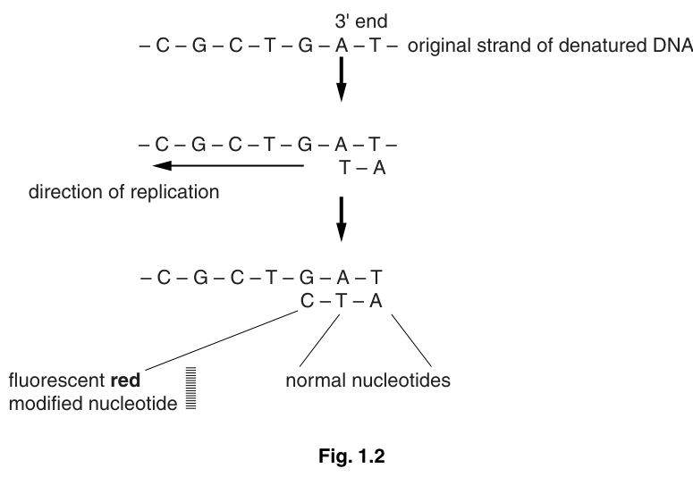
• Fragments of different lengths are produced depending on the position of the modified nucleotides.
• The DNA fragments synthesised are separated by gel electrophoresis and a laser light used to identify the different colours.
Fig. 1.3 shows the results of sequencing part of a DNA molecule.
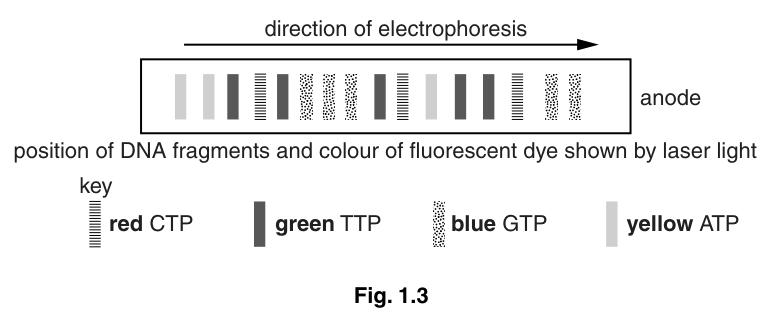
Use the information in Fig. 1.3 to work out the sequence of the DNA template, starting from the 3' end.
Answer
(3') CCGAATGACCCAGATT (5')
Background Concept
Sanger (chain-termination) sequencing works as follows. The DNA to be sequenced is denatured and used as a template. DNA polymerase synthesises a new strand in the 5'→3' direction, using normal deoxynucleotides (dATP, dTTP, dGTP, dCTP) plus a small proportion of modified (dideoxy) nucleotides that are each labelled with a different coloured fluorescent dye. The key property of the modified nucleotide is that, once it is incorporated, no further nucleotide can be added — synthesis terminates. This generates a set of fragments, each ending with a labelled modified nucleotide that reports the identity of the base at that position. The fragments are separated by gel (or capillary) electrophoresis; smaller fragments travel further towards the anode, so the shortest fragments are at the bottom of the gel / furthest from the wells. A laser reads the colour of the band as each fragment passes a detector, and the order of the colours (read from shortest fragment to longest) gives the sequence of the newly synthesised strand from 5' to 3'. The original template is then deduced by complementary base pairing — and because DNA strands are antiparallel, the template's 3' end corresponds to the 5' end of the synthesised strand (i.e. to the shortest fragment).
Understanding the Question
The question shows a sequencing gel (Fig. 1.3) with the direction of electrophoresis towards the anode (right) and asks the student to work out the template sequence, starting from the 3' end. This requires several linked steps: reading the colours from the band closest to the anode (the shortest fragment) towards the wells (the longest fragment); translating each colour into a base of the newly synthesised strand; then inverting and complementing to obtain the template. The polarity must be written explicitly (3' at the start, 5' at the end).
Approach
- Identify the shortest fragment (the band closest to the anode — i.e. the rightmost band in Fig. 1.3). That band tells us the first nucleotide added by the polymerase, which is complementary to the 3' end of the template.
- Use the key to convert each band's colour to a base: red = C, green = T, blue = G, yellow = A.
- Read the bands from the rightmost (shortest fragment) to the leftmost (longest fragment). The sequence of colours, in that order, is the synthesised strand in the 5'→3' direction.
- Because the new strand and the template are antiparallel and complementary, the 3' end of the template corresponds to the 5' end of the new strand (the shortest fragment), and the template sequence is the complement of the new strand, read in the opposite direction.
- Apply the base-pairing rules: C pairs with G, T pairs with A.
Step-by-Step Reasoning
Working through Fig. 1.3 from the right (anode end, shortest fragment) to the left (wells, longest fragment), the bands correspond (in 5'→3' order on the synthesised strand) to bases that, when complemented and reversed, give the template sequence:
- Shortest fragment (rightmost) → corresponds to the 3' end of the template.
- Reading the bands, applying the key, complementing and reversing yields the template read 3' to 5'.
- The full template, starting at the 3' end as the question requests, is:
Key Takeaways
- In Sanger sequencing, the shortest fragment is read first; it tells you the base at the 3' end of the template.
- The new strand is antiparallel to the template, so the template's 3' end is at the same end as the new strand's 5' end (the shortest fragment).
- The colour key converts each band's appearance into a base identity.
Common Mistakes
- Writing the new strand sequence instead of the template sequence: the question specifically asks for the template.
- Forgetting the polarity markers: the question asks for the sequence starting from the 3' end, and the 5' end must be marked at the other end.
- Reading the gel from left to right (longest to shortest) without reversing the order: this gives the sequence from 3'→5' of the new strand, not the template.
- Confusing the colour key (e.g. reading red as A instead of C): the key in Fig. 1.3 must be used exactly.
Things to Be Careful About
- The answer must explicitly show (3') at the start and (5') at the end.
- Read the gel from the anode end (right) towards the wells (left), then complement and reverse to obtain the template.
Explain how you worked out this sequence.
Answer
- The shortest fragment (the band closest to the anode) is read first; this is where the start of the sequence is located, and it corresponds to the 3′ end of the template.
- The colour of each band identifies the modified nucleotide that terminated that fragment; using the key (red C, green T, blue G, yellow A) gives the sequence of the newly synthesised strand.
- The new strand and the template pair by complementary base pairing (A–T, C–G).
- The two strands of DNA are antiparallel, so the template sequence is read in the opposite direction to the synthesised strand — giving the template from 3′ to 5′.
Read from the shortest fragment; identify bases by colour; apply complementary base pairing; remember that the template is antiparallel to the synthesised strand.
Background Concept
Sequencing by synthesis (Sanger / chain-termination) relies on four principles:
- DNA polymerase adds nucleotides in the 5'→3' direction of the new strand.
- Modified (dideoxy) nucleotides terminate synthesis when they are incorporated. Each is labelled with a different fluorescent dye, so the colour of the band = the identity of the base at the 3' end of that fragment.
- The two strands of DNA are antiparallel and complementary (A with T, C with G), so the template sequence is the reverse complement of the synthesised strand.
- Gel (or capillary) electrophoresis separates by size: smaller fragments travel further towards the anode. Therefore, the shortest fragment in the gel corresponds to the position closest to the 5' end of the new strand — and to the 3' end of the template.
Understanding the Question
The previous part (e)(i) gave the template sequence. The question now asks the student to explain how the sequence was worked out. The mark scheme gives any three of four specific points for three marks, so the explanation must cover at least three of the four principles above.
Approach
Lay out the logic in the order it is applied: (1) where do you start reading the gel; (2) how do you turn a band into a base; (3) how do you turn a list of new-strand bases into a template; (4) why you have to reverse the order (antiparallel strands).
Step-by-Step Reasoning
- Where to start reading. The shortest fragment is the one that has migrated furthest from the wells — i.e. the band closest to the anode (the right-hand end of Fig. 1.3). This fragment was the first one terminated and corresponds to the 5' end of the new strand, which is opposite the 3' end of the template. This is the "start" of the sequence.
- Identifying each base. Each band on the gel corresponds to a fragment whose last nucleotide is a modified (fluorescently labelled) nucleotide. The colour of the band, read with the key, tells you the identity of that terminal base. Reading the bands in order from shortest to longest therefore gives the sequence of the new strand, from its 5' end to its 3' end.
- Complementary base pairing. DNA polymerase makes the new strand by complementary base pairing with the template. A in the new strand means T in the template, T in the new strand means A in the template, C means G, and G means C. Applying these pair rules to the new strand gives the template sequence.
- Antiparallel strands. The two strands of DNA run in opposite directions: if the new strand runs 5'→3', the template runs 3'→5'. This means the template sequence is the reverse complement of the new strand — i.e. the order of the bases must be reversed as well as complemented. The question asks for the template starting at the 3' end, which is why the answer in (e)(i) is written 3'→5'.
Key Takeaways
- Start reading at the shortest fragment (closest to the anode); this corresponds to the 5' end of the new strand and the 3' end of the template.
- Band colour = base identity of the terminal modified nucleotide.
- The template is the reverse complement of the new strand (complementary base pairing + antiparallel orientation).
Common Mistakes
- Stating only that the new strand is complementary to the template without mentioning the antiparallel nature: this gives a wrong direction for the template.
- Saying "the colour tells you the base" without explaining that the colour identifies the modified nucleotide that terminated the fragment (the colour is not on every nucleotide, only on the terminating one).
- Saying "the shortest fragment corresponds to the 5' end of the template": incorrect — the shortest fragment corresponds to the 3' end of the template.
- Not making a link between the position of the start and the 3' end of the original / template DNA: the mark scheme explicitly requires this link for the first marking point.
Things to Be Careful About
- The four points are independent; the mark scheme accepts any three of the four.
- The word "complementary" alone is not enough — make the link to base pairing (A–T, C–G).
- The word "antiparallel" is required (or an equivalent explanation, e.g. "the template is read in the opposite direction to the fragment").
The rest of this paper
1 more questions- Q2Planning · Analysis, Conclusions and Evaluation11M