Biology 5090/22 — October/November 2025
Cambridge O-Level · Theory · worked solutions for every part, with the mark scheme
Topics Coordination and Control · Transport in Flowering Plants · Inheritance · Disease and Immunity · Coordination and Response in Plants · Classification · +9 more
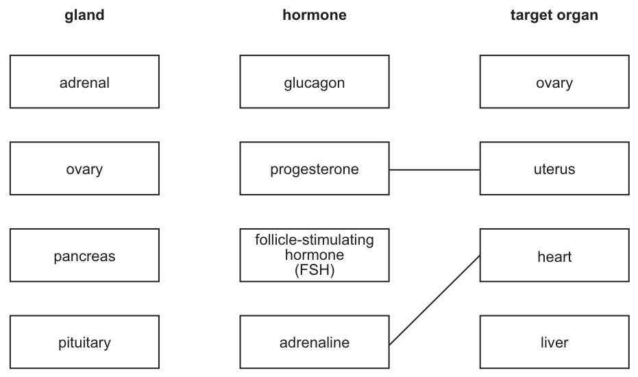
Hormones are produced by glands in the body. They alter the activity of specific target organs.
Draw lines to link:
- each gland to a hormone it produces
- each hormone to a specific target organ.
Two lines have been drawn for you. Draw six more lines.
Answer
The six correct lines to draw are:
- adrenal gland → adrenaline → heart
- ovary → progesterone → uterus
- pancreas → glucagon → liver
- pituitary → follicle-stimulating hormone (FSH) → ovary
This can be summarised in the following completed table:
| gland | hormone | target organ |
|---|---|---|
| adrenal | adrenaline | heart |
| ovary | progesterone | uterus |
| pancreas | glucagon | liver |
| pituitary | follicle-stimulating hormone (FSH) | ovary |
(Note: The links progesterone → uterus and adrenaline → heart were already given in the question.)
See working
Walkthrough
The question asks us to match four glands to the hormones they produce, and then to the target organs those hormones act on. Two connections are already provided: progesterone acts on the uterus, and adrenaline acts on the heart. We need to draw the remaining six lines to complete the chains.
- Adrenal gland: Produces adrenaline, which acts on the heart (and other organs like the liver and lungs, but heart is the only option here). This link is given as adrenaline → heart, so we draw adrenal → adrenaline.
- Ovary: Produces progesterone (and oestrogen), which acts on the uterus to prepare it for pregnancy. This link is given as progesterone → uterus, so we draw ovary → progesterone.
- Pancreas: Produces glucagon (and insulin), which acts on the liver to stimulate the breakdown of glycogen into glucose. We draw pancreas → glucagon, and glucagon → liver.
- Pituitary gland: Produces follicle-stimulating hormone (FSH), which acts on the ovary to stimulate the development of follicles. We draw pituitary → FSH, and FSH → ovary.
Key Takeaways
- Endocrine glands produce specific hormones that travel in the blood to target organs.
- The adrenal gland produces adrenaline; the ovary produces progesterone; the pancreas produces glucagon; the pituitary produces FSH.
- Target organs are specific: adrenaline acts on the heart, progesterone on the uterus, glucagon on the liver, and FSH on the ovary.
Common Mistakes
- Matching the pancreas to insulin instead of glucagon (insulin is not in the list).
- Matching the pituitary to growth hormone instead of FSH (growth hormone is not in the list).
- Matching FSH to the uterus instead of the ovary.
- Drawing extra lines from any box, which the mark scheme explicitly states will negate the mark for that box.
Things to Be Careful About
- The mark scheme awards marks arithmetically for correct lines on the left side of the diagram, but any additional lines to or from a box negate that mark. Only draw the six required lines.
- Ensure you draw exactly six lines, as the question asks. The two given lines (progesterone → uterus and adrenaline → heart) should not be redrawn.
- Use the exact terms from the boxes (e.g., "follicle-stimulating hormone (FSH)", not just "FSH" if writing it out).
Name a chemical that is produced in a plant shoot to coordinate the shoot's response to gravity.
______
Answer
auxin
auxin
Walkthrough
The question asks for the name of the chemical (hormone) produced in a plant shoot that coordinates its response to gravity. This response is called gravitropism (or geotropism). In shoots, the hormone auxin is produced in the tip of the shoot and moves downwards. When the shoot is placed horizontally, auxin accumulates on the lower side, causing cells on that side to elongate more than cells on the upper side, resulting in the shoot growing upwards (negative gravitropism).
Key Takeaways
- Auxin is the plant hormone responsible for coordinating growth responses to external stimuli like gravity and light.
- In shoots, auxin causes cell elongation on the side where it accumulates, leading to bending away from gravity (negative gravitropism) or towards light (positive phototropism).
Common Mistakes
- Naming other plant hormones like gibberellin, ethene (ethylene), or cytokinin.
- Confusing the response of shoots (negative gravitropism, growing up) with roots (positive gravitropism, growing down), though the hormone is still auxin in both cases.
- Spelling "auxin" incorrectly.
Things to Be Careful About
- The question specifically asks for a chemical produced in a plant shoot. Ensure the answer is a plant hormone, not an animal hormone.
- The exact term required is auxin. Other terms like "growth hormone" or "plant growth regulator" will not score.
A constant supply of water is essential for both animals and plants.
Fig. 2.1 is a diagram showing the average daily water intake and average daily water loss for an adult human.
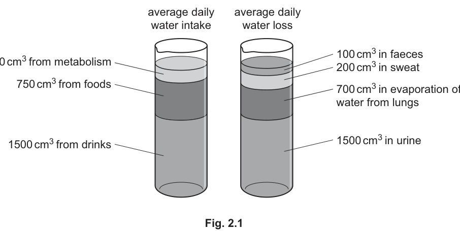
Calculate the percentage of average daily water intake that is lost in sweat from an adult human.
______ %
Working
Answer
8 %
8 %
Walkthrough
The figure gives three sources of water intake: 250 cm³ from metabolism, 750 cm³ from foods and 1500 cm³ from drinks. Add them to get the total daily intake of 2500 cm³. The question asks what percentage of this intake is lost in sweat, which is given as 200 cm³. Divide 200 by 2500 to get 0.08, then multiply by 100 to express it as a percentage: 8 %.
Key Takeaways
- Always find the total of the whole quantity before calculating a percentage.
- Percentage = (part ÷ total) × 100.
- Read all the component values carefully off a stacked diagram before adding them.
Common Mistakes
- Dividing by only one component (e.g. 200/1500) instead of the total intake — you must add all three sources first.
- Forgetting to multiply by 100, leaving the answer as 0.08.
- Confusing water loss in sweat (200 cm³) with another loss category such as urine (1500 cm³).
Things to Be Careful About
- The mark scheme awards one mark for the correct working ( or ) and one for the final answer, so show your substitution even if you can do it mentally.
- The answer line ends in %, so your final answer must be a percentage, not a fraction or decimal.
Answer
Sweating is part of homeostasis: it helps maintain a constant body temperature. When body temperature rises, e.g. during increased activity or in a hot environment, more sweat is produced; the evaporation of sweat from the skin removes heat energy, cooling the body. Also, if the volume lost by other routes (urine, faeces, lungs) changes, the percentage lost as sweat changes too.
See working
Walkthrough
The average figures in Fig. 2.1 are just that — averages. On any particular day the percentage lost as sweat varies. The main reason is thermoregulation: humans maintain a constant core temperature by negative feedback (homeostasis). When you exercise or the environment is hot, the body makes more sweat so that its evaporation carries away more heat energy; when cool or at rest, less sweat is made. A second valid reason is simply that the other routes of loss (urine, faeces, evaporation from the lungs) also vary day to day, so even if sweat volume stayed constant, its percentage of total loss would change.
Key Takeaways
- Sweating is a homeostatic mechanism for cooling the body by evaporation.
- Evaporation removes heat energy from the skin surface.
- More activity or higher environmental temperature → more sweat (and the reverse, ora).
- Percentages change if either the part or the total changes.
Common Mistakes
- Writing 'to get rid of excess water' as the purpose of sweating — the mark scheme wants the link to temperature control, not just water balance.
- Saying 'sweat cools the body' without mentioning evaporation — the cooling happens because evaporation takes heat energy away.
- Giving only an observation ('it changes') without any reason — an 'Explain why' needs because-clauses.
Things to Be Careful About
- This is marked 'max three' from five listed points, so give three distinct points and stop; padding with restatements earns nothing extra.
- Use the word homeostasis — it is a named marking point.
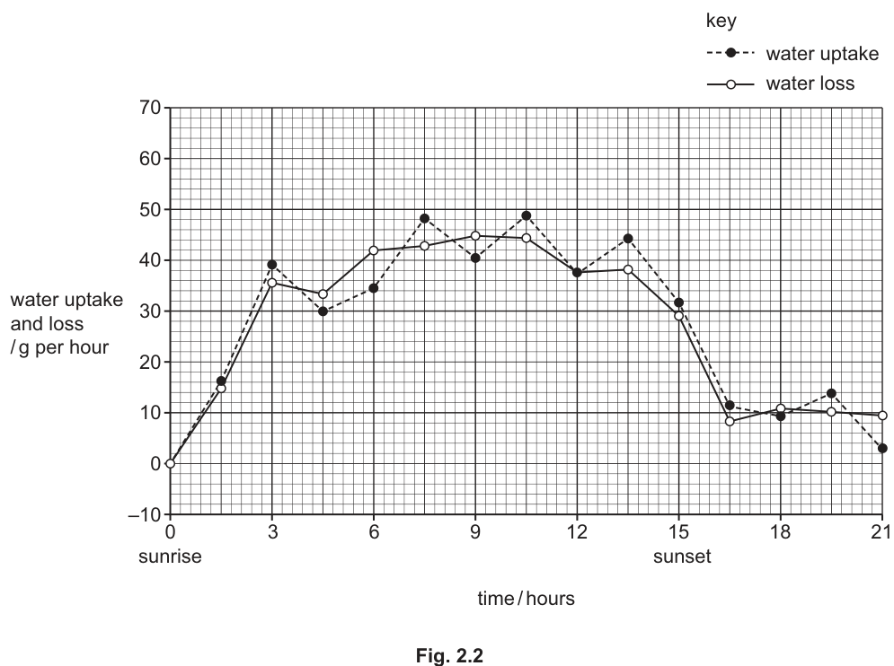
A scientist measured the mass of water taken up and lost by a plant over 21 hours. The plant was kept at a constant temperature of on a windowsill in a laboratory. Readings were taken every 90 minutes.
At 0 hours it was sunrise and at 15 hours it was sunset.
The results are shown in Fig. 2.2.
Plants lose water by evaporation from their leaves.
State the name of this process.
______
Answer
transpiration
transpiration
Walkthrough
Loss of water vapour by evaporation from the leaves of a plant is called transpiration. It occurs mainly through the stomata, which are pores on the underside of the leaf.
Key Takeaways
- Transpiration = loss of water vapour from leaves, mostly via the stomata.
Common Mistakes
- Writing 'evaporation' alone — that describes the physical process but not the named plant process the mark scheme requires.
- Writing 'condensation' or 'guttation'.
Things to Be Careful About
- Spell the word correctly: transpiration, not transperation or transportation.
Explain the shape of the curve for water loss from the plant over the 21 hours of the experiment.
Answer
During daylight hours (0–15 hours, between sunrise and sunset) the stomata are open, so water vapour diffuses out rapidly and water loss is high. After sunset (15–21 hours) it is dark, the stomata close, and water loss falls to a low level. Greater light intensity during the day also increases the rate of evaporation / diffusion of water from the leaves.
See working
Walkthrough
The graph shows water loss rising steeply after sunrise, staying high through the middle of the day, then falling sharply after sunset. The key variable here is light. In daylight the guard cells photosynthesise and the stomata open, giving a large surface for water vapour to diffuse out of the leaf, so transpiration is fast. In darkness the stomata close, cutting off most water loss, which matches the low flat section after hour 15. Higher light intensity during the day also warms the leaf and increases evaporation inside it, further raising the rate.
Key Takeaways
- Stomata open in light and close in darkness.
- Transpiration rate follows light intensity through the day.
- Water vapour diffuses out through open stomata down a water potential gradient.
Common Mistakes
- Describing the shape of the curve ('it goes up then down') without explaining it — an 'Explain' question needs reasons tied to stomata and light.
- Attributing the pattern to temperature alone; the experiment was at constant temperature, so light/stomata is the intended explanation.
- Ignoring the sunrise/sunset markers printed on the graph, which are there to anchor your explanation.
Things to Be Careful About
- Each credited point pairs an observation with its cause joined by '+': e.g. decreased water loss + dark + stomata close. Give both halves.
- Max two marks — two well-linked points are enough.
Explain why the shape of the curve for water loss is very similar to the shape of the curve for water uptake.
Answer
Water evaporates from the leaves (transpiration), which lowers the water potential of the leaf cells. Water therefore moves up through the xylem vessels from the roots to replace the water lost — the transpiration pull / transpiration stream. Forces of attraction between the water molecules help hold the column of water together as it is pulled up, so uptake always matches loss and the plant does not lose turgidity / wilt.
See working
Walkthrough
Why do the two curves track each other so closely? Because uptake is driven by loss. When water evaporates from the mesophyll cells and diffuses out of the stomata, the water content — and hence the water potential — of the leaf cells falls. Water then moves from the xylem into these cells, and to replace it, water is drawn up the continuous columns of water in the xylem vessels from the roots. This is the transpiration pull, or transpiration stream. The forces of attraction (cohesion) between neighbouring water molecules keep each column unbroken as it is pulled upwards. Because replacement keeps pace with loss, the plant stays turgid and does not wilt, and the uptake curve mirrors the loss curve throughout the day.
Key Takeaways
- Transpiration creates a water potential gradient from roots to leaves.
- Water moves up the xylem by transpiration pull.
- Attraction between water molecules maintains the unbroken water column.
- Uptake matches loss, keeping cells turgid.
Common Mistakes
- Saying water is 'sucked up' or 'pushed up' without naming the transpiration stream/pull.
- Omitting the term water potential — the mark scheme credits 'reduces water potential of leaves', not just 'leaf gets drier'.
- Saying water moves by active transport up the xylem — it is a passive process driven by evaporation.
Things to Be Careful About
- Max three from six listed points; give three clearly separated points.
- The exact terms rewarded are: water potential, xylem vessels, transpiration pull/stream, attraction between water molecules, turgidity/wilting.
The scientist repeated the experiment. All conditions were kept the same except the temperature of the laboratory, which was lower.
On Fig. 2.2, draw a new curve to show the expected results for water loss at the lower temperature.
Answer
Draw a curve with the same shape as the original water-loss curve (rising after sunrise, peaking mid-day, falling after sunset) but positioned below it, since a lower temperature reduces the rate of evaporation and so reduces water loss at every point.
Same-shaped curve drawn below the original water-loss curve
Walkthrough
Temperature affects how fast water molecules evaporate. At lower, the water molecules have less kinetic energy, so evaporation from the mesophyll surfaces and diffusion out of the stomata are both slower. Light still opens and closes the stomata on the same schedule, so the daily pattern — rise after sunrise, peak around midday, fall after sunset — is unchanged. Only the magnitude drops, so the new curve sits below the original one across the whole 21 hours.
Key Takeaways
- Lower temperature → lower rate of evaporation → lower transpiration rate.
- Changing one factor changes the size of the response, not necessarily its pattern.
Common Mistakes
- Drawing a completely flat line — the stomata still respond to light, so the daily rhythm remains.
- Drawing the new curve above the original, implying cold increases water loss.
- Changing the shape (e.g. shifting the peak to a different time) — the timing depends on light, which is unchanged.
Things to Be Careful About
- One mark only: the essential features are the same shape and a position below the original curve. Label the new curve clearly so the examiner can identify it.
Azolla is a genus of small ferns that float in slow-flowing or stationary fresh water.
Azolla has cavities in its leaves that contain nitrogen-fixing bacteria called Anabaena azollae.
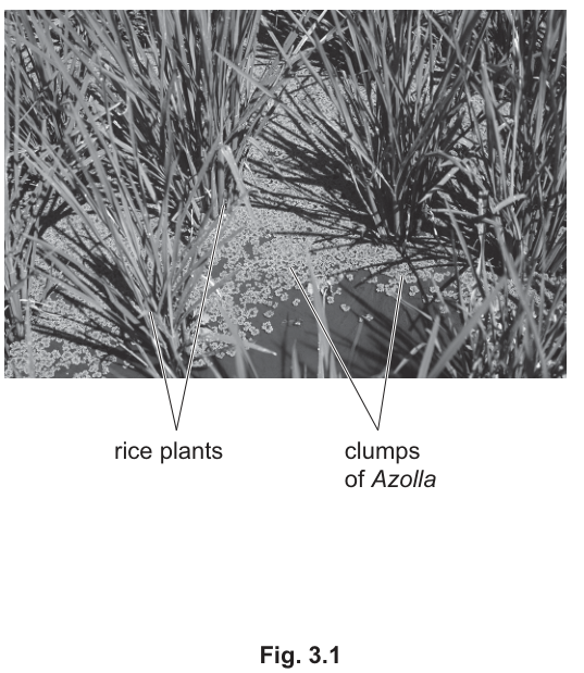
In some parts of the world, farmers grow Azolla in their rice fields. Rice fields are flooded with water so that rice plants grow half submerged.
Fig. 3.1 shows rice and Azolla growing together in a flooded field.
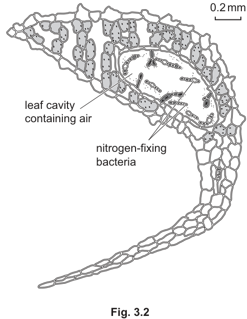
Fig. 3.2 shows a section through an Azolla leaf.
Azolla is a fern and rice is a flowering plant.
Ferns and flowering plants are two groups in the plant kingdom.
Using information from Fig. 3.1 and Fig. 3.2, state two structural features used to classify Azolla in the plant kingdom.
- ______
- ______
Answer
- Has chloroplasts (visible as shaded structures in the leaf cells of Fig. 3.2).
- Has cell walls (around every cell in Fig. 3.2).
chloroplasts; cell walls (also accepted: multicellular)
Walkthrough
The question asks for features that place Azolla in the plant kingdom, and it insists you use the figures. In Fig. 3.2 the leaf cells contain shaded bodies — these are chloroplasts — and every cell is surrounded by a distinct wall, which is a cellulose cell wall. Both are defining plant features. The scheme also accepts 'multicellular', since Azolla is clearly made of many cells. Any two of the three score.
Key Takeaways
Plant kingdom features: cellulose cell walls, chloroplasts, multicellular, and (for ferns and flowering plants) reproduction by spores or seeds.
Common Mistakes
Writing 'has leaves' or 'is green' — these are not kingdom-level features. Naming 'nitrogen-fixing bacteria' — that is a feature of the bacteria, not of the plant. Giving only one feature when two are asked for.
Things to Be Careful About
The question says 'using information from Fig. 3.1 and Fig. 3.2', so anchor each feature to something visible: chloroplasts as the shaded bodies in the cells, cell walls as the outlines around each cell. Give exactly two points.
Complete the sentences below.
Rice plants reproduce sexually and after pollination they produce ______ that are then dispersed.
Azolla reproduces sexually by producing ______ .
Answer
Rice plants reproduce sexually and after pollination they produce seeds that are then dispersed.
Azolla reproduces sexually by producing spores.
seeds; spores
Walkthrough
This gap-fill tests the key division within the plant kingdom: flowering plants (like rice) are seed-producing plants, while ferns (like Azolla) reproduce by spores. The scheme also accepts 'fruits' for rice and 'gametes' for Azolla, but the order matters — seeds/fruits must go with rice and spores/gametes with Azolla.
Key Takeaways
Ferns produce spores; flowering plants produce seeds (and fruits). This is a classic classification distinction.
Common Mistakes
Swapping the answers — putting 'spores' for rice and 'seeds' for Azolla scores zero because the scheme states 'must be in correct order'.
Things to Be Careful About
Both blanks must be correct for the single mark, and each must be in its correct sentence.
Farmers grow Azolla in rice fields to increase the rice yield.
Suggest why the rice yield increases.
Answer
- The nitrogen-fixing bacteria in the Azolla leaves fix nitrogen, producing nitrates.
- The nitrates enter the water / soil of the flooded field.
- The rice plants absorb the nitrates through their roots.
- The nitrates are used to make amino acids and proteins.
- The proteins are used for growth, so the rice plants grow larger and the yield increases.
See working — nitrogen fixation supplies nitrates, absorbed by rice, used to make proteins for growth
Walkthrough
This is a 'suggest' question, so you must build the biology chain yourself. The stem tells you Azolla leaves contain nitrogen-fixing bacteria. Nitrogen fixation converts nitrogen gas into nitrogen compounds — nitrates. Because the field is flooded, the nitrates dissolve and spread into the water and soil where the rice roots are. Rice absorbs nitrate ions (by active transport into root cells) and uses them to make amino acids, which are joined into proteins. Proteins are needed for growth, so bigger, healthier rice plants produce a higher yield.
Key Takeaways
Nitrates are needed by plants to make amino acids and proteins, which are used for growth. Nitrogen-fixing bacteria convert atmospheric nitrogen into usable nitrogen compounds. A 'suggest why' answer needs the full causal chain, not just the first step.
Common Mistakes
Stopping at 'the bacteria fix nitrogen' without saying the nitrates reach the rice. Saying rice 'absorbs nitrogen' instead of nitrates. Saying nitrates 'give energy' — nitrates make proteins, not energy. Omitting the link to growth, which is what 'yield' means.
Things to Be Careful About
The scheme gives 5 possible points for 3 marks, so any three clear links score — but the strongest answers include the protein/growth link, because that is what actually increases yield.
Azolla can grow very quickly. It can remove as much as of carbon from the atmosphere.
Answer
- Carbon dioxide diffuses into the leaf through the stomata.
- Carbon dioxide and water are used in photosynthesis, driven by light energy trapped by chlorophyll:
- The carbon from the carbon dioxide becomes part of the glucose formed.
- The glucose is converted to starch and stored, keeping the carbon in the plant.
See working — carbon dioxide enters through stomata and is fixed into glucose by photosynthesis, then stored as starch
Walkthrough
To remove carbon from the atmosphere, Azolla photosynthesises. Carbon dioxide enters the leaf through stomata by diffusion, down a concentration gradient. Inside the chloroplasts, carbon dioxide and water react using light energy absorbed by chlorophyll to make glucose; the carbon atom of the carbon dioxide is now part of an organic molecule. The glucose is converted to starch for storage, so the carbon stays locked in the plant tissue — that is the 1.5 kg per square metre per year being removed.
Key Takeaways
Photosynthesis is the only major route by which carbon dioxide is removed from the atmosphere by living organisms. The word equation and the roles of stomata, chlorophyll and starch storage are all examinable.
Common Mistakes
Saying carbon dioxide 'is breathed in' — plants do not breathe. Omitting water as a raw material. Saying glucose is 'used for respiration' and so returned to the air — the question asks how carbon is removed, so storage as starch is the credited end point.
Things to Be Careful About
The scheme notes a correct balanced symbol equation scores 2 of the 4 marks (the carbon dioxide + water point and the glucose point), so writing the equation is efficient — but adding the stomata and starch points secures all four.
Answer
- Removing carbon dioxide decreases the amount of this greenhouse gas in the atmosphere.
- This reduces the greenhouse effect, so less heat energy is trapped in the atmosphere.
- This reduces global warming and climate change (for example, reduced melting of ice caps and rising sea levels).
See working — less carbon dioxide means a reduced greenhouse effect, less global warming and climate change
Walkthrough
Carbon dioxide is a greenhouse gas: it absorbs and re-radiates heat energy that would otherwise escape from the atmosphere, warming the Earth. If photosynthesis removes carbon dioxide, the concentration of this greenhouse gas falls, the greenhouse effect is reduced, less heat is trapped, and global warming slows. Slower warming means less climate change — the scheme also credits a named example such as reduced sea-level rise or extreme weather.
Key Takeaways
Carbon dioxide is a greenhouse gas; the greenhouse effect traps heat; enhanced greenhouse effect causes global warming and climate change. Removing carbon dioxide reverses the chain.
Common Mistakes
Saying carbon dioxide 'damages the ozone layer' — that is a different problem caused by CFCs. Listing consequences without the word 'reduce/decrease' — the scheme requires the response to mention reduction at least once for marks 2, 3 and 4.
Things to Be Careful About
Each mark needs the reduction idea attached: reduces greenhouse gas, reduces heat trapped, reduces climate change. A bare list of climate-change effects without 'reduced' will not score.
Different species of Azolla are found naturally in many countries. Azolla has also been introduced by humans to some countries where it is not native. When Azolla is introduced to a country, it can have an impact on natural fresh water ecosystems.
Suggest how Azolla can have an impact on these fresh water ecosystems.
Answer
- Azolla grows very quickly and competes with native aquatic plants for resources such as light, space and mineral ions.
- Having no natural consumers or pathogens in the new country, its population grows unchecked.
- It can cover the water surface, blocking light so submerged plants die, and its decay plus the nitrates it releases cause eutrophication, reducing biodiversity / causing loss of species.
See working — rapid growth, competition for light/space, no natural consumers, eutrophication and reduced biodiversity
Walkthrough
An introduced species often thrives because nothing in the new ecosystem controls it: no natural consumers (herbivores) or pathogens feed on it. Azolla grows very quickly and can blanket the water surface, competing with native plants for light, space and mineral ions. When it dies and decomposes, bacteria use up oxygen, and the nitrates/ammonium it releases fertilise the water, causing eutrophication and algal or plant overgrowth. The overall effect is a reduction in biodiversity as native species are lost.
Key Takeaways
Introduced (non-native) species can become invasive because they lack natural predators and competitors; their effects include competition, eutrophication and loss of biodiversity.
Common Mistakes
Saying 'Azolla kills the rice' — the question is about natural fresh water ecosystems, not the rice field. Naming 'competition' without a named resource — the scheme requires the resource (light, space, minerals) for that mark. Confusing eutrophication with simple fertilisation of crops.
Things to Be Careful About
The scheme lists seven possible points for three marks, so pick the clearest three. If you use the competition point, name the resource competed for; if you use the eutrophication point, link the increased nitrates to increased plant growth.
Lipases are enzymes produced by animals, plants and microorganisms. Lipases from different species can have different chemical structures.
Answer
A catalyst speeds up a chemical reaction and is not used up or changed in the process.
Speeds up chemical reactions; is not changed or used up.
Walkthrough
The question asks for the definition of a catalyst. A catalyst is a substance that increases the rate of a chemical reaction without being consumed or permanently altered in the process. In biology, enzymes act as catalysts to allow metabolic reactions to occur at rates compatible with life.
Key Takeaways
- A catalyst lowers the activation energy of a reaction, speeding it up.
- A catalyst is not used up or permanently changed during the reaction; it can be reused.
Common Mistakes
- Stating that a catalyst "provides energy" for the reaction (it does not; it lowers the activation energy).
- Saying a catalyst is "used up" or "changes" during the reaction.
Things to Be Careful About
- Ensure both parts of the definition are included: speeding up the reaction AND not being changed/used up. Missing either half will cost a mark.
Lipase is a digestive enzyme in humans.
Answer
pancreas
pancreas
Walkthrough
Lipase is an enzyme that breaks down lipids. In humans, it is secreted into the duodenum (the first part of the small intestine) by the pancreas. The pancreas also secretes other digestive enzymes such as amylase and proteases.
Key Takeaways
- The pancreas is a major accessory organ of the digestive system, producing alkaline fluid and digestive enzymes.
- Lipase specifically acts on lipids in the duodenum.
Common Mistakes
- Naming the liver (which produces bile, not lipase).
- Naming the stomach (which produces pepsin, not lipase).
Things to Be Careful About
- The question specifically asks for the organ that secretes lipase into the duodenum. The pancreas is the correct answer. Intestinal glands also secrete some lipase, but the pancreas is the primary source expected at this level.
Answer
substrate = lipid (or fat / oil)
products = fatty acids + glycerol
substrate: lipid; products: fatty acids and glycerol
Walkthrough
Lipase is an enzyme that hydrolyses lipids. The substrate it acts on is a lipid (commonly referred to as fat or oil). The hydrolysis reaction breaks the ester bonds in triglycerides, producing fatty acids and glycerol as the end products.
Key Takeaways
- Lipase substrate: lipid / fat / oil.
- Lipase products: fatty acids + glycerol.
Common Mistakes
- Naming the wrong substrate (e.g., starch for amylase, or proteins for protease).
- Naming incorrect products (e.g., glucose for amylase, or amino acids for protease).
- Forgetting to pluralise "fatty acids".
Things to Be Careful About
- Ensure the terms match the mark scheme: "lipid / fat / oil" for substrate and "fatty acid(s) + glycerol" for products. "Glycerol" is sometimes accepted as "glycerin", but "glycerol" is the standard biological term.
Some scientists collected species of bacteria from three different sources, A, B and C.
The scientists investigated how temperature affected the activity of the lipase produced by the bacteria collected from the three different sources. They wanted to find a source of lipase that could be used in biological washing powders.
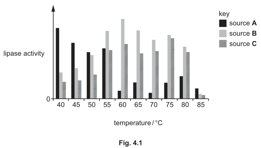
Fig. 4.1 shows the results.
Answer
- Lipases from sources A, B and C are all active between 40 and 85 °C.
- Each source has a different optimum temperature: A at 40 °C, B at 60 °C, and C at 75 °C.
- Source B is most suitable for biological washing powders as it has the highest activity at 60 °C, which is a typical washing temperature (or source A is most suitable for low-temperature washing).
See working
Walkthrough
The bar chart shows lipase activity (y-axis) against temperature (x-axis) for three bacterial sources: A (black), B (light grey), and C (medium grey).
- Range of activity: All three lipases show activity across the tested range, from 40 °C to 85 °C. Activity drops to near zero at the extremes.
- Optimum temperatures: The peak activity for each source occurs at a different temperature. Source A peaks at 40 °C, source B peaks at 60 °C, and source C peaks at 75 °C.
- Suitability: Biological washing powders typically operate at moderate to high temperatures (around 60 °C) to effectively clean clothes. Source B has the highest activity at 60 °C, making it the most suitable for this application. Alternatively, source A could be suitable for low-temperature washing powders.
Key Takeaways
- Enzymes from different organisms can have different optimum temperatures based on their natural habitat.
- When selecting an enzyme for an industrial application, consider the operating temperature of that application.
Common Mistakes
- Failing to state the range of temperatures over which activity is observed.
- Not identifying the specific optimum temperatures for each source.
- Making a judgment about suitability without linking it to the activity at a relevant temperature (e.g., stating B is best without mentioning 60 °C or washing temperatures).
Things to Be Careful About
- Read the graph carefully: the x-axis starts at 40 °C, not 0 °C.
- The mark scheme awards marks for naming the optimum temperatures. Naming all three correctly can earn 2 marks for this point alone.
- Ensure the discussion covers both the description of the results and the application to biological washing powders.
The lipase molecules produced by different species of bacteria have different chemical structures.
Explain:
- what determines the different chemical structures of lipase molecules
- why having different chemical structures can affect their activity at different temperatures.
Answer
- Different species have different DNA / sequences of bases in their genes.
- This codes for different sequences of amino acids in the lipase proteins.
- The amino acid sequence determines the 3D shape (or active site shape) of the enzyme.
- Different shapes mean the active sites denature at different temperatures (some are more stable at higher temperatures than others).
See working
Walkthrough
This question has two parts: what determines the chemical structure, and why does this affect temperature activity.
- What determines the structure: The chemical structure of a protein (like lipase) is determined by its amino acid sequence. The amino acid sequence is coded for by the sequence of bases in DNA. Different species of bacteria have different DNA sequences in the genes that code for lipase.
- Why this affects activity at different temperatures: The sequence of amino acids determines how the protein folds into its 3D shape, including the shape of the active site. The 3D shape is maintained by various bonds (e.g., hydrogen bonds, ionic bonds). Different amino acid sequences will have different arrangements of these bonds, making the protein more or less stable at different temperatures. Therefore, some lipases will denature (lose their shape and function) at lower temperatures, while others with more stable structures can remain active at higher temperatures.
Key Takeaways
- DNA -> RNA -> protein (amino acid sequence).
- Amino acid sequence determines protein folding and 3D shape.
- Protein shape (especially the active site) is essential for enzyme function.
- Denaturation is the loss of 3D shape due to the breaking of bonds maintaining the structure, which occurs at different temperatures for different proteins.
Common Mistakes
- Stating that DNA determines the shape directly without mentioning amino acids.
- Saying enzymes "melt" or "break down" instead of "denature".
- Failing to link the different structures to different stability or denaturation temperatures.
Things to Be Careful About
- The mark scheme requires a logical chain: DNA/sequence of bases -> sequence of amino acids -> shape/function -> denaturation at different temperatures.
- Ensure both parts of the question are answered: what determines the structure, and why it affects activity at different temperatures.
A sample of human blood is put in a small tube. The tube is placed in a centrifuge, which spins it around very fast, to separate the components of the blood. The denser components of the blood collect at the bottom of the tube.
Fig. 5.1 shows the composition of this blood sample.
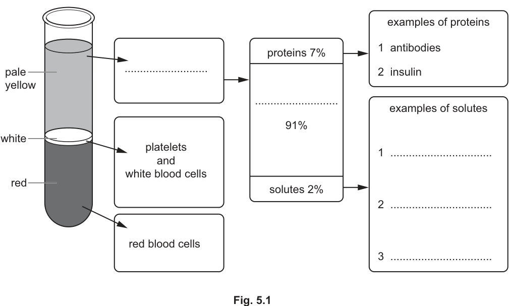
Complete Fig. 5.1 by filling in the names of five components shown by the blank spaces.
Answer
- Main top component: plasma
- 91% component of plasma: water
- Examples of solutes (any three from):
- glucose
- amino acids
- urea
plasma; water; glucose; amino acids; urea
Walkthrough
When whole blood is centrifuged, it separates by density into three distinct layers:
- The topmost pale yellow liquid layer is plasma.
- The thin middle buffy coat contains white blood cells and platelets.
- The bottom dense red layer contains red blood cells.
Looking at the breakdown of plasma:
- About of plasma is water.
- About consists of dissolved plasma proteins (such as antibodies, fibrinogen, and hormones like insulin).
- About consists of dissolved solutes transported around the body. Valid examples of these solutes include nutrients (such as glucose, amino acids, fatty acids, glycerol, vitamins), waste products of metabolism (such as urea), and mineral ions (such as sodium, potassium, chloride, or calcium).
Key Takeaways
- Blood consists of plasma () and formed cellular elements ().
- Plasma is predominantly water () containing proteins () and dissolved transport solutes ().
Common Mistakes
- Confusing proteins with small solutes (e.g. listing insulin or antibodies as solutes under the section).
- Giving cellular components (e.g. red blood cells) inside the plasma breakdown.
Things to Be Careful About
- Ensure you provide exactly three distinct solutes for the section as requested by the blank lines.
Answer
Any two from:
- Red blood cells are densely packed with haemoglobin.
- Haemoglobin contains iron (a dense metal).
- Red blood cells contain less water than white blood cells.
Red blood cells are densely packed with haemoglobin, which contains iron.
Walkthrough
In a centrifuge, denser components sink to the bottom. Red blood cells collect at the bottom because they have a higher physical density than white blood cells. This is due to:
- Haemoglobin content: Red blood cells lack a nucleus and most organelles, allowing them to be densely packed with the protein haemoglobin.
- Iron content: Each haemoglobin molecule contains haem groups with iron atoms, which are relatively heavy/dense.
- Water content: Red blood cells contain a higher concentration of dense solids/solutes and less water compared to white blood cells.
Key Takeaways
- The absence of a nucleus allows red blood cells to pack maximum haemoglobin.
- Iron-rich haemoglobin increases the overall density of the red blood cell compared to other cells.
Common Mistakes
- Stating simply that red blood cells are "heavier" without explaining why (i.e. omitting haemoglobin, iron, or packing).
Things to Be Careful About
- Make sure to give two distinct reasons (e.g. naming haemoglobin/iron and packing/water content).
The ABO blood groups in humans are determined by three alleles, , and .
There are four blood groups (phenotypes) but six different genotypes for the ABO blood groups.
Complete Table 5.1 to match the blood groups to their possible genotypes.
Table 5.1
| blood group (phenotype) | possible genotype(s) |
|---|---|
| A | |
| B | |
| AB | |
| O |
Answer
| blood group (phenotype) | possible genotype(s) |
|---|---|
| A | and |
| B | and |
| AB | |
| O |
A: IA IA and IA IO; B: IB IB and IB IO; AB: IA IB; O: IO IO
Walkthrough
The ABO blood group system is controlled by a single gene with three alleles:
- : codes for antigen A (dominant to , codominant with )
- : codes for antigen B (dominant to , codominant with )
- : recessive allele (codes for no antigen)
Matching the phenotypes to genotypes:
- Group A: Individuals can be homozygous () or heterozygous (). Both must be stated for the mark.
- Group B: Individuals can be homozygous () or heterozygous (). Both must be stated for the mark.
- Group AB: Codominance means both antigens are expressed, giving genotype .
- Group O: The recessive phenotype requires two recessive alleles, giving genotype .
Key Takeaways
- Alleles and are codominant with each other and dominant over .
- A dominant phenotype (A or B) can arise from two different genotypes (homozygous dominant or heterozygous).
Common Mistakes
- Omitting the heterozygous genotype ( or ) for blood groups A and B.
- Writing alleles without proper superscripts (e.g. "IA", "IB", "IO").
Things to Be Careful About
- Always use the standard genetic notation , , as specified in the syllabus.
A pregnant woman has blood group O.
The fetus in the woman's uterus developed from one of her fertilised eggs and has blood group B.
Answer
IB IO
Walkthrough
- The mother has blood group O, so her genotype must be .
- Consequently, all her egg cells carry only the allele.
- The fetus develops from one of her eggs and has blood group B (phenotype B).
- To be blood group B, the fetus must have received an allele from the father and an allele from the mother.
- Therefore, the genotype of the fetus must be .
Key Takeaways
- An offspring of a blood group O mother must inherit an allele from her.
- Therefore, an offspring with blood group B can only be heterozygous ().
Common Mistakes
- Writing , which is impossible because the mother can only pass on .
Things to Be Careful About
- Keep the base letter with the superscript and .
The woman and the fetus have different blood groups so they have different antigens on their red blood cell membranes.
The woman has antibodies to the antigens on the red blood cell membranes of the fetus.
Explain why the woman's antibodies are not usually able to come into contact with the antigens of the fetus.
Answer
Any three from:
- Maternal and fetal blood streams are separate / do not mix.
- The placenta acts as a barrier.
- Maternal anti-A and anti-B antibodies are too large to pass across the placenta / through capillary walls into the fetal circulation.
Maternal and fetal blood systems are kept separate by the placenta, so maternal antibodies cannot cross the placental barrier/capillary walls.
Walkthrough
- Separation of blood systems: In the uterus, the mother's blood and the fetal blood do not mix directly. They are kept separated by the placental membrane and capillary walls.
- Placental barrier: The placenta allows the diffusion of small molecules (such as oxygen, glucose, amino acids, and urea) between maternal and fetal blood, but acts as a barrier preventing direct contact between red blood cells.
- Antibody size/permeability: Naturally occurring anti-A and anti-B antibodies (which are large IgM pentamers) cannot readily diffuse across the placental barrier into the fetal blood vessels, protecting the fetal red blood cells from agglutination.
Key Takeaways
- Maternal and fetal blood circulations remain completely separate to protect the fetus from maternal blood pressure and immunological attack.
- Exchange occurs across thin placental membranes via diffusion without direct mixing of blood cells.
Common Mistakes
- Claiming that the mother and fetus share the same blood circulation.
- Forgetting to mention the placenta as the structure maintaining this separation.
Things to Be Careful About
- Clearly identify the placenta and the fact that the two blood supplies do not mix directly.
Cholera is a disease caused by the pathogen Vibrio cholerae.
Outbreaks of cholera often occur after natural disasters such as earthquakes.
Answer
bacterium
bacterium
Walkthrough
Vibrio cholerae is a prokaryote — a single-celled organism with no nucleus, a cell wall and a curved rod shape. In the 5090 scheme of classification it belongs to the kingdom of bacteria, so the type of microorganism is a bacterium. One word earns the mark; no reasoning is needed.
Key Takeaways
- Vibrio cholerae is a bacterial pathogen.
- Bacteria are prokaryotes: no true nucleus, a cell wall, and genetic material loose in the cytoplasm.
Common Mistakes
- Writing "virus" — viruses are non-cellular and much smaller; Vibrio is a living cell.
- Writing "pathogen" — that describes its role, not its type of microorganism.
Things to Be Careful About
- The mark scheme accepts "bacterium" or "bacteria" — either singular or plural scores. Give the precise term, not "germ" or "microbe".
Suggest and explain reasons why outbreaks of cholera are more likely after natural disasters.
Answer
- Cholera is transmitted through drinking water contaminated with Vibrio cholerae.
- After a natural disaster, sanitation breaks down: sewage removal is disrupted, so water supplies become contaminated.
- Difficulties with waste disposal, personal hygiene and food preparation allow the bacteria to spread more easily.
See working
Walkthrough
This is a "suggest" question: the answer is not pure recall but the application of what you know about cholera transmission to the disaster scenario.
Cholera is a waterborne disease — the bacteria leave the body in faeces and enter a new host when that faecal matter contaminates drinking water. So the question becomes: why does an earthquake make faecal contamination of water more likely?
- Water pipes and sewage systems are damaged, so clean water and dirty sewage mix — water supplies are contaminated.
- Sewage treatment and removal stop working, so faeces are not safely disposed of.
- People displaced from their homes lose access to clean facilities, so waste disposal, hand-washing (personal hygiene) and food preparation all become difficult, and each is a route by which the bacteria reach the mouth.
Any three of these linked ideas score the three marks. The key link is: disaster → sanitation fails → faeces contaminate drinking water → cholera spreads.
Key Takeaways
- Cholera is transmitted by drinking water contaminated with faeces containing Vibrio cholerae.
- Anything that disrupts sanitation — damaged sewers, no clean water, poor hygiene — increases transmission.
- "Suggest" questions are answered by applying a known principle to the new situation, not by recalling a list.
Common Mistakes
- Writing only "people are injured" or "buildings collapse" — these describe the disaster, not the route of transmission of the pathogen.
- Saying cholera spreads "through the air" — it is waterborne/foodborne, not airborne.
- Giving one idea three times in different words — the three marks need three distinct points.
Things to Be Careful About
- Each mark is a separate point: transmission route, contaminated water/sanitation, and hygiene/waste disposal. Make each point explicitly rather than writing one long sentence.
An infection with Vibrio cholerae can cause severe diarrhoea. This can be treated by giving the patient a solution to drink.
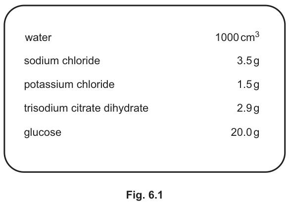
Fig. 6.1 shows the ingredients of a standard solution.
Answer
- The bacteria produce a toxin.
- The toxin stimulates the release of chloride ions into the small intestine.
- Water therefore passes into the intestine, from high to low water potential.
- This movement of water is by osmosis, making the faeces watery (diarrhoea).
See working
Walkthrough
The question asks you to explain the mechanism of diarrhoea in cholera, and the mark scheme rewards a precise chain of cause and effect:
- Toxin. The bacteria themselves do not physically damage the gut lining — they secrete a toxin (called choleragen). Naming the toxin is the first mark.
- Chloride ions released. The toxin causes cells lining the small intestine to secrete chloride ions () into the lumen of the intestine. This is the second mark.
- Water follows. The chloride ions lower the water potential of the fluid inside the intestine. Water now moves from high to low water potential — from the body tissues and blood, across the gut wall, into the intestine. Stating the direction in terms of water potential is a mark in its own right.
- Osmosis and watery faeces. Movement of water down a water potential gradient through a partially permeable membrane is osmosis — the exact term earns the mark. The result is a large volume of water in the gut, so the faeces are watery: this is the diarrhoea.
Notice how the biology hangs together: ions first, water second. Water always moves passively in response to the water potential gradient that the ions create.
Key Takeaways
- Cholera toxin causes intestinal cells to secrete chloride ions into the gut.
- Ions in the gut lower the water potential there, so water moves in by osmosis from high to low water potential.
- The loss of water in the faeces is the diarrhoea, and it can cause severe dehydration.
Common Mistakes
- Saying the bacteria "eat away" the gut lining — the damage is chemical, caused by the toxin.
- Writing "water moves by diffusion" — the movement of water down a water potential gradient is osmosis; the mark scheme requires that word.
- Saying water moves "from low to high concentration" — 5090 wants the argument in terms of water potential, from high to low.
- Omitting the ions and jumping straight to water — without the chloride ions there is no gradient, and the explanation loses its logic.
Things to Be Careful About
- The mark scheme is "max four from" six listed points, so four distinct points are needed. "From high to low water potential" and "osmosis" are separate marks — give both, using both exact terms.
Using Fig. 6.1, suggest why giving the patient this solution to drink is an effective treatment for diarrhoea.
Answer
- The solution contains water, so it replaces the water lost in the diarrhoea and rehydrates the patient.
- It contains salts (sodium chloride, potassium chloride, trisodium citrate), so it replaces the ions lost from the intestine.
See working
Walkthrough
Part (b)(i) established that cholera causes the loss of water and ions from the body. Treatment must therefore replace both. Read Fig. 6.1 with that in mind:
- Water, 1000 cm³ — replaces the water lost in the watery faeces, rehydrating the patient. This is the first mark.
- Sodium chloride, potassium chloride, trisodium citrate dihydrate — these are all salts, so they replace the chloride and other ions secreted into the intestine. This is the second mark.
The glucose in the solution also helps: glucose is absorbed with the ions and improves uptake of sodium, but the mark scheme only asks for the water and the ions, so those two points are what you must give.
Key Takeaways
- Oral rehydration therapy works because it replaces exactly what cholera removes: water and ions.
- Always link the treatment back to the mechanism of the disease — the ingredients of the solution mirror the losses.
Common Mistakes
- Saying "it gives the patient energy from the glucose" — the question is about treating diarrhoea, so the credited points are water and ions.
- Naming the salts without saying they replace ions — the link to the losses in (b)(i) is the mark.
- Writing "it cures the cholera" — the solution treats the dehydration; it does not kill the bacteria.
Things to Be Careful About
- Two marks, two distinct points: water/rehydration and ions. Use the word "replaces" — it makes the link to the losses explicit and matches the mark scheme's wording.
The mammalian nervous system is essential for coordination and control.
It is an organ system that contains the eyes.
Explain the differences between organs, tissues and cells, using the eye as an example.
Answer
- A cell is the basic unit of life; in the eye the light receptor cells are the rods and cones.
- A tissue is a group of cells with similar structure and function; in the eye the retina (or cornea / iris / sclera) is a tissue.
- An organ is a group of different tissues working together to carry out a specific function; the eye is an organ.
Cell = basic unit of life (rods/cones in the eye); tissue = group of similar cells (retina); organ = group of tissues with a specific function (the eye).
Walkthrough
This question tests the hierarchy of biological organisation: cell → tissue → organ → organ system. Each level must be defined AND illustrated with a part of the eye, because each mark in the scheme comes as a pair: definition + eye example.
- The cell is the smallest basic unit of life. In the eye, the light receptor cells are the rods and cones in the retina — they detect light and convert it into electrical impulses.
- A tissue is a group of cells with a similar structure working together to do the same job. The retina is a tissue made of receptor cells; other valid examples are the cornea, iris, sclera or conjunctiva.
- An organ is made of several different tissues coordinated to perform one specific function. The eye combines nervous tissue (retina), muscle tissue (iris), connective/protective tissue (sclera, cornea) and so on, all serving the function of vision.
Notice the pattern: each level groups together units from the level below, and each gains a new collective function.
Key Takeaways
- Cell → tissue → organ → organ system is a hierarchy of increasing complexity.
- Every definition at this level pairs 'a group of …' with 'similar structure/function' (tissue) or 'different tissues with a specific function' (organ).
- Examiners award marks in linked pairs here: always attach a named eye example to each definition.
Common Mistakes
- Giving only the definitions without eye examples, or only examples without definitions — each mark needs both halves.
- Defining a tissue as 'any group of cells' without stating similar structure/function.
- Calling the optic nerve a tissue instead of an organ (it is accepted as an organ example in the scheme).
- Confusing organ with organ system — the eye is one organ within the nervous system.
Things to Be Careful About
- Use the word function in both the tissue and organ definitions — it is the marking point.
- Four marks means four credited points: pick three definitions plus their examples, presented clearly.
- Do not describe the eye's parts in detail (lens accommodation etc.) — that earns nothing here.
Synapses are found in the mammalian nervous system.
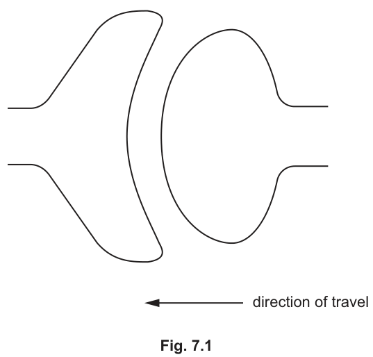
Explain how the structure of a synapse ensures that electrical impulses travel in one direction only.
You may use the outline of a synapse in Fig. 7.1 to illustrate your answer.
Answer
- Vesicles in the presynaptic neurone contain neurotransmitter.
- These vesicles are found only on one side of the synaptic gap (in Fig. 7.1, the neurone on the right).
- When an impulse arrives, the vesicles release neurotransmitter into the gap by diffusion.
- The neurotransmitter diffuses across the synaptic cleft.
- It binds to receptors (proteins) in the membrane of the postsynaptic neurone.
- These receptors are found only on the opposite side of the gap, so the impulse can only pass in one direction.
Neurotransmitter in vesicles occurs only on the presynaptic side and receptors only on the postsynaptic side, so impulses can travel in one direction only.
Walkthrough
The question asks WHY transmission across a synapse is one-way. The answer lies in an asymmetry: the two sides of the synapse are not built the same.
In Fig. 7.1 the arrow shows the impulse travelling from the neurone on the right (the presynaptic terminal bulb) towards the neurone on the left (postsynaptic membrane), across the synaptic cleft between them.
The chain of events:
- The terminal bulb of the presynaptic neurone contains vesicles filled with neurotransmitter, a signalling chemical.
- Crucially, these vesicles exist only on this one side of the gap.
- When an electrical impulse reaches the terminal bulb, the vesicles fuse with the membrane and release neurotransmitter into the synaptic cleft.
- The neurotransmitter molecules are small and diffuse quickly across the narrow cleft.
- On the far side, the postsynaptic membrane carries receptor proteins shaped to fit the neurotransmitter (lock-and-key). Binding triggers a new impulse in the postsynaptic neurone.
- These receptors occur only on the postsynaptic side.
So even if an impulse somehow arrived at the postsynaptic side, there would be no vesicles there to release transmitter, and no receptors on the presynaptic side to detect any — transmission physically cannot run backwards. That is the full explanation of unidirectionality.
Key Takeaways
- A synapse is a junction where an electrical impulse is converted into a chemical signal and back again.
- Directionality is explained by structural asymmetry: vesicles + neurotransmitter on one side, receptors on the other.
- Neurotransmitter crosses the cleft by diffusion — no energy is needed for the crossing itself.
- The lock-and-key idea applies: the receptor protein has a complementary shape to the neurotransmitter molecule.
Common Mistakes
- Saying the impulse 'jumps' the gap electrically — the crossing is chemical, by neurotransmitter diffusing.
- Naming the chemical but never explaining why the direction cannot reverse — the marks sit in 'vesicles only on one side' and 'receptors only on the other'.
- Writing 'neurotransmitter moves' instead of diffuses across the gap.
- Confusing synapse with neurone, or saying the whole neurone contains vesicles rather than just the terminal bulb.
- Describing the reflex arc instead of the synapse — off-topic.
Things to Be Careful About
- Six marks = six separate creditable points; write them as distinct statements covering vesicles, their one-sided position, release, diffusion, binding to receptors, and the one-sided position of receptors.
- The scheme underlines only twice — the words 'only on one side' and 'only on the other side' carry two of the six marks, so make the asymmetry explicit.
- If you use Fig. 7.1, orient your answer to match it: the bulb with vesicles is on the right, the receptors on the left membrane.
- Say 'binds to receptors/proteins', not 'attaches to the neurone'.