Biology 5090/21 — October/November 2025
Cambridge O-Level · Theory · worked solutions for every part, with the mark scheme
Topics Coordination and Control · Transport in Humans · Sexual Reproduction in Humans · Inheritance · Human Nutrition · Excretion · +8 more
Fig. 1.1 shows the female human reproductive system as viewed from the front.
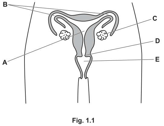
Identify the letter on Fig. 1.1 that has been used to label the area where each of the following happens:
where sperm is deposited ______
where oestrogen is produced ______
where implantation occurs. ______
Answer
- where sperm is deposited: E (vagina)
- where oestrogen is produced: A (ovary)
- where implantation occurs: C (uterus)
E; A; C
Walkthrough
Fig. 1.1 shows the female reproductive system from the front: A is an ovary, B is an oviduct, C is the uterus, D is the cervix and E is the vagina.
- Sperm are deposited in the vagina (E) during sexual intercourse.
- Oestrogen is a female sex hormone made by the ovaries (A) — the ovaries also produce eggs and progesterone.
- After fertilisation in the oviduct, the embryo travels to the uterus (C) and implants into its lining, where it develops.
Key Takeaways
- Know the positions and functions of ovary, oviduct, uterus, cervix and vagina.
- Oestrogen is produced by the ovaries; implantation happens in the uterus lining; sperm are deposited in the vagina.
Common Mistakes
- Writing the structure name when the question asks for the letter (or vice versa) — here the letter is required.
- Confusing the oviduct (B) with the uterus (C): fertilisation happens in the oviduct, but implantation happens in the uterus.
- Confusing the cervix (D) with the vagina (E).
Things to Be Careful About
- The question asks to identify the letter, so give E, A and C — naming the structure as well is fine but the letter is what scores.
- Keep the three answers in the order asked: deposition, oestrogen, implantation.
Some women can have blockages in the tubes labelled B.
Explain why this would prevent them becoming pregnant.
Answer
- The sperm cannot travel up the oviduct to meet the egg (which passes down the oviduct after ovulation).
- Therefore fertilisation cannot occur, so the woman cannot become pregnant.
Sperm cannot meet the egg, so fertilisation cannot occur.
Walkthrough
The oviduct (B) is the tube along which the egg travels from the ovary to the uterus, and it is where fertilisation normally happens: sperm deposited in the vagina swim up through the uterus into the oviduct and meet the egg there.
If the oviduct is blocked, the sperm and the egg physically cannot reach each other. Without the sperm nucleus and egg nucleus fusing, there is no fertilisation, no zygote, and therefore no pregnancy. This is exactly why IVF helps these women — fertilisation is carried out outside the body and the embryo is placed directly into the uterus, bypassing the blocked tubes.
Key Takeaways
- Fertilisation in humans occurs in the oviduct, not the uterus.
- A blocked oviduct prevents the gametes meeting, so fertilisation cannot happen.
Common Mistakes
- Saying only "the egg cannot get through" without linking to fertilisation — the mark sits in the because-clause.
- Saying the sperm "cannot reach the uterus" — the destination that matters is the egg in the oviduct.
- Confusing fertilisation with implantation; blocking the tube stops fertilisation first.
Things to Be Careful About
- This is an explain question: each point needs its reason. "Fertilisation cannot occur" alone is only half the answer — state that the sperm and egg cannot meet.
In vitro fertilisation (IVF) can be used to help women who have blocked tubes to get pregnant.
It involves several steps.
- step 1 The ovaries are stimulated to produce a large number of eggs.
- step 2 The eggs are removed from the ovaries before they are released naturally.
- step 3 The eggs are fertilised with sperm.
- step 4 The fertilised eggs are replaced into the uterus.
In step 1, the patient is injected with a hormone.
Name a hormone that could be used.
______
Answer
follicle-stimulating hormone (FSH)
follicle-stimulating hormone / FSH
Walkthrough
Step 1 of IVF needs the ovaries to produce many eggs at once. In the natural menstrual cycle, follicle-stimulating hormone (FSH), secreted by the pituitary gland, stimulates follicles in the ovary to mature and produce eggs. Injecting extra FSH makes several follicles mature simultaneously, so several eggs can be collected.
Key Takeaways
- FSH stimulates the maturation of eggs (follicles) in the ovary.
- LH triggers the release of the egg (ovulation) — that distinction is tested in the next part.
Common Mistakes
- Naming LH, which causes egg release, not egg production.
- Naming oestrogen or progesterone, which do not stimulate egg production.
Things to Be Careful About
- Give the full name or the accepted abbreviation FSH; "follicle stimulating hormone" without hyphens is accepted.
In step 2, a drug is used to stop the action of luteinising hormone (LH).
Explain why it is important to stop the action of LH.
Answer
- LH stimulates the release of an egg from the ovary (ovulation).
- If LH acted, the eggs would be released before they could be collected/removed, so they would be lost.
LH causes ovulation; the eggs would be released before they could be collected.
Walkthrough
Luteinising hormone (LH) is the hormone that triggers ovulation — the bursting of a mature follicle and the release of the egg from the ovary into the oviduct.
In IVF, step 2 requires the doctor to remove the eggs from the ovaries surgically, before they are released naturally. If LH were allowed to act, ovulation would happen at the wrong time: the eggs would be released into the oviduct before the collection procedure, and they would be lost (or fertilisation timing would be impossible to control). Blocking LH keeps the eggs in the ovaries until they can be collected.
Key Takeaways
- LH stimulates ovulation (egg release); FSH stimulates egg maturation.
- IVF needs eggs collected from the ovary, so natural ovulation must be prevented.
Common Mistakes
- Saying LH "stimulates egg production" — that is FSH; LH causes egg release.
- Giving only the role of LH without the consequence for the IVF procedure — both halves are needed for the two marks.
Things to Be Careful About
- This is an explain question: point 1 is the role of LH, point 2 is why stopping it matters for step 2 (the eggs can then be collected). Link the two.
When a man and a woman are unable to have children, they are said to be an infertile couple.
There are estimated to be 90 million infertile couples in the world.
There are many causes of infertility but 17% of cases are due to blocked tubes in women.
Calculate the number of couples in the world whose infertility is due to blocked tubes in women.
Give your answer in standard form.
number of couples = ______
Working
In standard form:
Answer
number of couples =
1.53 × 10^7
Walkthrough
17% of 90 million means 17 out of every 100 of the 90 000 000 infertile couples. Multiply 90 000 000 by 17/100 to get 15 300 000 couples.
Standard form writes a number as a value between 1 and 10 multiplied by a power of ten. Move the decimal point in 15 300 000 seven places to the left to get 1.53, so the answer is .
The mark scheme awards marks in stages: 15 300 000 alone scores 2 of the 3 marks; only the correct standard form earns all 3.
Key Takeaways
- "Percentage of a quantity" = percentage ÷ 100 × quantity.
- Standard form is where — so is not standard form.
Common Mistakes
- Leaving the answer as 15 300 000 — this loses a mark because the question demands standard form.
- Writing or — the first number must be between 1 and 10.
- Miscounting the power of ten (e.g. ).
Things to Be Careful About
- The blank asks for the number in standard form, so the final line must be , not the ordinary number.
- Show the working: the scheme gives credit for 1.53 or 15 300 000 in the working even if the final form is wrong.
Fig. 2.1 shows a section through the human eye.
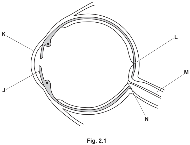
Identify the letter on Fig. 2.1 that has been used to label the part of the eye that:
contains circular and radial muscles ______
has the highest concentration of light receptors. ______
Answer
- Contains circular and radial muscles: J (iris)
- Highest concentration of light receptors: L (fovea / yellow spot)
J; L
Walkthrough
The question asks to identify two structures on the eye diagram based on their function. Label J points to the iris, which contains circular and radial muscles that control the size of the pupil. Label L points to the fovea (yellow spot), which is the region of the retina with the highest concentration of light receptors (cones) for sharp central vision.
Key Takeaways
Understanding the functions of the iris (muscle control of pupil size) and the fovea (high receptor density for detailed vision).
Common Mistakes
Confusing the iris with the ciliary body, or the fovea with the blind spot. Naming the letter alone without the biological name if required (though here just the letter is asked, it is good practice to know the name).
Things to Be Careful About
Ensure the letters match the exact structures asked for. J is the iris, L is the fovea. Read the diagram labels carefully as they can be placed near multiple structures.
Answer
See working
Walkthrough
The diagram omits the lens and suspensory ligaments. The lens is a biconvex structure located behind the iris and pupil. The suspensory ligaments (zonules of Zinn) connect the ciliary bodies (the small muscular structures on either side of the lens) to the equator of the lens. Drawing them requires placing the lens in the correct anatomical position and attaching the ligaments from the ciliary bodies to the lens edges.
Key Takeaways
The anatomical position of the lens and suspensory ligaments relative to the iris, ciliary body, and retina.
Common Mistakes
Drawing the lens too far forward or backward. Attaching the suspensory ligaments to the cornea or retina instead of the ciliary bodies. Using shading or colour.
Things to Be Careful About
Labels must be clear and horizontal. Lines must not cross the structures they label. The lens must be biconvex. Do not add shading, stippling, or ruled lines.
Answer
- Suspensory ligaments are attached to the ciliary bodies (or ciliary muscles).
- They can become slack (loose) or taut (tight).
- When they pull on the lens, they change the shape of the lens, which allows accommodation (focusing on objects at different distances).
See working
Walkthrough
The function of the suspensory ligaments is to hold the lens in place and modify its shape. They are anchored to the ciliary bodies. When the ciliary muscles contract or relax, the tension in the suspensory ligaments changes (taut or slack). This pulling force alters the curvature of the lens, a process called accommodation, which focuses light from objects at varying distances onto the retina.
Key Takeaways
The mechanical relationship between the ciliary body, suspensory ligaments, and lens in achieving accommodation.
Common Mistakes
Saying the ligaments 'move the lens' instead of 'change its shape'. Forgetting to mention the ciliary bodies. Not explaining the link between shape change and accommodation.
Things to Be Careful About
Give exactly three linked points. Use precise terms like 'taut', 'slack', 'ciliary bodies', and 'accommodation'. Avoid saying 'the muscles move the lens'—it is the ligaments that pull on the lens.
Some people have a genetic condition called cornea plana. This condition causes the cornea to be flatter than usual.
Answer
- A flatter cornea does not bend (refract) light enough.
- Therefore, light is not focused on the retina.
See working
Walkthrough
The cornea is the main refracting surface of the eye, bending incoming light to help focus it on the retina. If the cornea is abnormally flat (cornea plana), its curvature is reduced, meaning it cannot bend (refract) the light rays sufficiently. As a result, the focal point falls behind the retina, and the image is not clear.
Key Takeaways
The cornea's role in refracting light and how its curvature affects focusing.
Common Mistakes
Saying the cornea 'doesn't let light in' or 'blocks light'. Confusing the cornea with the lens. Not linking the lack of refraction to the failure to focus on the retina.
Things to Be Careful About
Use the terms 'refract' or 'bend' and 'focus on the retina'. Give both the cause (insufficient refraction) and the effect (not focused on the retina) for full marks.
One type of cornea plana is caused by a recessive allele.
Two people who do not have cornea plana have a child who has the condition.
Explain how this is possible.
Answer
- Both parents must be heterozygous (carriers), meaning they each carry one affected recessive allele but do not show the condition.
- Each parent passes the recessive allele to their child, resulting in a homozygous recessive genotype that expresses the condition.
See working
Walkthrough
Cornea plana is caused by a recessive allele. For a child to express a recessive condition, they must inherit two copies of the recessive allele (homozygous recessive). If both parents do not have the condition but have an affected child, both parents must be carriers (heterozygous). They each possess one dominant normal allele (masking the recessive one) and one recessive affected allele. During fertilisation, both parents can pass on their recessive allele to the same child.
Key Takeaways
How recessive traits can skip a generation and appear in offspring of unaffected parents through carrier status.
Common Mistakes
Saying the parents 'have the gene but don't show it' without using the term 'heterozygous' or 'carrier'. Not explaining that both parents must contribute the allele. Confusing recessive with dominant inheritance.
Things to Be Careful About
Use precise genetic terminology: 'heterozygous', 'carrier', 'recessive allele'. Explain that both parents pass the allele on. Do not draw a full Punnett square unless asked, but the reasoning must clearly show both parents contributing the recessive allele.
Fig. 3.1 shows a summary of protein metabolism in the body.
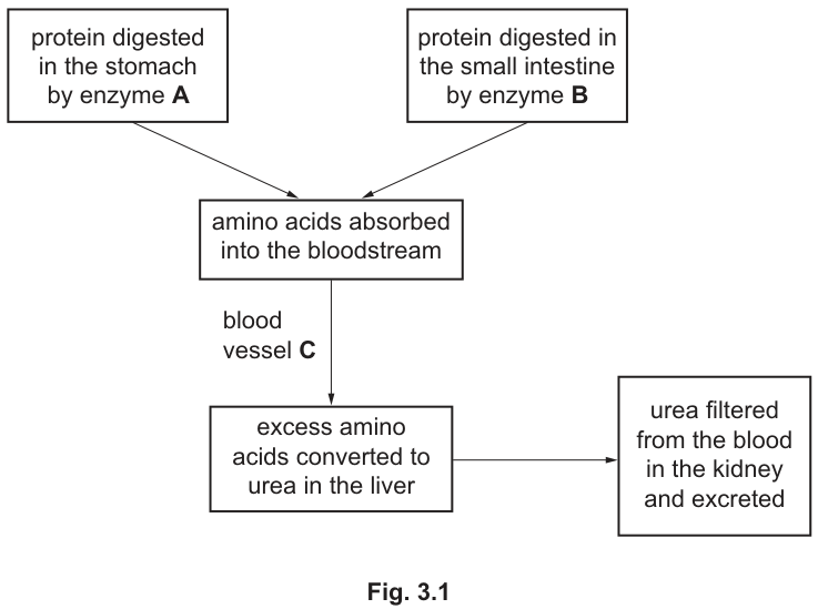
Use Fig. 3.1 to answer these questions.
Answer
enzyme A = pepsin
enzyme B = trypsin
enzyme A = pepsin; enzyme B = trypsin
Walkthrough
The flow chart tells you where each enzyme works, and that is all you need. Enzyme A digests protein in the stomach — the stomach's protease is pepsin, which works best in the acidic conditions produced by hydrochloric acid there. Enzyme B digests protein in the small intestine — that protease is trypsin, secreted by the pancreas into the small intestine where conditions are closer to neutral. Both enzymes break proteins down into amino acids, which are then absorbed.
Key Takeaways
- Pepsin = stomach protease; trypsin = small intestine (pancreatic) protease.
- Both catalyse protein → amino acids; they differ in location and optimum pH.
Common Mistakes
- Writing 'protease' alone — the question asks for the name of the enzyme, so a class name does not score.
- Swapping the two: pepsin belongs to the stomach, trypsin to the small intestine.
- Naming amylase or lipase — these digest starch and fats, not protein.
Things to Be Careful About
- Spell both names correctly: p-e-p-s-i-n and t-r-y-p-s-i-n.
- The mark scheme awards one mark per enzyme, so both blanks must be filled.
Answer
hepatic portal vein
hepatic portal vein
Walkthrough
Blood vessel C carries the absorbed amino acids from the gut (small intestine) to the liver, where excess amino acids are deaminated. The vessel that runs from the gut to the liver is the hepatic portal vein — 'hepatic' means relating to the liver, and 'portal' describes a vessel joining two capillary beds. It carries nutrient-rich blood straight from the intestines for processing before it reaches the general circulation.
Key Takeaways
- The hepatic portal vein transports absorbed food molecules (amino acids, glucose) from the small intestine directly to the liver.
Common Mistakes
- Writing 'hepatic artery' — the artery carries blood from the heart to the liver, not nutrients from the gut.
- Writing 'aorta' or 'vena cava' — these are general circulation vessels, not the gut-to-liver link.
- Confusing ureter with any blood vessel — the ureter carries urine, not blood.
Things to Be Careful About
- Use the full name 'hepatic portal vein'; 'portal vein' alone may not be credited as the precise term.
Answer
glomerulus
glomerulus
Walkthrough
Urea is removed from the blood at the start of each nephron. Blood enters a knot of capillaries called the glomerulus, held inside the cup-shaped Bowman's capsule. High blood pressure in the glomerulus forces water, urea, glucose and salts out of the blood through the capillary walls into the capsule — this is filtration. Useful substances are later reabsorbed, but urea stays in the tubule and leaves in the urine.
Key Takeaways
- The glomerulus is the capillary network where filtration of urea (and other small molecules) from the blood takes place.
Common Mistakes
- Naming the whole nephron, the Bowman's capsule, or the collecting duct — the question asks specifically for the capillary structures that filter.
- Writing 'kidney' or 'nephron' — too vague for this precise request.
Things to Be Careful About
- Plural is fine ('glomeruli') but the singular 'glomerulus' matches the scheme exactly.
Two groups of students, X and Y, investigate the effect of different diets on the concentration of urea in their urine.
For one day, the two groups eat different foods:
- group X eat bread, jam, orange juice, lettuce, fruit
- group Y eat nuts, milk, bread, cheese, meat.
Early the next morning, they collect and analyse their urine to measure the concentration of urea.
Table 3.1 shows their results.
Table 3.1
| concentration of urea/ per of urine | ||
|---|---|---|
| individual students | mean | |
| group X | 1.5, 1.7, 2.3, 1.6, 2.1, 1.4 | 1.8 |
| group Y | 1.8, 1.9, 2.3, 2.5, 1.9, 2.4 |
Calculate the mean concentration of urea for the students in group Y.
Give your answer to two significant figures.
mean concentration of urea = ______ per of urine
Working
Rounded to two significant figures:
Answer
mean concentration of urea = 2.1 per of urine
2.1 g per 100 cm³ of urine
Walkthrough
Add the six individual readings for group Y: . Divide by the number of students, six: . The question demands two significant figures, so keep only the first two digits: 2.1. Note that group X's printed mean of 1.8 confirms the method — their values also average to about 1.77, rounded to 1.8.
Key Takeaways
- Mean = sum of values ÷ number of values.
- Two significant figures means two digits counting from the first non-zero digit: 2.133… → 2.1.
Common Mistakes
- Dividing by 5 instead of 6 — count the readings carefully.
- Giving 2.13 (three significant figures) or 2 (one significant figure) — the rounding instruction is part of what is marked.
- Adding up wrongly; check the total against the scheme's accepted intermediate value of 12.8.
Things to Be Careful About
- Round only at the end, never during the calculation.
- Include the unit with the answer since the answer line names one.
Explain the difference in the mean concentration of urea for the two groups of students.
Answer
- Group Y ate more protein / ate protein-rich foods (nuts, milk, cheese, meat).
- So more amino acids were deaminated in the liver to produce urea.
- So more urea was filtered from the blood in the kidney and passed into the urine.
Group Y ate more protein, so more amino acids were deaminated to urea in the liver, so more urea was filtered into the urine.
Walkthrough
Compare the two diets. Group X ate bread, jam, orange juice, lettuce and fruit — almost entirely carbohydrate, with very little protein. Group Y ate nuts, milk, cheese and meat — all rich in protein. More protein eaten means more protein digested into amino acids and absorbed. The body cannot store excess amino acids, so the liver removes the nitrogen-containing amino group from them — deamination — converting them to carbohydrate and producing urea as the waste product. That urea travels in the blood to the kidneys, where the glomeruli filter it out into the urine. So group Y's urine contains more urea per : mean 2.1 compared with 1.8 g.
Each link in the chain earns a mark: the dietary difference, the deamination step naming urea, and the filtration into urine. You could equally argue the reverse for group X (ora), but the forward argument for group Y is the clearest.
Key Takeaways
- Excess amino acids cannot be stored; they are deaminated in the liver, yielding urea.
- Urea is transported in the blood plasma and excreted by the kidneys.
- A high-protein diet therefore raises the urea concentration of the urine.
Common Mistakes
- Saying only 'group Y ate more protein' without following the consequence through — the marks sit in the because-clauses.
- Confusing deamination with digestion: digestion breaks protein into amino acids in the gut; deamination happens later, in the liver.
- Saying urea is made in the kidney — it is made in the liver and only filtered in the kidney.
- Describing the numbers without explaining them — an 'explain' command word requires reasons.
Things to Be Careful About
- Use the exact terms 'deamination', 'liver' and 'filtered' — they carry the marks.
- Anchor the answer in the actual foods listed, not a generic statement about diets.
One of the students said: 'To make a fair comparison, we should have made sure that the students in each group drank the same volume of water'.
Explain why this student is correct.
Answer
- The volume of water drunk affects the volume of water used to form urine.
- This changes the concentration of urea in the urine — drinking more water dilutes the urea, giving a lower concentration even if the same mass of urea is excreted.
Water intake affects urine volume, which affects the concentration of urea in the urine.
Walkthrough
The students measured the concentration of urea — grams of urea per of urine. Concentration depends on two things: how much urea is excreted AND how much water it is dissolved in. If a student drinks a lot of water, the kidneys produce a larger volume of dilute urine, so the same amount of urea spread through more water gives a lower reading. A student who drank little would produce concentrated urine and a higher reading. So unless every student drank the same volume of water, differences in the results could be due to water intake rather than diet — the comparison would not be fair.
Key Takeaways
- In a fair test, all variables except the independent variable must be controlled.
- Urine concentration reflects both solute load and water balance, so water intake must be standardised when comparing urea concentrations.
Common Mistakes
- Stopping at 'water dilutes the urine' without stating that water intake changes the urine volume/concentration — both marking points need the link made explicit.
- Vague answers like 'to make it fair' with no reason — the scheme wants the mechanism.
- Suggesting water changes how much urea is produced — it changes the concentration by changing the volume, not the production rate.
Things to Be Careful About
- Give both halves: water intake → urine volume, and urine volume → urea concentration. Each is worth a mark.
Chlorophyll is found in all organisms that photosynthesise.
Complete these sentences about chlorophyll by writing suitable words in the gaps.
A chlorophyll molecule contains carbon, hydrogen, oxygen, nitrogen and an atom of ______ .
Chlorophyll molecules are found in plant cells in structures called ______ .
The type of leaf cells that contain the most chlorophyll are the ______ cells.
Chlorophyll molecules trap sunlight energy and this is used by plants to produce glucose.
Glucose can then be converted to ______ to be transported around the plant.
Glucose can also be converted to ______ to make cell walls.
Answer
- magnesium
- chloroplasts
- palisade (mesophyll)
- sucrose
- cellulose
magnesium; chloroplasts; palisade (mesophyll); sucrose; cellulose
Walkthrough
The question is a straightforward gap-fill testing five distinct facts about chlorophyll and the products of photosynthesis.
- Chlorophyll is a magnesium porphyrin, so the missing atom is magnesium.
- Chlorophyll is located inside the chloroplasts, the organelles where photosynthesis occurs.
- In a leaf cross-section, the palisade mesophyll cells are columnar and packed with the most chloroplasts, hence the most chlorophyll.
- Glucose produced in photosynthesis is converted to sucrose for transport through the phloem.
- Glucose is also polymerised into cellulose, the main structural component of plant cell walls.
Key Takeaways
- Chlorophyll requires magnesium to form its central ring structure.
- Chloroplasts are the specific organelles that house chlorophyll.
- Palisade mesophyll cells are the primary photosynthetic cells in a leaf.
- Sucrose is the main transport sugar in plants, while cellulose provides structural support.
Common Mistakes
- Writing "magnesium ion" instead of just "magnesium" (the question asks for the atom).
- Naming "chlorophyll" instead of "chloroplasts" for the second gap (chlorophyll is the pigment, chloroplasts are the structures).
- Writing "starch" instead of "sucrose" for transport (starch is for storage, sucrose is for transport).
- Writing "glucose" for cell walls (glucose is the monomer, cellulose is the polymer used in cell walls).
Things to Be Careful About
- Ensure exact spelling: palisade (not palisade), sucrose (not sucrose), cellulose (not cellulose).
- The mark scheme accepts "palisade mesophyll" or just "palisade". Both are fine, but "palisade" alone is the most direct answer.
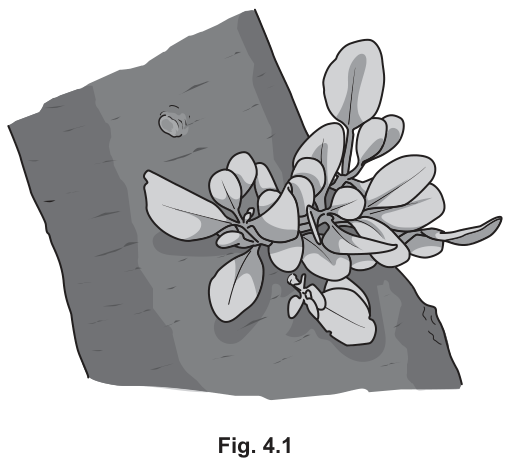
Mistletoe is a parasitic green plant that can often be seen growing attached to trees.
Fig. 4.1 shows some mistletoe growing on the branch of a tree.
Most plants have about of chlorophyll in of their leaves.
Mistletoe, however, has about of chlorophyll in of its leaves.
Mistletoe seeds are stuck to the branches of trees by birds. The seeds germinate to produce special roots that grow into the phloem of the tree branch.
Suggest why mistletoe grows these special roots.
Answer
- Mistletoe has less chlorophyll than typical plants.
- Therefore it performs less photosynthesis and produces less sugar.
- It needs to obtain sugars / sucrose from the host tree.
- The special roots grow into the phloem, which transports sugars / sucrose.
See working
Walkthrough
This question asks for a logical chain of reasoning to explain a biological adaptation (special roots in mistletoe).
- Observation: The text states mistletoe has only of chlorophyll per of leaves, compared to in most plants.
- Inference 1: Less chlorophyll means mistletoe can trap less light energy, so it performs less photosynthesis.
- Inference 2: Less photosynthesis means it makes less sugar (glucose/sucrose) than it needs to grow.
- Solution/Adaptation: To survive, it must obtain sugars / sucrose from the tree (the host).
- Mechanism: The roots grow into the phloem of the tree branch. The phloem is the tissue responsible for translocating sugars / sucrose from sources (like leaves) to sinks (like roots or growing parts). By tapping into the phloem, the mistletoe directly accesses the tree's sugar supply.
Key Takeaways
- Photosynthesis rate is limited by chlorophyll concentration.
- Parasitic plants like mistletoe are partial or full heterotrophs that supplement or replace their own photosynthesis.
- The phloem translocates sugars (sucrose) throughout the plant, making it the ideal target for parasitic roots seeking carbohydrates.
Common Mistakes
- Saying the roots absorb "water and minerals" (xylem does this; the question specifically mentions the phloem, which carries sugars).
- Forgetting to link the low chlorophyll to reduced photosynthesis (just saying "it's a parasite" is not enough; you must explain why it needs the roots based on the data given).
- Writing "glucose" instead of "sucrose" when referring to translocation in the phloem (glucose is converted to sucrose for transport).
Things to Be Careful About
- The mark scheme awards up to 4 marks for a logical chain. Ensure each step is connected: less chlorophyll → less photosynthesis → less sugar → needs to obtain sugar from phloem.
- Use the exact terms from the mark scheme: chlorophyll, photosynthesis, sugars / sucrose, and phloem.
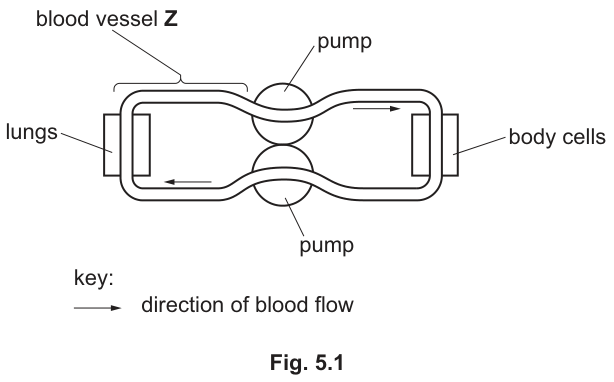
Fig. 5.1 shows a model of the human circulatory system.
Answer
pulmonary vein
pulmonary vein
Walkthrough
In Fig. 5.1 the upper loop is the pulmonary circuit: blood leaves the heart, passes through the lungs, and returns to the heart. Vessel Z is drawn carrying blood towards the lungs, so it must be the vessel leaving the heart on the way to the lungs — the pulmonary artery. But look carefully at the arrows: the arrow on Z points from the lungs back to the pump, so Z carries blood from the lungs to the heart. A vessel carrying oxygenated blood from the lungs to the heart is the pulmonary vein — the only vein in the body that carries oxygenated blood.
Key Takeaways
- The pulmonary artery carries deoxygenated blood from the heart to the lungs; the pulmonary vein carries oxygenated blood from the lungs to the heart.
- Trace the arrows on the diagram before naming a vessel — direction of flow is the discriminating feature.
Common Mistakes
- Writing "pulmonary artery" — this is the classic trap; the artery goes heart → lungs, but Z carries blood lungs → heart.
- Writing just "vein" — the mark scheme requires the named vessel.
Things to Be Careful About
- Read the arrowheads on Fig. 5.1 carefully; the direction of blood flow tells you whether the vessel is going to or coming from the lungs.
The system is described as a double circulatory system.
Describe what is meant by a double circulation.
Answer
- Blood passes through the heart twice for each complete circuit of the body.
- One circuit goes from the heart to the lungs and back (pulmonary circulation); the other goes from the heart to the body cells and back (systemic circulation).
Blood passes through the heart twice for each complete circuit of the body — once through the pulmonary circuit and once through the systemic circuit.
Walkthrough
A single circulation (like in a fish) sends blood through the heart once per circuit. In humans the blood makes a figure-of-eight: the right side of the heart pumps blood to the lungs and back (pulmonary circulation), and then the left side pumps that same blood around the body and back (systemic circulation). So for one complete trip around the whole system, the blood passes through the heart twice. That is the entire meaning of "double circulation" — the mark scheme credits exactly this: blood travels through the pump/heart twice for each full circuit.
Key Takeaways
- Double circulation = two circuits (pulmonary and systemic) joined at the heart; blood passes through the heart twice per complete circuit.
- The advantage, worth knowing, is that blood is pumped to the body at high pressure after being re-pressurised by the left side of the heart.
Common Mistakes
- Saying "blood goes to the lungs and the body" without stating that it passes through the heart twice — the defining feature is the double pass through the heart.
- Confusing "double circulation" with "two hearts" — there is one heart with two sides.
Things to Be Careful About
- Both marking points are needed for the two marks: "through the heart twice" AND "for each full circuit". One alone scores one mark.
Explain how the structure of the human heart is adapted to be the pump in a double circulatory system.
Answer
- The heart has four chambers and a septum, which keeps the blood on the left and right sides completely separate.
- This means deoxygenated blood and oxygenated blood do not mix, so the blood sent to the body is fully oxygenated.
- The wall of the left ventricle is thicker (more muscular) than the right, because it must pump blood at higher pressure all around the body, while the right ventricle only pumps blood a short distance to the lungs.
Four chambers with a septum keep deoxygenated and oxygenated blood separate; the thicker-walled left ventricle pumps blood around the whole body at high pressure while the right ventricle pumps only to the lungs.
Walkthrough
The question asks how the heart's structure suits it to be the pump in a double circulation — so every point must pair a structure with its function.
-
Four chambers and a septum. The septum is the muscular wall dividing the left and right sides. Because a double circulation has two separate circuits, the two sides must be separate: the right side handles deoxygenated blood going to the lungs, the left side handles oxygenated blood going to the body. If the sides were not divided, the two bloods would mix.
-
Separation of blood. This is the reason the septum matters — deoxygenated and oxygenated blood are kept apart, so the blood delivered to the body cells carries the maximum oxygen.
-
Thicker left ventricle wall. The left ventricle pumps blood the long way round — to the toes and back — so it needs a thick, muscular wall to generate high pressure. The right ventricle only pushes blood to the nearby lungs, so its wall is thinner. This contrast (the "ora" in the mark scheme — either direction of the comparison scores) is the classic structure-function link.
Key Takeaways
- Structure-function pairing is what earns the marks: septum → separation; thick left ventricle wall → high-pressure systemic pump.
- The left ventricle has the thickest wall of all four chambers.
Common Mistakes
- Naming structures without functions — "it has a septum" alone does not score; you must say why (keeps the two bloods separate).
- Saying the left ventricle has a thicker wall "because it pumps more blood" — the volume is the same; the reason is the greater pressure and distance needed for the systemic circuit.
- Confusing atria and ventricles — the wall-thickness point is about the ventricles.
Things to Be Careful About
- The mark scheme allows a maximum of three points from a longer list — give three clean, complete structure-function pairs and stop.
- Use "deoxygenated" and "oxygenated" precisely; "dirty" and "clean" blood score nothing.
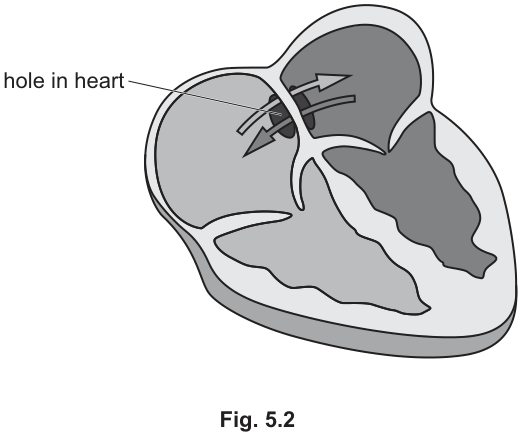
Some people have a heart defect called a 'hole in the heart', as shown in Fig. 5.2.
People with a hole in their heart may develop pain in their muscles and quickly become tired when they exercise.
Explain why this happens.
Answer
- The hole in the septum allows blood from the right and left sides of the heart to mix, so deoxygenated blood mixes with oxygenated blood.
- The blood leaving the heart therefore carries less oxygen, so less oxygen reaches the muscles during exercise.
- The muscles cannot respire aerobically enough to meet the energy demand, so they respire anaerobically.
- Anaerobic respiration produces lactic acid, which builds up in the muscles and causes pain and fatigue.
The hole lets deoxygenated and oxygenated blood mix, so less oxygen reaches the muscles; they respire anaerobically, producing lactic acid that builds up and causes pain and tiredness.
Walkthrough
This is a causal chain — each link earns a mark, so write all four links.
-
The hole lets the two bloods mix. The septum normally keeps the right side (deoxygenated blood) apart from the left side (oxygenated blood). A hole in the septum breaks that separation.
-
Less oxygen reaches the muscles. Once the bloods mix, the blood pumped to the body is a mixture — its oxygen content is diluted by the deoxygenated blood. During exercise, when muscles need maximum oxygen, they receive less than normal.
-
Muscles respire anaerobically. Aerobic respiration needs oxygen to release energy from glucose:
With too little oxygen, the muscles switch to anaerobic respiration:
This releases much less energy per glucose molecule, which is why the person quickly becomes tired.
- Lactic acid builds up. The lactic acid produced accumulates in the muscles, and this is what causes the pain. (It is later broken down in the liver during recovery, using extra oxygen — the "oxygen debt".)
Key Takeaways
- A hole in the heart defeats the whole point of a double circulation — the separation of the two bloods.
- Anaerobic respiration in muscle: glucose → lactic acid; less energy released, lactic acid causes fatigue and pain.
Common Mistakes
- Stopping at "less oxygen reaches the muscles" — the question says explain, so the chain must continue to anaerobic respiration and lactic acid to earn all four marks.
- Saying "less oxygen in the blood" without linking it to the mixing caused by the hole.
- Writing the wrong anaerobic equation — in human muscle the product is lactic acid, not ethanol and carbon dioxide (that is yeast).
- Saying the muscles "run out of energy" without naming anaerobic respiration or lactic acid — vague statements score nothing.
Things to Be Careful About
- The mark scheme lists five points for a maximum of four — any four of them score, so give the four in the logical chain above.
- Use "deoxygenated" and "oxygenated" exactly; "used blood" and "fresh blood" are not accepted terms.
- Note the mark scheme's "max four" — do not pad with irrelevant extra material.
Biofuels are made from plant material or animal waste. They are used as a replacement for fossil fuels such as petrol and oil.
Fossil fuels were made over long periods of time, millions of years ago.
Biofuels may or may not be a sustainable resource.
Answer
A sustainable resource is one that is produced as rapidly as it is removed/used, so that it does not run out.
Produced as rapidly as it is removed, so it does not run out.
Walkthrough
The definition has two linked halves, and the mark scheme credits each separately. A sustainable resource is one that is replaced (produced, regrown, replenished) at least as fast as humans take it away. The consequence of this balance is that the resource never becomes exhausted — it does not run out. Both the balance and the consequence are needed for full marks.
Applied to biofuels: if sugar cane is replanted and regrows as fast as it is harvested for ethanol production, the fuel supply is sustainable. If forest is cleared faster than anything regrows, it is not.
Key Takeaways
- Sustainability is a rate comparison: rate of production versus rate of removal.
- The defining outcome is that the resource is not exhausted.
- Biofuels are only sustainable if the crops are regrown at the rate they are harvested.
Common Mistakes
- Writing only 'it does not run out' without the rate-balance idea — that is one mark, not two.
- Saying 'it can be used again' or 'it is renewable' as a vague catch-all; the mark scheme wants the production/removal comparison.
- Confusing sustainability with recycling — recycling is a different concept.
Things to Be Careful About
- Give both halves of the definition; the second mark sits in 'so that it does not run out'.
- Use the mark scheme's own wording: 'produced as rapidly as it is removed'.
Alcohol (ethanol) is used as a biofuel. It can be made from plants, such as sugar cane, which grow in tropical countries such as Brazil.
Describe how a living organism can be used to convert sugar found in plants into alcohol.
Answer
Yeast carries out fermentation (anaerobic respiration) of the sugar (glucose), producing ethanol and carbon dioxide.
Fermentation by yeast.
Walkthrough
The question asks how a living organism converts plant sugar into alcohol. The organism is yeast — a single-celled fungus. The process is fermentation, which is yeast's anaerobic respiration: it breaks down glucose without oxygen to give ethanol and carbon dioxide.
The word equation worth remembering:
The mark scheme credits the process name ('fermentation') and the organism name ('yeast') — one mark each. In practice, sugar cane juice provides the glucose, yeast is added, and the mixture is kept warm and anaerobic so the yeast ferments the sugar into ethanol, which is then distilled as biofuel.
Key Takeaways
- Fermentation is anaerobic respiration by yeast: glucose → ethanol + carbon dioxide.
- Yeast is the named organism for ethanol production in biotechnology.
- Both the process name and the organism name are usually required for the marks.
Common Mistakes
- Saying 'respiration' alone without 'anaerobic' or 'fermentation' — the mark scheme wants the specific term.
- Naming bacteria instead of yeast; bacteria are used for yoghurt and insulin, not ethanol.
- Writing the equation backwards or omitting carbon dioxide.
- Saying the yeast 'converts sugar into alcohol' without naming the process — the process name is the marking point.
Things to Be Careful About
- The question says 'living organism' — you must name yeast explicitly, not just describe the process.
- 'Fermentation' is the exact term; 'anaerobic respiration' is accepted as an alternative but plain 'respiration' is too vague.
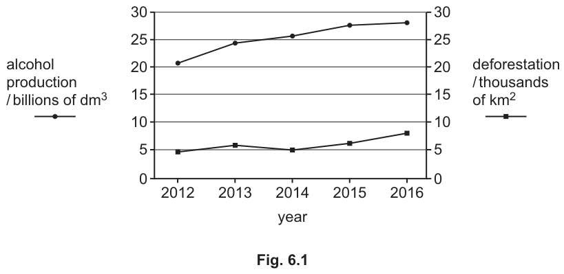
Fig. 6.1 gives some information about alcohol production and deforestation in Brazil from 2012 to 2016.
Suggest an explanation for the changes shown in Fig. 6.1 and describe the possible impact of these changes.
Answer
As alcohol production increased from 2012 to 2016, deforestation also increased (sharply in 2016).
Impacts:
- Forest is cut down to plant more sugar cane for alcohol production.
- Habitats are destroyed, so organisms lose their homes.
- Biodiversity decreases; species may become endangered or extinct.
- Fewer trees means less carbon dioxide is removed from the atmosphere by photosynthesis, and burning the felled trees releases more carbon dioxide, contributing to global warming / the greenhouse effect.
- Deforestation can cause soil erosion and flooding.
- Positive impact: using more ethanol as a biofuel means less fossil fuel is burned, so less carbon dioxide is released from fossil fuels.
See working
Walkthrough
Step 1 — read the graph. Fig. 6.1 plots two variables on one pair of axes: alcohol production (circles, left axis) rises steadily from about 21 to about 28 billion dm³ between 2012 and 2016, while deforestation (squares, right axis) stays around 4–6 thousand km² until 2015 and then jumps to about 8 in 2016. Both trends go up together — that is the correlation the first mark rewards.
Step 2 — explain the link. More ethanol production needs more sugar cane, so more land is cleared. In Brazil that land comes from forest, so deforestation rises with alcohol production. This is a 'suggest' question: you apply the syllabus idea that expanding crop land causes deforestation.
Step 3 — impacts. The mark scheme allows a maximum of three impact marks, and they can be negative or positive:
- Negative: forest cut down to grow sugar cane; loss of habitats; loss of biodiversity, extinction or endangerment of species; more carbon dioxide in the atmosphere (less photosynthesis removing it, more released when trees are burned or decompose) causing global warming; soil erosion and flooding.
- Positive: more ethanol use means less fossil fuel burned, so less carbon dioxide from fossil fuels — a lower greenhouse effect.
Notice the carbon dioxide point can be earned from either direction — deforestation adds carbon dioxide, but replacing fossil fuels with ethanol avoids some. Both are valid; the graph alone does not tell you which effect dominates.
Key Takeaways
- When two curves on a dual-axis graph rise together, describe the correlation explicitly before explaining it.
- Deforestation impacts cluster into four themes: habitat loss, biodiversity loss, the carbon cycle/global warming, and soil erosion/flooding.
- Biofuels have a positive side: they substitute for fossil fuels and so reduce carbon dioxide release from that source.
Common Mistakes
- Describing only one curve instead of linking the two — the first mark requires the relationship ('as alcohol production increased, so did deforestation').
- Reading the wrong axis: the two curves use different scales, so never compare their raw numbers.
- Saying 'deforestation causes global warming' with no mechanism — the mark needs carbon dioxide named.
- Giving only negative impacts when a positive impact (less fossil fuel burned) is also creditable.
- Writing more than the required number of impact points and running out of time; the scheme caps impacts at three.
Things to Be Careful About
- The question has two commands: 'suggest an explanation' AND 'describe the possible impact' — both must be answered.
- Quote the graph trend with its direction and rough values to anchor the explanation.
- 'Biodiversity' is the precise term; 'fewer animals' alone is too vague for the mark.
- The mark scheme caps the impacts at three, so choose the clearest three and stop.
HIV and MRSA are two different microorganisms which are pathogens.
Put ticks (✓) in Table 7.1 to show the characteristics of each pathogen.
Table 7.1
| characteristic | HIV | MRSA |
|---|---|---|
| is a living cell | ||
| contains cytoplasm | ||
| contains genetic material | ||
| has a protein coat |
Answer
| characteristic | HIV | MRSA |
|---|---|---|
| is a living cell | ✓ | |
| contains cytoplasm | ✓ | |
| contains genetic material | ✓ | ✓ |
| has a protein coat | ✓ |
See table
Walkthrough
HIV is a virus. Viruses are not cells: they have no cytoplasm and no cell structures, only a core of genetic material (RNA in HIV) surrounded by a protein coat (capsid). MRSA (Methicillin-resistant Staphylococcus aureus) is a bacterium, so it is a living cell with cytoplasm, a cell wall, a cell membrane and genetic material (DNA), but it has no protein coat. The mark scheme says 'mark by column', so both ticks in a column must be correct for the marks to be awarded — a single wrong tick in a column loses that column's marks.
Key Takeaways
- A virus is not a cell: genetic material + protein coat only, no cytoplasm.
- A bacterium is a living cell: cytoplasm, membrane, cell wall, genetic material, no protein coat.
- HIV stores its genetic information as RNA; bacteria use DNA.
Common Mistakes
- Ticking 'is a living cell' for HIV — viruses are not cells and are often described as being on the boundary of life.
- Ticking 'has a protein coat' for MRSA — bacteria have a cell wall, not a capsid.
- Confusing the protein coat with a cell wall: only viruses have a protein coat.
Things to Be Careful About
- The scheme marks by column: both ticks in the HIV column must be right, and both in the MRSA column must be right, so check each column as a whole.
- 'Contains genetic material' is the one row ticked for both — every organism and virus carries genetic material.
Answer
- HIV weakens the immune system;
- it decreases the number of lymphocytes;
- so the body has a reduced ability to produce antibodies.
HIV weakens the immune system by decreasing lymphocyte numbers, reducing the ability to produce antibodies.
Walkthrough
HIV attacks and destroys lymphocytes (specifically the helper T lymphocytes that coordinate the immune response). As lymphocyte numbers fall, the immune system is weakened, and because lymphocytes are the cells that produce antibodies against antigens, the body's ability to make antibodies is reduced. That is why people with AIDS die of opportunistic infections — the pathogens themselves are often ordinary microbes the immune system would normally destroy. The three mark points form a logical chain: weakens immune system → fewer lymphocytes → fewer antibodies.
Key Takeaways
- HIV targets lymphocytes, the white blood cells of active immunity.
- Lymphocytes produce antibodies, so losing them removes antibody-mediated defence.
- AIDS is the syndrome of infections that follow once immunity has collapsed.
Common Mistakes
- Writing 'destroys red blood cells' or 'destroys all white blood cells' — HIV attacks lymphocytes specifically.
- Saying HIV 'causes AIDS' without explaining the mechanism — the question asks how it affects the immune system, so the lymphocyte/antibody chain is required.
- Confusing antibodies with antibiotics in the answer.
Things to Be Careful About
- Use 'lymphocytes' — 'white blood cells' alone is vaguer than the scheme wants.
- Give all three points; the chain is worth one mark each.
For different reasons, HIV and MRSA are not destroyed by antibiotics such as penicillin.
Give these reasons.
HIV ______
MRSA ______
Answer
HIV — it is a virus, and antibiotics do not work against viruses.
MRSA — it is resistant to (many) antibiotics such as penicillin.
HIV: it is a virus; MRSA: it is resistant to antibiotics/penicillin.
Walkthrough
Antibiotics such as penicillin work by attacking structures found only in bacteria — for example penicillin interferes with bacterial cell wall formation. Viruses like HIV have no cell wall, no cell membrane and no metabolism of their own, so antibiotics have nothing to attack; this is why HIV is a virus and antibiotics are useless against it. MRSA, on the other hand, is a bacterium that antibiotics would normally kill, but this strain has evolved resistance (through natural selection of resistant bacteria) and is not destroyed by penicillin and many other antibiotics. Two different reasons: one is about the type of organism, the other about resistance.
Key Takeaways
- Antibiotics act against bacteria only — they have no effect on viruses.
- MRSA stands for methicillin-resistant Staphylococcus aureus; resistance is the whole point of its name.
Common Mistakes
- Saying HIV is 'too small' for antibiotics to work — size is not the reason; the absence of bacterial structures is.
- Saying MRSA is a virus, or that HIV is resistant to antibiotics — both reverse the logic.
- Writing 'antibodies' instead of 'antibiotics'.
Things to Be Careful About
- Give a separate reason for each pathogen — the blanks ask for two distinct answers.
- For MRSA, use the word 'resistant'; 'the antibiotics don't work' without naming resistance may not score.
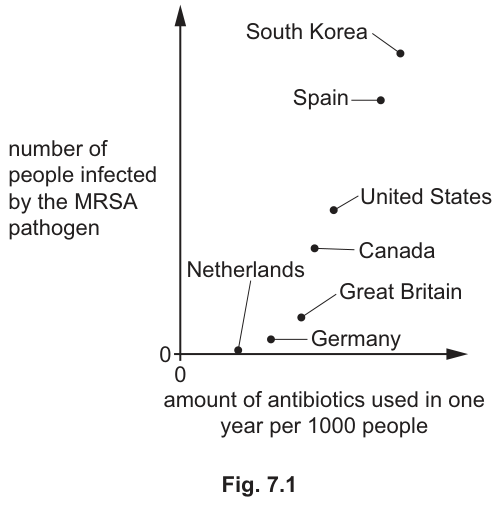
Fig. 7.1 shows the number of people infected by the MRSA pathogen in different countries.
It also shows the total amount of antibiotics used in each country in one year per 1000 people.
Describe and explain the relationship shown between antibiotic use and the number of people infected by the MRSA pathogen.
Answer
- The more antibiotics used, the more people are infected with MRSA (positive correlation);
- more antibiotics are used in an attempt to kill the pathogen;
- but the antibiotics do not kill the resistant MRSA;
- (the use of antibiotics) increases the number of resistant bacteria.
Positive correlation: the more antibiotics used, the more people infected with MRSA, because antibiotic use selects for and increases resistant bacteria, which the antibiotics cannot kill.
Walkthrough
The scatter graph plots antibiotic use per 1000 people against MRSA infections. The points (Netherlands and Germany low on both axes; South Korea and Spain high on both) rise from bottom-left to top-right — a positive correlation. Describe that first: as antibiotic use increases, the number of MRSA infections increases. Then explain it. Countries with more MRSA use more antibiotics trying to control it. But antibiotics do not kill resistant bacteria like MRSA; worse, using antibiotics kills the non-resistant bacteria, leaving the resistant ones to survive, reproduce and spread — natural selection in action. So heavy antibiotic use actively increases the resistant population, which is why high-use countries have more MRSA.
Key Takeaways
- Correlation must be described with both variables named and the direction stated.
- Antibiotic use selects for resistant bacteria: susceptible bacteria die, resistant ones survive and multiply.
- This is the standard 5090 example of natural selection in bacteria.
Common Mistakes
- Describing the graph as 'countries with more antibiotics have more disease' without naming MRSA or stating the positive direction.
- Saying antibiotics 'cause' MRSA directly — the mechanism is selection of already-resistant bacteria.
- Confusing correlation with causation without giving the selection explanation.
- Writing 'more antibiotics kill more bacteria so fewer infections' — that contradicts the graph.
Things to Be Careful About
- The scheme offers four points for a maximum of three, so any three of the listed points score; give the description plus the two explanation points.
- Use the word 'resistant' — it is the key term linking the graph to the biology.
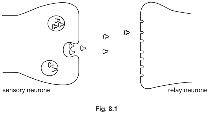
Fig. 8.1 shows a synapse between a sensory neurone and a relay neurone.
The sensory neurone receives information from a pain receptor.
Answer
- An impulse stimulates the release of neurotransmitter molecules from vesicles in the sensory neurone.
- The neurotransmitter molecules are released into the synaptic gap / cleft.
- The neurotransmitter molecules pass across the gap by diffusion.
- The neurotransmitter molecules bind with receptor proteins / sites on the post-synaptic membrane of the relay neurone.
- This binding causes an impulse to be initiated in the relay neurone.
See working
Walkthrough
Synaptic transmission is the chemical process by which a nerve impulse is passed from one neurone to the next across a synapse. It cannot happen by direct electrical contact, which is why a gap exists between the two neurones.
-
Impulse arrival and vesicle release: When an electrical impulse (action potential) reaches the synaptic terminal of the pre-synaptic sensory neurone, it stimulates the synaptic vesicles to fuse with the pre-synaptic membrane. This releases their contents — neurotransmitter molecules — into the synaptic gap (also called the synaptic cleft).
-
Diffusion across the gap: The neurotransmitter molecules do not cross the gap by electrical transmission or active transport. They move by diffusion down their concentration gradient from the pre-synaptic side, where they are highly concentrated, to the post-synaptic side, where the concentration is lower.
-
Binding to receptors: On the post-synaptic membrane of the relay neurone, there are specific receptor proteins (or receptor sites). The neurotransmitter molecules fit into these receptors and bind to them. This is often compared to a lock-and-key mechanism, where the neurotransmitter is the key and the receptor is the lock.
-
Initiation of a new impulse: The binding of the neurotransmitter to the receptors changes the permeability of the post-synaptic membrane, which triggers a new electrical impulse in the relay neurone (the post-synaptic neurone).
Key Takeaways
- Synapses rely on chemical messengers (neurotransmitters) rather than direct electrical connections.
- Vesicles store neurotransmitters, the synaptic cleft is the gap they cross, and diffusion is the mechanism of crossing.
- Receptor proteins on the post-synaptic membrane are required to convert the chemical signal back into an electrical impulse.
Common Mistakes
- Saying "the impulse jumps across the gap" or "electrical transmission" — synapses are chemical, not electrical.
- Saying "neurotransmitter is pumped across" or "active transport" — it crosses by diffusion.
- Forgetting to mention vesicles or the synaptic gap / cleft.
- Saying "nerve" instead of "neurone" — 5090 strictly rewards the term neurone.
- Failing to state that the impulse is initiated in the relay / post-synaptic neurone (not just "sent to the brain" at this stage).
Things to Be Careful About
- Use the exact terms required by the mark scheme: neurotransmitter, vesicles, synaptic gap / cleft, diffusion, receptor proteins / sites.
- The mark scheme awards up to 6 marks for this explanation. Ensure all six logical stages are covered: release from vesicles, into the gap, diffusion across, binding to receptors, and initiation of a new impulse.
- Do not add extra information that is not credited, such as the role of calcium ions or the breakdown of neurotransmitters by enzymes, as these are not required for full marks and may distract from the core points.
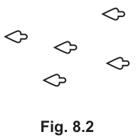
Analgesics are drugs that help to stop a person from feeling pain.
Fig. 8.2 shows the shape of some analgesic drug molecules.
Suggest how this analgesic drug can help to stop a person from feeling pain.
Use information from Fig. 8.1 and Fig. 8.2 in your answer.
Answer
- The analgesic / drug molecules have a similar shape to the neurotransmitter molecules (Fig. 8.2).
- They fit into and bind with the receptor proteins / sites on the relay neurone (Fig. 8.1).
- This blocks the receptors, stopping the actual neurotransmitter from binding.
- As a result, no impulse is initiated in the relay neurone, and information from the pain receptor cannot reach the brain.
See working
Walkthrough
This question asks you to explain how an analgesic drug stops pain using the principles of synaptic transmission and the lock-and-key model.
-
Shape similarity: Look at Fig. 8.2. The analgesic drug molecules have a specific shape. Look at Fig. 8.1. The neurotransmitter molecules have a similar shape that fits into the receptor sites on the relay neurone. The drug molecules mimic the shape of the neurotransmitter.
-
Binding to receptors: Because of this similar shape, the drug molecules can fit into and bind with the receptor proteins / sites on the post-synaptic membrane of the relay neurone. This is a classic example of competitive inhibition at a synapse.
-
Blocking the signal: By occupying the receptor sites, the drug molecules block the receptors. This means the natural neurotransmitter molecules released from the sensory neurone cannot bind to the receptors.
-
Stopping the pain signal: If the neurotransmitter cannot bind, it cannot trigger a change in the post-synaptic membrane. Therefore, no impulse is initiated in the relay neurone. Since the impulse is not passed on, the information from the pain receptor cannot reach the brain, and the person does not feel pain.
Key Takeaways
- Drugs can act as competitive inhibitors at synapses by mimicking the shape of natural neurotransmitters.
- Binding to receptors without triggering a response blocks the normal signal transmission.
- Understanding the pathway (receptor -> synapse -> brain) helps explain how blocking one step stops the entire sensation.
Common Mistakes
- Saying the drug "destroys" or "breaks down" the neurotransmitter — it blocks the receptor, it does not affect the neurotransmitter itself.
- Saying the drug "stops the nerve impulse in the sensory neurone" — the impulse still travels down the sensory neurone; the block happens at the synapse.
- Forgetting to mention that the pain information cannot reach the brain — this is the final physiological consequence that explains why the person "doesn't feel pain".
- Not referencing the shape of the drug molecules in relation to the neurotransmitter or receptors.
Things to Be Careful About
- The mark scheme specifically asks to "Use information from Fig. 8.1 and Fig. 8.2". You must explicitly mention that the drug has a similar shape to the neurotransmitter (from Fig. 8.2) and that it binds to the receptor sites (from Fig. 8.1).
- Ensure all four marking points are covered: shape similarity, binding to receptors, blocking the receptors, and the consequence (no impulse / no brain signal).
- Use the term analgesic or drug interchangeably, but be precise about what it does (blocks receptors, does not destroy neurotransmitters).