Biology 5090/11 — October/November 2025
Cambridge O-Level · Multiple Choice · answer key with instant marking and worked solutions
Topics Movement Into and Out of Cells · Plant Nutrition · Disease and Immunity · Coordination and Control · Organisms and Their Environment · Cell Division and Reproduction · +14 more
Tap an option under each question to check it — your score builds as you go.
What is a function of a plant cell wall?
Options
A to control loss of ions from the cell
B to control movement of glucose into the cell
C to prevent evaporation of water from the cell
D to prevent the cell bursting when water enters
Working
The plant cell wall is made of cellulose and is freely permeable, so it cannot control which substances enter or leave the cell — that is the job of the cell membrane (options A and B). It is not waterproof, so it does not prevent evaporation (option C). Its main function is to provide strength and support, and to prevent the cell bursting when water enters by osmosis.
Answer
D
D
Walkthrough
This question asks for the function of the plant cell wall. Let's think about what the cell wall actually does.
The plant cell wall is a rigid layer made mainly of cellulose, lying outside the cell membrane. It is freely permeable, meaning water and dissolved substances can pass straight through it. Because it is freely permeable, it cannot control the movement of ions (option A) or glucose (option B) into the cell — that selective control is done by the cell membrane, which is partially permeable and uses active transport where needed.
The cell wall is not waterproof, so it does not stop water evaporating from the cell (option C). Preventing water loss from a plant is the job of the waxy cuticle on leaves and stems.
Option D is correct. When a plant cell is surrounded by a solution with a higher water potential (more dilute) than its own cytoplasm, water enters the cell by osmosis. The cytoplasm and vacuole swell and push against the cell wall. The strong, rigid cell wall resists this expansion, so the cell becomes turgid rather than bursting. This is exactly what would happen to an animal cell, which has no cell wall, and which would burst (lyse) under the same conditions.
Key Takeaways
- The plant cell wall is freely permeable — it lets everything through, so it does not control what enters or leaves the cell.
- The cell wall provides strength and support, and prevents the cell bursting when water enters by osmosis.
- The cell membrane is the structure that controls movement of substances in and out of the cell.
- A turgid plant cell is firm because the cell wall resists the expansion caused by osmosis.
Common Mistakes
- Choosing A or B: these confuse the cell wall with the cell membrane. The membrane is partially permeable and controls what enters and leaves; the wall is freely permeable.
- Choosing C: this confuses the cell wall with the cuticle, a waxy waterproof layer on leaves and stems that reduces water loss.
- Thinking the cell wall is waterproof — it is not; water passes freely through it.
Things to Be Careful About
- The question asks for a function of the cell wall — answer with what the wall actually does, not what other structures do.
- Remember the key word turgid: a plant cell with water entering becomes turgid, not burst, because of the cell wall.
- In the exam, if you see options about controlling movement of substances, those point to the cell membrane, not the cell wall.
The international system for naming organisms is the binomial system.
Which two parts form the scientific name?
Options
A kingdom and genus
B genus and species
C species and variety
D variety and kingdom
Working
The binomial system gives every organism a two-part scientific name. The two parts are the genus and the species.
For example, Homo sapiens is made up of the genus Homo and the species sapiens.
- A is incorrect because kingdom and genus are not the two parts that form the scientific name.
- C is incorrect because variety is not one of the two binomial parts.
- D is incorrect because variety and kingdom are not the two binomial parts.
Answer
B
B
Walkthrough
The question tests the binomial system, the internationally agreed way of naming living organisms.
A binomial name literally means a name with "two parts". Both parts are needed to identify the organism precisely:
- Genus – the first word, written with a capital letter.
- Species – the second word, written with a lower-case letter.
Together they give the scientific name, such as Homo sapiens.
Now look at the options:
- A: kingdom and genus – kingdom is far too broad a group and is not part of the binomial name.
- B: genus and species – this is exactly what the binomial name is made of.
- C: species and variety – variety is not part of the binomial name.
- D: variety and kingdom – neither of these is part of a two-part scientific name.
Therefore the correct answer is B.
Key Takeaways
The binomial system gives every species a unique two-part name (genus and species). It is used worldwide so scientists can be sure they are talking about the same organism, even if local common names differ. You should remember that the genus name is written first with a capital letter, and the specific name is written second with a lower-case letter.
Common Mistakes
- Choosing kingdom and genus – kingdom is a very large and broad classification group; it is definitely not part of the binomial name.
- Choosing species and variety – variety is not one of the two binomial parts, and the specific name alone is not the whole binomial.
- Thinking that the order does not matter – the genus always comes first and the species second; sapiens Homo is not correct.
Things to Be Careful About
The exact wording is "genus and species". Since the question asks for the two parts of the scientific name, only the option containing those two ranks is correct. No analysis or calculation is needed; it is direct recall of a definition.
A student draws six different arthropods found in woodland.
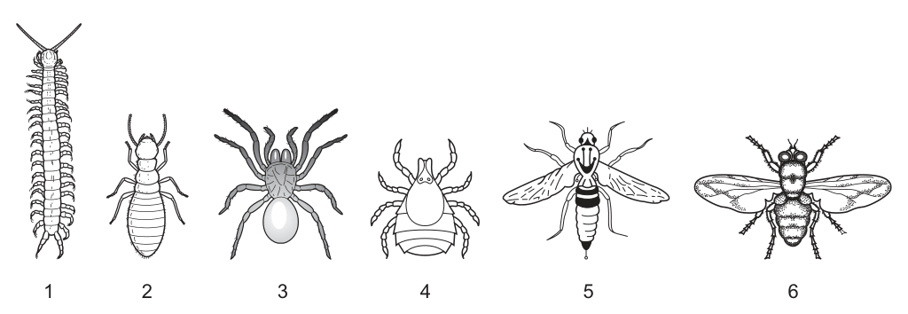
When constructing a dichotomous key, which question could be used to separate arthropods 2, 5 and 6 from arthropods 1, 3 and 4?
Options
A Does it have a segmented body?
B Does it have antennae?
C Does it have six legs?
D Does it have wings?
Working
The question requires a feature that is present in arthropods 2, 5, and 6, but absent in arthropods 1, 3, and 4.
- Arthropods 2, 5, and 6 are insects. Insects are defined by having three body regions and exactly six legs.
- Arthropod 1 is a myriapod (many legs).
- Arthropods 3 and 4 are arachnids (eight legs).
Evaluating the options:
- A Segmented body: present in 1, 2, 5, 6. Does not match the split.
- B Antennae: present in 1, 2, 5, 6. Does not match the split.
- C Six legs: present in 2, 5, 6; absent in 1 (many legs), 3 and 4 (eight legs). Matches the split perfectly.
- D Wings: present in 5 and 6 only; absent in 2, 1, 3, 4. Does not match the split.
Only option C separates the two groups as required.
C
Walkthrough
- Identify the two groups: The question asks for a dichotomous key question that separates arthropods 2, 5, and 6 from arthropods 1, 3, and 4. We must find a feature that is present in all of 2, 5, 6 and absent in all of 1, 3, 4.
- Identify the arthropod groups:
- Arthropods 2, 5, and 6 are insects (they have three body parts: head, thorax, abdomen).
- Arthropod 1 is a myriapod (centipede or millipede), characterised by a long segmented body and many pairs of legs.
- Arthropods 3 and 4 are arachnids (a spider and a tick), characterised by two body parts and eight legs.
- Evaluate Option A (Segmented body): Arthropods 1, 2, 5, and 6 have clearly segmented bodies. Arthropods 3 and 4 have two fused body segments. This would split the organisms into {1, 2, 5, 6} and {3, 4}, which does not match the required groups.
- Evaluate Option B (Antennae): Arthropods 1, 2, 5, and 6 have antennae. Arachnids (3 and 4) do not have antennae. This would also split the organisms into {1, 2, 5, 6} and {3, 4}, which is incorrect because arthropod 1 belongs in the second group.
- Evaluate Option C (Six legs): Insects (2, 5, 6) have exactly six legs. Myriapods (1) have many legs, and arachnids (3, 4) have eight legs. This question correctly places 2, 5, 6 on the "yes" side and 1, 3, 4 on the "no" side.
- Evaluate Option D (Wings): Only arthropods 5 and 6 have wings. Arthropod 2 is an insect but lacks wings. This would split the organisms into {5, 6} and {1, 2, 3, 4}, which is incorrect.
- Conclusion: Option C is the only question that produces the exact grouping required by the question.
Key Takeaways
- Insects are distinguished from other arthropods by having exactly six legs and three body regions (head, thorax, abdomen).
- Arachnids have eight legs and two body regions, and lack antennae.
- Myriapods have many body segments and many pairs of legs.
- A valid dichotomous key question must split the organisms into the two exact groups specified; it cannot leave any organism from the target groups on the wrong side.
Common Mistakes
- Choosing "Does it have antennae?" (Option B): Students often remember that insects have antennae and arachnids do not, but they forget to check all the organisms. Arthropod 1 is a myriapod and does have antennae, so it would be grouped with 2, 5, and 6, breaking the required split.
- Choosing "Does it have wings?" (Option D): Students see that 5 and 6 have wings and assume this separates the insects. However, not all insects have wings (e.g., fleas, lice, or the insect shown as number 2), so this question fails to group 2 with 5 and 6.
- Confusing leg counts: Forgetting that arachnids have eight legs and insects have six legs is a common error that leads to incorrect grouping.
Things to Be Careful About
- Check every member: Always verify that the proposed key question places every member of the first group on one side and every member of the second group on the other side. A single organism on the wrong side makes the key question invalid.
- Not all insects have wings: When using "wings" or "flight" as a key feature, remember that some insects are wingless. Use leg number or body segments instead to identify insects reliably.
- Read the groups carefully: The question asks to separate {2, 5, 6} from {1, 3, 4}. Pay close attention to which numbers are in which group so you do not accidentally answer for a different split.
Contact lenses are placed on the surface of the eye to correct problems with vision.
One type of contact lens is partially permeable and is normally stored in a sterile sodium chloride solution.
The sodium chloride solution has the same concentration as the fluid that covers the surface of the eye.
A person makes a mistake and stores their contact lenses in distilled water rather than sodium chloride solution.
They then place a contact lens on the surface of their eye.
Which pair of statements describe what then happens?
- Water from the eye fluid enters the contact lens by osmosis.
- Water leaves the contact lens and enters the eye fluid by osmosis.
- The contact lens shrinks.
- The contact lens swells.
Options
A 1 and 3
B 1 and 4
C 2 and 3
D 2 and 4
Working
The contact lens is partially permeable, so water can pass through it by osmosis. Distilled water is pure water with no dissolved solutes, so it has a very high water potential. The fluid covering the eye contains salts, so it has a lower water potential.
When the lens (filled with distilled water) is placed on the eye, water moves by osmosis from the higher water potential inside the lens to the lower water potential of the eye fluid. So water leaves the contact lens — statement 2 is correct.
As water leaves the lens, the lens loses water and shrinks — statement 3 is correct.
Statements 1 and 4 describe the opposite direction of water movement and are incorrect.
Answer
C
C
Walkthrough
This question tests osmosis: the net movement of water from a region of higher water potential to a region of lower water potential, across a partially permeable membrane.
The contact lens is partially permeable — water can pass through it, but dissolved substances cannot (or only very slowly). Normally the lens is stored in a sodium chloride solution with the same concentration as the fluid covering the eye. That means the lens is in equilibrium with its surroundings — there is no net movement of water in or out.
The mistake: the person stores the lens in distilled water. Distilled water is pure water with no dissolved solutes, so it has the highest possible water potential. The lens soaks up this distilled water while it is stored.
Now the lens is placed on the eye. The fluid covering the eye has salts (sodium chloride) dissolved in it, so it has a lower water potential than the distilled water inside the lens. Water therefore moves by osmosis from the lens (higher water potential) into the eye fluid (lower water potential). So statement 2 is correct: "Water leaves the contact lens and enters the eye fluid by osmosis."
As water leaves the lens, the lens loses water and shrinks. So statement 3 is correct: "The contact lens shrinks."
Statements 1 and 4 describe the opposite — water entering the lens and the lens swelling. That would happen if the lens were stored in a solution more concentrated than the eye fluid, not in distilled water. So the correct pair is 2 and 3, which is option C.
Key Takeaways
- Osmosis is the net movement of water from a region of higher water potential to a region of lower water potential, across a partially permeable membrane.
- Pure water (distilled water) has the highest possible water potential.
- Adding solute lowers the water potential of a solution.
- Water always moves from the more dilute solution to the more concentrated solution.
- When a structure loses water by osmosis it shrinks; when it gains water it swells.
Common Mistakes
- Confusing the direction of water movement. Water always moves from higher water potential to lower water potential — from the more dilute solution to the more concentrated one. Here the distilled water inside the lens is more dilute than the eye fluid, so water leaves the lens.
- Picking statements 1 and 4 (option B) by thinking the lens swells because it was stored in distilled water. The swelling would have happened during storage in distilled water, not when the lens is placed on the eye — and the question asks specifically what happens when the lens is on the eye.
- Forgetting that the lens is partially permeable — this is the property that allows osmosis to occur at all.
Things to Be Careful About
- Read the question carefully: it asks what happens when the lens is placed on the eye, not what happened during storage.
- The eye fluid has the same concentration as the normal storage solution (sodium chloride), so it has a lower water potential than distilled water.
- The statements come in matching pairs: statement 2 (water leaves) must pair with statement 3 (shrinks), because losing water is what causes the shrinking.
Which process involves the movement of molecules against a concentration gradient?
Options
A active transport
B diffusion
C osmosis
D transpiration
Working
Active transport is the only process listed that moves molecules or ions against the concentration gradient, using energy from respiration.
- Diffusion and osmosis both move substances down the concentration gradient.
- Transpiration is the loss of water vapour from the leaves, not movement against a gradient.
Answer
A
A
Walkthrough
Active transport is a process in which a cell uses energy to move particles against their concentration gradient — that is, from a region where they are less concentrated to a region where they are more concentrated. For example, root hair cells use active transport to take in mineral ions from the very dilute solution in the soil even when the concentration of those ions inside the root is already higher.
The other processes move particles in the opposite direction:
- Diffusion is the net movement of particles from a higher concentration to a lower concentration, down the concentration gradient.
- Osmosis is the diffusion of water through a partially permeable membrane, again from a higher water potential to a lower water potential — down the gradient, never against it.
- Transpiration is the loss of water vapour from the leaves of a plant through the stomata. It does not involve active movement against a gradient.
So the only option that fits the definition is A, active transport.
Key Takeaways
- Active transport requires energy from respiration and can move substances against the concentration gradient.
- Diffusion and osmosis are both passive processes — they move substances down the concentration gradient without the cell using energy.
- Recognising the phrase “against a concentration gradient” should immediately point to active transport.
Common Mistakes
- Choosing osmosis because it involves water moving into cells — but osmosis is still down the water potential gradient, not against it.
- Confusing transpiration with active transport because both involve movement in plants — transpiration is evaporation of water, not transport against a gradient.
- Thinking diffusion can sometimes go against the gradient if the molecules are small — it cannot; diffusion always goes down the concentration gradient.
Things to Be Careful About
- The question asks for the process involving movement against a concentration gradient. Use the exact mark-scheme term “active transport” if writing an explanation.
- In an exam, underline or circle the correct option letter on the answer sheet — here the correct answer is A.
Many reactions in cells are dependent on enzyme activity.
Which statement about enzymes is not correct?
Options
A They are protein molecules.
B They catalyse biological reactions.
C They remain unchanged at the end of a reaction.
D They have active sites which are the same shape as their substrate molecules.
Working
A is correct: enzymes are proteins.
B is correct: enzymes catalyse biological reactions.
C is correct: enzymes are not used up and remain unchanged at the end of a reaction.
D is not correct: the active site has a shape complementary to the substrate molecule, not the same shape as the substrate.
Answer
D
D
Walkthrough
The question asks which statement about enzymes is not correct. Read each option carefully.
- A says enzymes are protein molecules. This is correct. Enzymes are biological catalysts and they are made of protein.
- B says they catalyse biological reactions. This is also correct. Catalysing means speeding up a reaction without being used up.
- C says they remain unchanged at the end of a reaction. This is correct too. An enzyme can be used again and again because it is not destroyed by the reaction it speeds up.
- D says they have active sites which are the same shape as their substrate molecules. This is the incorrect statement. In the lock-and-key model, the active site has a shape that is complementary to the substrate, like a lock matching a key. The active site is not the same shape as the whole substrate molecule; it is shaped so that the substrate can fit into it.
Therefore, the answer is D.
Key Takeaways
- Enzymes are proteins that act as biological catalysts.
- Enzymes are not used up during a reaction, so they can be reused.
- Each enzyme has an active site with a specific shape.
- The substrate fits into the active site because the shapes are complementary, not identical.
- This is called the lock-and-key hypothesis.
Common Mistakes
- Confusing "same shape" with "complementary shape". The active site is not the same shape as the substrate; it is shaped to fit the substrate.
- Thinking that enzymes are changed or used up in a reaction. They are not.
- Assuming all enzymes are proteins but forgetting that enzymes are also catalysts.
Things to Be Careful About
- The question asks for the statement that is not correct, so read every option before choosing.
- Use the precise term "complementary" when describing the relationship between active site and substrate.
- Do not confuse the active site with the whole enzyme molecule; only a small part of the enzyme is the active site.
Which changes occur in the air spaces of a green leaf on a sunny day?
Options
| carbon dioxide concentration | oxygen concentration | |
|---|---|---|
| A | decreases | decreases |
| B | decreases | increases |
| C | increases | decreases |
| D | increases | increases |
Working
On a sunny day, the leaf photosynthesises. Photosynthesis uses carbon dioxide and produces oxygen. Therefore, in the air spaces of the leaf, carbon dioxide concentration decreases and oxygen concentration increases.
Answer
B
B
Walkthrough
On a sunny day, the leaf is carrying out photosynthesis. The word equation for photosynthesis is:
This means the leaf is taking in carbon dioxide from the air spaces and releasing oxygen into them. So carbon dioxide concentration decreases and oxygen concentration increases.
Key Takeaways
- Photosynthesis requires light, so it occurs during the day.
- Photosynthesis removes CO₂ from the air spaces and adds O₂ to them.
- The gas exchange in a leaf during the day is the reverse of respiration.
Common Mistakes
- Confusing photosynthesis with respiration: respiration would increase CO₂ and decrease O₂, which is option C, not B.
- Thinking both gases change in the same direction (options A and D).
Things to Be Careful About
- The question specifies "on a sunny day" — this signals photosynthesis is occurring.
- Both gases change in opposite directions: one increases while the other decreases.
Which diagram correctly links the carbohydrates made as a result of photosynthesis with their uses?
Options
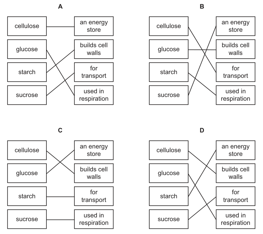
Working
Recall the biological roles of the four carbohydrates produced from photosynthesis:
- Cellulose is a structural carbohydrate used to build plant cell walls.
- Glucose is the primary respiratory substrate used in respiration to release energy.
- Starch is an insoluble storage carbohydrate used as an energy store.
- Sucrose is a soluble transport carbohydrate moved through the phloem.
Examining the diagrams:
- Diagram A incorrectly links cellulose to an energy store.
- Diagram B incorrectly links cellulose to transport.
- Diagram C incorrectly links glucose to an energy store.
- Diagram D correctly matches cellulose to builds cell walls, glucose to used in respiration, starch to an energy store, and sucrose to for transport.
Answer
D
D
Walkthrough
The question asks to match four carbohydrates produced during photosynthesis with their correct biological uses in the plant. We evaluate each carbohydrate based on its known function:
- Cellulose: A structural polysaccharide. Its primary role is to provide rigidity and strength to plant cell walls. It is not used for energy storage or transport.
- Glucose: A simple sugar (monosaccharide) produced directly in the photosynthetic reactions. It is immediately used as the respiratory substrate in cells to release energy via aerobic or anaerobic respiration.
- Starch: An insoluble polysaccharide formed from glucose monomers. Because it is insoluble, it does not affect the water potential of the cell, making it ideal for long-term energy storage in roots, tubers, and seeds.
- Sucrose: A soluble disaccharide formed from glucose and fructose. It is the main carbohydrate transported through the phloem from source leaves to sink organs (roots, fruits, growing tips).
By matching these functions to the right-hand column, we find that cellulose connects to "builds cell walls", glucose to "used in respiration", starch to "an energy store", and sucrose to "for transport". This exact set of connections is shown in Diagram D.
Key Takeaways
- Photosynthesis produces glucose, which is immediately converted into other carbohydrates for specific roles.
- Glucose is for respiration (energy release).
- Starch is for storage (energy store).
- Sucrose is for transport (via phloem).
- Cellulose is for structure (cell walls).
Common Mistakes
- Confusing glucose and starch: Glucose is the immediate respiratory substrate (used in respiration), while starch is the long-term storage form (an energy store). Students often link glucose to an energy store because it contains energy, but the plant converts it to starch for storage.
- Confusing sucrose and cellulose: Sucrose is soluble and used for transport in the phloem. Cellulose is insoluble and structural, used for cell walls.
- Linking cellulose to respiration or energy storage: Cellulose cannot be broken down by most animals (including humans) because we lack the enzyme cellulase, and plants do not use it for energy.
Things to Be Careful About
- Read the diagrams carefully. The lines cross in different ways in each option. Verify every single connection, not just one.
- Remember that glucose is used in respiration, not as a store. The word "store" specifically applies to starch.
- Ensure you match the correct diagram by checking all four links, as a single wrong link eliminates an option.
An investigation is carried out on the rate of photosynthesis in different environmental conditions.
The graph shows the results.
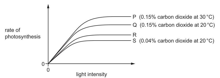
Which conditions could have produced the results for curve R?
Options
| percentage carbon dioxide in air | environmental temperature / | |
|---|---|---|
| A | 0.004 | 20 |
| B | 0.04 | 30 |
| C | 0.4 | 20 |
| D | 4.0 | 30 |
Working
The graph plots the rate of photosynthesis against light intensity for four different sets of conditions. The rate is limited by carbon dioxide concentration and temperature at high light intensities.
- Curve P (0.15% CO, 30°C) has the highest plateau.
- Curve Q (0.15% CO, 20°C) is lower than P, showing that a lower temperature reduces the rate.
- Curve S (0.04% CO, 20°C) is the lowest, showing that a lower carbon dioxide concentration reduces the rate.
- Curve R lies between Q and S. It must therefore have a higher rate than S but a lower rate than Q.
Evaluating the options for Curve R:
- A (0.004% CO, 20°C): Lower CO than S, so the rate would be lower than S.
- B (0.04% CO, 30°C): Same CO as S but a higher temperature (30°C > 20°C), so the rate is higher than S. It has lower CO than Q (0.15%), so the rate is lower than Q. This matches Curve R.
- C (0.4% CO, 20°C): Higher CO than Q, so the rate would be higher than Q.
- D (4.0% CO, 30°C): Higher CO and temperature than P, so the rate would be higher than P.
Answer
B
B
Walkthrough
The question asks us to identify the environmental conditions (carbon dioxide concentration and temperature) that produced Curve R in a graph of photosynthesis rate against light intensity.
First, we analyze the labeled curves to understand how carbon dioxide and temperature affect the rate of photosynthesis. Curve P (0.15% CO, 30°C) has the highest plateau, meaning these conditions support the fastest rate. Curve Q (0.15% CO, 20°C) is lower than P. Since the CO is the same, the drop in rate must be due to the lower temperature (20°C instead of 30°C). Curve S (0.04% CO, 20°C) is lower than Q. Since the temperature is the same, the drop in rate must be due to the lower CO concentration (0.04% instead of 0.15%).
Curve R is positioned between Q and S. This means its plateau is higher than S's (so it must have either higher CO or higher temperature than S) and lower than Q's (so it must have either lower CO or lower temperature than Q).
We test each option:
- Option A (0.004% CO, 20°C): This has less CO than S at the same temperature, so it would produce a curve below S.
- Option B (0.04% CO, 30°C): This has the same CO as S but a higher temperature (30°C > 20°C), so it produces a higher rate than S. It has less CO than Q (0.04% vs 0.15%), so it produces a lower rate than Q. This perfectly fits Curve R.
- Option C (0.4% CO, 20°C): This has more CO than Q at the same temperature, so it would produce a curve above Q.
- Option D (4.0% CO, 30°C): This has more CO and the same temperature as P, so it would produce a curve above P.
Key Takeaways
- The rate of photosynthesis is limited by several factors, including light intensity, carbon dioxide concentration, and temperature.
- At high light intensities, the rate levels off at a plateau determined by the limiting factor (CO or temperature).
- Higher CO concentrations and higher temperatures (up to the optimum) increase the rate of photosynthesis.
- To deduce conditions for an unlabeled curve, compare its position relative to the labeled curves and apply the principles of limiting factors.
Common Mistakes
- Assuming that a higher curve always means higher temperature, ignoring the effect of CO concentration.
- Misreading the graph and thinking Curve R is higher than Q or lower than S.
- Forgetting that 0.04% is less than 0.15%, or 0.004% is less than 0.04%.
- Confusing the axes: the x-axis is light intensity, so the curves show how rate changes as light increases, but the plateau is determined by CO and temperature.
Things to Be Careful About
- Always compare the unlabeled curve to the labeled curves to establish an upper and lower bound for its conditions.
- Pay close attention to the decimal places in the percentages: 0.004% is ten times less than 0.04%, and 0.4% is more than double 0.15%.
- Remember that both temperature and CO can act as limiting factors; a change in either will shift the plateau of the curve.
Hydrogencarbonate indicator changes colour according to the concentration of carbon dioxide present.
The table shows these changes.
| colour of hydrogencarbonate indicator | concentration of carbon dioxide |
|---|---|
| orange | atmospheric carbon dioxide concentration |
| yellow | higher than atmospheric carbon dioxide concentration |
| purple | lower than atmospheric carbon dioxide concentration |
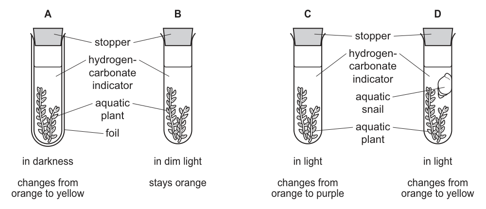
Four test-tubes were set up under different conditions. The colour of hydrogencarbonate indicator in each test-tube at the start of the experiment and after one hour is also shown.
Which test-tube shows that the rates of photosynthesis and respiration are the same?
Options
Answer
B
The hydrogencarbonate indicator stays orange in test-tube B, which means the carbon dioxide concentration is unchanged. This happens when the rate of photosynthesis equals the rate of respiration, so there is no net uptake or release of carbon dioxide.
B
Walkthrough
The hydrogencarbonate indicator changes colour based on the carbon dioxide concentration in the test-tube: orange indicates atmospheric levels, yellow indicates a higher concentration, and purple indicates a lower concentration.
- In test-tube A, the plant is in darkness, so it can only respire, releasing carbon dioxide. The concentration rises and the indicator turns yellow.
- In test-tube B, the plant is in dim light. The light intensity is low enough that the rate of photosynthesis exactly matches the rate of respiration. Carbon dioxide is consumed and produced at the same rate, so the concentration remains at atmospheric levels and the indicator stays orange.
- In test-tube C, the plant is in bright light, so photosynthesis is faster than respiration. Carbon dioxide is taken up from the solution, the concentration falls, and the indicator turns purple.
- In test-tube D, both a plant and a snail are present in the light. The snail respires and releases carbon dioxide in addition to what the plant produces. The overall carbon dioxide concentration rises and the indicator turns yellow.
The question asks for the test-tube where the rates of photosynthesis and respiration are the same. This is test-tube B, where the indicator stays orange.
Key Takeaways
- Hydrogencarbonate indicator is a useful tool for showing net changes in carbon dioxide concentration during photosynthesis and respiration experiments.
- When photosynthesis and respiration are balanced, the carbon dioxide concentration remains constant and the indicator stays orange.
Common Mistakes
- Confusing the colour changes: remembering that yellow means higher carbon dioxide (respiration dominant) and purple means lower carbon dioxide (photosynthesis dominant).
- Forgetting that respiration occurs continuously in all living cells, regardless of light conditions.
- Assuming that "dim light" means no photosynthesis is occurring, rather than photosynthesis being limited to match respiration.
Things to Be Careful About
- Read the final colour change carefully: "stays orange" is the key phrase indicating no net change in carbon dioxide.
- Remember that the plant in test-tube A is wrapped in foil to ensure complete darkness, preventing any photosynthesis.
- In test-tube D, the addition of the snail means there is an extra source of carbon dioxide from respiration, which is why it turns yellow even in the light.
The diagrams show sections through a dicotyledonous root, stem and leaf.
The vascular tissues are labelled using numbers 1–6.
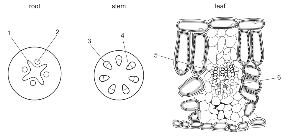
The ends of pieces of roots, stems and leaf stalks are placed in water containing a red dye.
After several hours, the coloured water has travelled along some of the vascular tissues.
Which vascular tissues appear red?
Options
| root | stem | leaf | |
|---|---|---|---|
| A | 1 | 3 | 6 |
| B | 1 | 4 | 5 |
| C | 2 | 4 | 6 |
| D | 2 | 3 | 5 |
Answer
B
B
Walkthrough
The red dye is dissolved in water. In plants, water and dissolved mineral ions are transported upwards from the roots through the stem and into the leaves via the xylem vessels. Therefore, the vascular tissues that will appear red are the xylem in the root, stem, and leaf.
We must identify the xylem in each of the three cross-sectional diagrams:
- Root: In a dicot root, the vascular tissue is arranged with the xylem in the centre, forming a solid star or cross shape. The phloem is found in patches between the arms of the xylem. Looking at the diagram, label 1 points to the central star-shaped xylem, and label 2 points to the phloem. Thus, the xylem in the root is 1.
- Stem: In a dicot stem, vascular bundles are arranged in a ring. Within each individual bundle, the xylem is towards the inside (closer to the centre of the stem) and the phloem is towards the outside. Label 4 points to the inner xylem, and label 3 points to the outer phloem. Thus, the xylem in the stem is 4.
- Leaf: In a leaf vein, the vascular bundle is arranged with the xylem on the upper side (towards the upper epidermis) and the phloem on the lower side. Label 5 points to the upper xylem, and label 6 points to the lower phloem. Thus, the xylem in the leaf is 5.
The xylem tissues are 1, 4, and 5, which corresponds to option B.
Key Takeaways
- Water and dissolved minerals are transported in the xylem, so any dye dissolved in water will travel along the xylem.
- The position of xylem and phloem differs in roots, stems, and leaves:
- Root: xylem in the centre (star/cross shape), phloem between the arms.
- Stem: vascular bundles in a ring; xylem inside, phloem outside.
- Leaf: xylem on the upper side of the vein, phloem on the lower side.
Common Mistakes
- Confusing xylem and phloem positions: Students often assume xylem is always on the outside or always on the inside. Remember that in the root, xylem is central, whereas in the stem and leaf vein, it is towards the inside/upper side respectively.
- Forgetting what the dye travels in: The red dye is carried by water. Water is transported in the xylem, not the phloem. Phloem transports dissolved sugars (translocation), not bulk water flow from the soil.
- Misreading the diagram labels: Carefully trace the label lines to see exactly which tissue they point to, especially in the root where the central star (xylem) and the surrounding patches (phloem) can be easily swapped if not looked at closely.
Things to Be Careful About
- Always check which tissue carries water. The question states the dye is in the water, which is a direct clue to select the xylem.
- When identifying vascular tissues in cross-sections, use the standard dicot patterns: root xylem is central and solid; stem xylem is inner in a ring of bundles; leaf xylem is upper in the vein.
- Do not assume the labels are in numerical order (1, 2, 3...) from inside to outside or top to bottom; always follow the line to the specific structure.
Which nutrient deficiency causes scurvy?
Options
A calcium
B iron
C vitamin C
D vitamin D
Working
Scurvy is caused by a lack of vitamin C (ascorbic acid), which is needed for the maintenance of connective tissue and healthy skin and gums. Calcium and vitamin D deficiencies cause rickets, and iron deficiency causes anaemia.
Answer
C
C
Walkthrough
The question asks for the nutrient whose deficiency causes scurvy. This is a direct recall item from the Human Nutrition topic on deficiency diseases. Vitamin C (ascorbic acid) is required for the formation and maintenance of connective tissue; without it, capillaries become fragile and the classic symptoms of scurvy appear — bleeding gums, poor wound healing and bruising. Citrus fruits and fresh vegetables are good sources.
Checking the distractors confirms the answer:
- Calcium deficiency causes weak bones and teeth (rickets in children, alongside vitamin D deficiency).
- Iron deficiency reduces haemoglobin production, causing anaemia.
- Vitamin D deficiency causes rickets in children and osteomalacia in adults, because vitamin D is needed for calcium absorption.
So option C is correct.
Key Takeaways
- Scurvy = vitamin C deficiency; rickets = vitamin D (and calcium) deficiency; anaemia = iron deficiency.
- Vitamin C is needed for healthy connective tissue, skin and gums.
- Learn the three classic deficiency pairings — they are examined repeatedly.
Common Mistakes
- Confusing vitamin C with vitamin D: both are vitamins, but only vitamin C deficiency causes scurvy; vitamin D deficiency causes rickets.
- Choosing iron: iron deficiency causes anaemia, not scurvy.
- Choosing calcium: calcium deficiency affects bones and teeth, causing rickets (with vitamin D), not scurvy.
Things to Be Careful About
- Match the disease to the nutrient exactly — a one-mark MCQ leaves no room for a near-miss.
- Remember that rickets involves BOTH vitamin D and calcium, so an option table pairing 'vitamin D with rickets' is correct, while pairing either with scurvy is not.
Which sets of muscles contract to cause the body to breathe in?
Options
A diaphragm and external intercostal muscles
B diaphragm and internal intercostal muscles
C diaphragm muscles only
D external and internal intercostal muscles
Working
Breathing in (inspiration) is an active process. The diaphragm muscle contracts and flattens, and the external intercostal muscles contract to raise the ribs upwards and outwards. Both are needed for normal inspiration.
Answer
A
A
Walkthrough
The question asks which sets of muscles contract to cause the body to breathe in (inspiration). Inspiration is an active process: the diaphragm muscle contracts, flattening and moving downwards, which increases the volume of the chest cavity. At the same time, the external intercostal muscles contract, pulling the ribs upwards and outwards. Both of these together increase the volume of the thoracic cavity, reducing pressure inside, so air is drawn into the lungs. The internal intercostal muscles, by contrast, are used mainly in forced expiration: they pull the ribs downwards and inwards to reduce chest volume. The diaphragm alone (option C) could not increase chest volume enough for normal breathing, as the intercostal muscles are also needed. Option A is the only one that names both muscles that contract during inspiration.
Key Takeaways
- Inspiration is active: diaphragm contracts (flattens) and external intercostal muscles contract (ribs up and out).
- The external intercostal muscles are specifically the ones that lift the ribs during inspiration.
- The internal intercostal muscles act during forced expiration, pulling the ribs down and in.
- Knowing the effect of each muscle on chest volume is essential for answering breathing questions.
Common Mistakes
- Confusing the internal intercostal muscles with the external ones. The internal intercostals are used for forced and deep expiration, not for normal breathing in.
- Thinking the diaphragm alone is enough for raising the chest. Normal inspiration requires both the diaphragm and the external intercostals.
Things to Be Careful About
- The question asks for the sets of muscles that contract to cause the body to breathe in, not out. Ensure you identify the inspiratory muscles, not the expiratory ones.
- Remember that inspiration is an active process requiring the diaphragm and the external intercostal muscles.
Which processes use energy obtained from respiration?
- contraction of intercostal muscles during breathing
- nitrate ions entering a root hair cell against the concentration gradient
- oxygen moving from the lungs into blood capillaries
- production of antibodies by lymphocytes
- water entering a bacterial cell by osmosis
Options
A 1, 2 and 3
B 1, 2 and 4
C 2, 3 and 4
D 3, 4 and 5
Working
Judge each numbered statement in turn.
- Contraction of intercostal muscles during breathing — muscle contraction needs energy released by respiration. Uses energy. ✓
- Nitrate ions entering a root hair cell against the concentration gradient — this is active transport, which requires energy from respiration. Uses energy. ✓
- Oxygen moving from the lungs into blood capillaries — this is diffusion down a concentration gradient, a passive process needing no energy. ✗
- Production of antibodies by lymphocytes — making antibodies is protein synthesis, which uses energy from respiration. Uses energy. ✓
- Water entering a bacterial cell by osmosis — osmosis is the passive movement of water, needing no energy. ✗
So the processes that use energy are 1, 2 and 4, which is option B.
Answer
B
B
Walkthrough
This question asks you to sort five everyday processes into those that need energy from respiration and those that do not. The trick is that many processes in the body look as though they "do something", but only some of them actually spend energy.
Statement 1 — contraction of intercostal muscles during breathing. Muscles can only contract if they get energy from respiration: the energy released from glucose drives the sliding of the muscle filaments. So this uses energy.
Statement 2 — nitrate ions entering a root hair cell against the concentration gradient. Moving any substance against its concentration gradient is active transport, and active transport always needs energy from respiration — the carrier proteins use it to pump the ions across the membrane. So this uses energy.
Statement 3 — oxygen moving from the lungs into blood capillaries. Oxygen moves from a high concentration in the alveoli to a lower concentration in the blood. That is diffusion, a passive process, so no energy is spent. This does not use energy.
Statement 4 — production of antibodies by lymphocytes. Antibodies are proteins, and building any protein (protein synthesis) requires energy from respiration. So this uses energy.
Statement 5 — water entering a bacterial cell by osmosis. Osmosis is the passive movement of water through a partially permeable membrane, driven by the water potential gradient rather than by energy. This does not use energy.
The three that use energy are 1, 2 and 4, which matches option B. Each distractor includes a wrong statement: A includes 3 (diffusion), C includes 3 and drops 1, and D includes 3 and 5 while dropping 1 and 2.
Key Takeaways
- Respiration releases energy that is used for muscle contraction, active transport, protein synthesis and other life processes.
- Active transport moves substances against the concentration gradient and always needs energy.
- Diffusion and osmosis are passive processes that move substances down their gradients and need no energy.
- Making any protein, including antibodies, is an energy-requiring process.
Common Mistakes
- Thinking any movement into a cell needs energy — diffusion and osmosis are passive and cost nothing.
- Confusing active transport with diffusion — active transport goes against the gradient and needs energy; diffusion goes down the gradient and does not.
- Forgetting that protein synthesis needs energy — making antibodies is not free; it uses energy from respiration.
- Assuming breathing is automatic so it needs no energy — any muscle contraction, including the intercostal muscles, needs energy from respiration.
Things to Be Careful About
- The phrase "against the concentration gradient" is the signal for active transport — that is what tells you energy is needed.
- Osmosis is often wrongly listed as needing energy; it is driven by the water potential gradient, not by respiration.
- The correct option is B — processes 1, 2 and 4.
During vigorous exercise, lactic acid builds up in the muscles. Blood leaving these muscles will have a high concentration of lactic acid.
Which process removes most of the lactic acid from the blood?
Options
A It is broken down in the liver.
B It is excreted during sweating.
C It is expired during faster breathing.
D It is oxidised to release energy in muscles.
Working
Lactic acid produced during vigorous exercise is carried in the blood to the liver, where it is broken down (converted back to glucose / glycogen) using oxygen. This is what repays the oxygen debt.
- B is wrong: sweating removes water, salts and small amounts of urea, not significant amounts of lactic acid.
- C is wrong: faster breathing removes carbon dioxide, not lactic acid.
- D is wrong: lactic acid is not oxidised in the muscles; it is removed from the muscles to the liver.
Answer
A
A
Walkthrough
During vigorous exercise the muscles may not receive enough oxygen to respire aerobically, so they respire anaerobically. In humans, anaerobic respiration in muscles produces lactic acid:
This lactic acid builds up in the muscles and causes muscle fatigue. It then diffuses into the blood and is carried away.
The question asks which process removes most of the lactic acid from the blood. The correct answer is A: it is broken down in the liver. After exercise, the body continues to breathe deeply and quickly for a while, taking in extra oxygen. This extra oxygen is the oxygen debt — the oxygen needed to break down the lactic acid. The lactic acid is transported to the liver, where it is converted back to glucose or glycogen. This is the main way lactic acid is removed.
Now check the other options:
- B — excreted during sweating. Sweat is mainly water with some salts and a little urea. It does not remove significant amounts of lactic acid, so this is not the main removal process.
- C — expired during faster breathing. Faster breathing removes carbon dioxide, which is a waste product of aerobic respiration. Lactic acid is not a gas and is not breathed out.
- D — oxidised to release energy in muscles. Lactic acid is not oxidised in the muscles themselves. It is carried away from the muscles to the liver. Also, oxidising lactic acid would not be a way of removing it from the blood — it would keep it in the muscles.
So A is the only correct answer.
Key Takeaways
- Anaerobic respiration in human muscle produces lactic acid, not ethanol and carbon dioxide (that is the yeast version).
- Lactic acid is removed from the blood mainly by being broken down in the liver, where it is converted back to glucose or glycogen.
- The oxygen debt is the extra oxygen needed after exercise to break down the lactic acid.
- Sweating removes water, salts and urea; breathing out removes carbon dioxide — neither removes lactic acid.
Common Mistakes
- Choosing D because the candidate thinks lactic acid is oxidised in the muscles. In fact lactic acid is removed from the muscles to the liver; it is not oxidised in the muscle cells.
- Choosing C because the candidate confuses lactic acid with carbon dioxide. Lactic acid is not a gas and cannot be expired.
- Choosing B because the candidate thinks sweating removes all waste products. Sweat removes water, salts and small amounts of urea, not significant lactic acid.
Things to Be Careful About
- The mark scheme requires the liver as the site of breakdown — naming any other organ loses the mark.
- Note the word most in the question: even if tiny amounts of lactic acid might be lost in other ways, the liver is the main site of removal.
- Remember the distinction between the two anaerobic respiration equations: humans produce lactic acid, while yeast produces ethanol and carbon dioxide.
The table shows the mean rate of blood flow through some blood vessels.
| blood vessel | mean rate of blood flow / per |
|---|---|
| W | 0.4 |
| X | 5.0 |
| Y | 15.0 |
| Z | 1200.0 |
Which row correctly identifies the blood vessels?
Options
| W | X | Y | Z | |
|---|---|---|---|---|
| A | main artery | small artery | small vein | capillary |
| B | small vein | small artery | main artery | capillary |
| C | capillary | small vein | small artery | main artery |
| D | capillary | small artery | small vein | main artery |
Working
Blood flows fastest where the total cross-sectional area is smallest, and slowest where it is largest.
- The main artery has the smallest total cross-sectional area, so blood flows fastest there: Z = 1200 mm/s.
- Capillaries have the largest total cross-sectional area, so blood flows slowest there: W = 0.4 mm/s.
- A small artery carries blood faster than a small vein, so Y = 15.0 mm/s is the small artery and X = 5.0 mm/s is the small vein.
This matches option C.
Answer
C
C
Walkthrough
The table lists mean rates of blood flow through four vessels. The key idea is that the speed of blood flow depends on the total cross-sectional area of all vessels of that type together. Blood must deliver the same volume per second through every part of the circulation, so where the total cross-sectional area is large, the blood must move slowly, and where it is small, the blood moves fast.
Capillaries (W = 0.4 mm/s): there are millions of them, so their combined cross-sectional area is enormous. Blood creeps through very slowly. This slow speed is essential because it gives time for oxygen, carbon dioxide, glucose and waste products to diffuse between the blood and the tissues.
Main artery (Z = 1200 mm/s): a single large vessel, so the total cross-sectional area is small. Blood surges through quickly.
Small artery vs small vein (Y = 15 mm/s vs X = 5 mm/s): the total cross-sectional area of the small arteries is smaller than that of the small veins, so blood flows faster in the small artery than in the small vein.
So W = capillary, X = small vein, Y = small artery, Z = main artery — option C.
Key Takeaways
Blood flow speed is not the same in every vessel. It is fastest in the main artery and slowest in the capillaries because of the total cross-sectional area. The slow flow in capillaries is an adaptation for efficient exchange of substances by diffusion.
Common Mistakes
- Thinking blood flows fastest in capillaries because each capillary is narrow. Each one is narrow, but there are so many that the total cross-sectional area is huge, so the speed is actually slow.
- Swapping the small artery and small vein: the small artery carries blood faster than the small vein because its total cross-sectional area is smaller.
Things to Be Careful About
- Read the table carefully: W has the lowest rate (0.4) and Z the highest (1200).
- Match each rate to the correct vessel type in the options table — the question is testing the relative order, not the exact values.
What happens during blood clotting?
Options
A Insoluble fibrin is converted to soluble fibrinogen.
B Insoluble fibrinogen is converted to soluble fibrin.
C Soluble fibrin is converted to insoluble fibrinogen.
D Soluble fibrinogen is converted to insoluble fibrin.
Working
During blood clotting, soluble fibrinogen is converted into insoluble fibrin. This is option D.
A, B and C all state the conversion in the wrong direction or with the wrong solubility.
Answer
D
D
Walkthrough
Blood clotting is one of the functions of the blood that prevents excessive bleeding when a blood vessel is damaged. The key protein names and their state are easy to mix up, so anchor them:
- Fibrinogen is a soluble plasma protein.
- Fibrin is the insoluble protein fibre formed during clotting.
When a vessel is damaged, platelets (and damaged tissues) cause a cascade of reactions that end with the enzyme thrombin converting soluble fibrinogen into insoluble fibrin. The fibrin forms a mesh that traps red blood cells and platelets, making a clot that seals the wound.
So the correct statement is: soluble fibrinogen is converted to insoluble fibrin.
Check each option:
- A says insoluble fibrin is converted to soluble fibrinogen. This travels backwards and swaps the solubilities.
- B says insoluble fibrinogen is converted to soluble fibrin. Both the name and the solubility are wrong.
- C says soluble fibrin is converted to insoluble fibrinogen. Again, both the protein name and the solubility are reversed.
- D says soluble fibrinogen is converted to insoluble fibrin. This is exactly what happens.
Key Takeaways
- Fibrinogen is the soluble precursor; fibrin is the insoluble result.
- The clot forms when insoluble fibrin fibres trap blood cells.
- Once you know the correct name-solubility pair, the four options collapse instantly.
Common Mistakes
- Reversing the two proteins: fibrinogen is the starting protein, fibrin is the product.
- Forgetting which one is soluble: fibrinogen is dissolved in plasma, fibrin is not.
- Thinking the clot is made of platelets alone; platelets trigger clotting, but the mesh itself is fibrin.
Things to Be Careful About
- The mark scheme wants the exact terms: soluble fibrinogen and insoluble fibrin.
- In a multiple-choice question it is enough to pick the option that matches this correct pair — do not add extra reasoning on the answer line.
Which statement explains the association between smoking tobacco and emphysema?
Options
A Tobacco smoke can cause mutations in lung cells.
B Tobacco smoke can lead to the breakdown of alveoli.
C Tobacco smoke may reduce the growth of a fetus.
D Tobacco smoke reduces the amount of oxygen carried by red blood cells.
Working
Emphysema is a lung disease in which the walls of the alveoli break down, reducing the surface area for gas exchange. Option B states this directly.
- A describes mutations in lung cells, which is associated with lung cancer, not emphysema.
- C describes reduced fetal growth, an effect of smoking during pregnancy.
- D describes carbon monoxide reducing the oxygen-carrying capacity of red blood cells.
Answer
B
B
Walkthrough
This question asks which statement explains the link between smoking and emphysema. Emphysema is a disease of the lungs. In emphysema, the walls of the alveoli (the tiny air sacs where gas exchange happens) are broken down and destroyed. This reduces the surface area available for gas exchange, so the person cannot get enough oxygen into their blood. Option B states exactly this: tobacco smoke can lead to the breakdown of alveoli.
The other options describe other effects of smoking but not emphysema:
- A: Tobacco smoke can cause mutations in lung cells. This is true — the chemicals in tobacco smoke are carcinogens that can cause mutations, which can lead to lung cancer. But mutations and cancer are not emphysema.
- C: Tobacco smoke may reduce the growth of a fetus. This is an effect of smoking during pregnancy — carbon monoxide and other chemicals reduce the oxygen supply to the fetus, slowing its growth. This is not emphysema.
- D: Tobacco smoke reduces the amount of oxygen carried by red blood cells. This is because carbon monoxide in tobacco smoke binds to haemoglobin more strongly than oxygen does, so less oxygen can be carried. This is a real effect of smoking but it is not emphysema.
So B is the only option that correctly explains the association between smoking and emphysema.
Key Takeaways
- Emphysema is a lung disease caused by the breakdown of the walls of the alveoli, reducing the surface area for gas exchange.
- Tobacco smoke has many harmful effects: it can cause mutations leading to cancer, reduce fetal growth during pregnancy, and reduce oxygen carriage because carbon monoxide binds to haemoglobin.
- Each disease associated with smoking has a specific mechanism; match the disease to its mechanism.
Common Mistakes
- Confusing the different effects of smoking: mutations lead to cancer, carbon monoxide reduces oxygen carriage, and reduced fetal growth affects pregnancy. Emphysema specifically involves the breakdown of alveoli.
- Choosing A because both smoking and lung damage are involved, but mutations cause cancer, not emphysema.
Things to Be Careful About
- The question asks for the statement that explains the association between smoking and emphysema specifically — not just any harmful effect of smoking.
- Emphysema is about the physical breakdown of alveoli walls, reducing the surface area for gas exchange.
What prevents pathogens from entering the human body?
Options
A antibodies and lymphocytes
B antigens and memory cells
C red blood cells and lymphocytes
D stomach acid and mucus
Working
Pathogens must be stopped at the body's surfaces. Stomach acid and mucus are barrier defences: mucus traps pathogens and stomach acid kills many of them. Antibodies and lymphocytes act only after pathogens have already entered the body, so they are immune responses, not barriers. Antigens are not defences, and red blood cells carry oxygen rather than preventing pathogen entry.
Answer
D
D
Walkthrough
The question asks what prevents pathogens from entering the human body, not what fights them after they have already got in. The body has several barriers at its surfaces. Mucus lines parts of the gas exchange and digestive systems and traps pathogens, while stomach acid creates conditions that kill many of them. These are true barrier defences.
Option A, antibodies and lymphocytes, describes the immune response that happens once a pathogen has already entered the body. Antibodies are produced after infection, and lymphocytes help destroy pathogens, but they do not stop entry at the surface. Option B is wrong because antigens are not defences; they are usually substances that trigger an immune response. Memory cells also act after infection. Option C is wrong because red blood cells are concerned with oxygen transport, not with preventing pathogen entry.
Therefore the only option that names actual barriers is D: stomach acid and mucus.
Key Takeaways
- The body has several barriers to pathogen entry, including skin, mucus, stomach acid and ciliated cells.
- Barriers act before infection; the immune response acts after the pathogen has entered.
- Antibodies, lymphocytes and memory cells are part of the immune response, not barriers.
Common Mistakes
- Choosing A because antibodies and lymphocytes sound like defences. They do help, but only after the pathogen has entered, so they do not prevent entry.
- Choosing B because antigens are mentioned, but antigens are not defences and memory cells act after infection.
- Choosing C because lymphocytes are included, but red blood cells are not a defence against pathogens.
Things to Be Careful About
- Read the wording carefully: the question asks what prevents pathogens from entering, not what removes them once they are inside.
- Stomach acid and mucus are both barrier defences, and both are needed for option D to be correct.
- Do not add extra ideas such as skin unless the options require it; the correct option is clear from the list given.
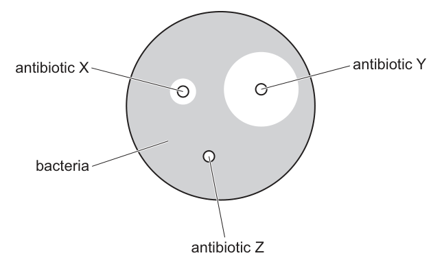
A doctor obtains some bacteria from an infected patient.
The bacteria are grown in a Petri dish containing nutrient agar jelly.
The bacteria produce a grey coating on the agar jelly.
The doctor then adds three paper discs, each containing a different antibiotic.
The diagram shows the Petri dish 24 hours later.
What can the doctor conclude from these results?
Options
A The bacteria causing this patient's infection are not resistant to antibiotic Y.
B Antibiotic X would be more effective than antibiotic Y for treating this patient's infection.
C This patient is resistant to antibiotic Z.
D Antibiotic X is the least effective treatment for this patient's infection.
Working
Antibiotic X shows no clear zone, so the bacteria are resistant to X. Antibiotic Y has the largest clear zone (zone of inhibition), so the bacteria are most sensitive to Y — Y is the most effective, not the least. Antibiotic Z has a small clear zone, so the bacteria are slightly sensitive to Z.
- A: bacteria not resistant to Y — correct, they are killed near Y.
- B: wrong — Y is more effective than X, not the reverse.
- C: wrong — it is the bacteria, not the patient, that show resistance/sensitivity.
- D: wrong — X is the least effective; Y is the most effective.
Answer
A
A
Walkthrough
This is an antibiotic susceptibility test (a disc diffusion test). Paper discs soaked in different antibiotics are placed on agar that has been evenly coated with the patient's bacteria. Each antibiotic diffuses outwards into the jelly, setting up a concentration gradient around its disc. Wherever the antibiotic concentration is high enough to kill or stop the bacteria, the agar stays clear — this is the zone of inhibition.
Read each disc from Fig. 1:
- Antibiotic X — bacteria grow right up to the disc; there is no clear zone at all. The bacteria are resistant to X.
- Antibiotic Y — a very large clear circle surrounds the disc. The bacteria are highly sensitive to Y, so Y would be the most effective treatment.
- Antibiotic Z — a small clear circle. The bacteria are only slightly sensitive to Z.
Now test each option:
- A says the bacteria are not resistant to Y — true, since Y kills them over a wide area. Correct.
- B claims X beats Y — the opposite of what the plate shows.
- C says the patient is resistant to Z. Resistance belongs to the bacteria, never to the person infected; this is a classic trap.
- D calls X the least effective treatment — but the question asks about treating the infection, and X is useless while Y is best; D inverts the finding.
Key Takeaways
- The larger the zone of inhibition around an antibiotic disc, the more sensitive the bacteria are to that antibiotic.
- No clear zone means the bacteria are resistant to that antibiotic.
- Resistance is a property of the bacteria, not of the patient.
- This type of test lets a doctor choose the antibiotic most likely to work before prescribing.
Common Mistakes
- Saying "the patient is resistant" (option C) — mark schemes always reject attributing resistance to the host; it is the pathogen that is resistant.
- Reversing the logic and thinking a big clear zone means the antibiotic is weak — the clear zone is where bacteria have been killed, so bigger = more effective.
- Misreading disc X: bacteria touching the disc means resistance, not effectiveness.
- Confusing "least effective" with "most effective" when comparing X and Y.
Things to Be Careful About
- Always compare clear-zone sizes relative to each other on the same plate before ranking antibiotics.
- Keep the subject straight: the bacteria are resistant or sensitive; the doctor concludes about the bacteria causing the infection.
- Option B and D both hinge on the X-versus-Y comparison — get the direction right: larger zone = better treatment.
The table shows the concentration of five substances in the blood entering the kidney, in fluid entering the nephron and in the urine.
| substance | concentration / blood entering the kidney | concentration / fluid entering the nephron | concentration / urine |
|---|---|---|---|
| urea | 0.4 | 20.0 | 20.0 |
| glucose | 1.5 | 1.5 | 0.0 |
| amino acids | 0.8 | 0.8 | 0.0 |
| ions | 8.0 | 8.0 | 16.5 |
| protein | 82.0 | 0.0 | 0.0 |
Which substances are totally reabsorbed into the blood from the nephron?
Options
A amino acids and glucose
B glucose and ions
C protein and amino acids
D protein and glucose
Working
A substance totally reabsorbed from the nephron back into the blood appears in the fluid entering the nephron but has a concentration of 0.0 in the urine.
- Glucose: 1.5 in nephron fluid, 0.0 in urine — totally reabsorbed.
- Amino acids: 0.8 in nephron fluid, 0.0 in urine — totally reabsorbed.
- Protein: 0.0 in the nephron fluid (too large to be filtered), so it is not reabsorbed — it was never in the nephron.
- Ions: present in urine at 16.5, so not totally reabsorbed.
Answer
A
A
Walkthrough
The kidney filters blood at the glomerulus: water, glucose, amino acids, urea and ions all pass into the nephron, but large protein molecules are too big to cross the filter and stay in the blood — that is why protein reads 0.0 in the fluid entering the nephron.
As the filtrate flows along the nephron, useful substances are reabsorbed back into the blood by selective reabsorption. To find which substances are totally reabsorbed, compare the 'fluid entering the nephron' column with the 'urine' column:
- Glucose enters the nephron at 1.5 mg per dm³ but is absent from urine (0.0) — all of it was reabsorbed.
- Amino acids enter at 0.8 mg per dm³ but are absent from urine — all reabsorbed.
- Ions are still in the urine (16.5), so only some ions are reabsorbed, not all.
- Protein never entered the nephron at all, so it cannot have been reabsorbed — this is the trap option C and D rely on.
So the substances totally reabsorbed are amino acids and glucose → A.
Key Takeaways
- Total reabsorption means: present in the filtrate, concentration 0.0 in the urine.
- Proteins are not filtered at the glomerulus because they are too large — absence from urine does not mean they were reabsorbed.
- Glucose and amino acids are normally completely reabsorbed; their presence in urine signals a problem (e.g. diabetes).
Common Mistakes
- Choosing an option containing protein: protein's 0.0 in urine is because it was never filtered, not because it was reabsorbed. The mark scheme's correct answer excludes protein.
- Confusing "absent from urine" with "reabsorbed" without checking it was present in the nephron fluid first.
- Including ions: they appear in urine at a higher concentration than in blood, so they are only partly reabsorbed.
Things to Be Careful About
- Always check both columns: presence in the nephron fluid AND absence from urine are both needed for "totally reabsorbed".
- Note that ion concentration rises in urine relative to blood — this reflects water being reabsorbed, concentrating what remains.
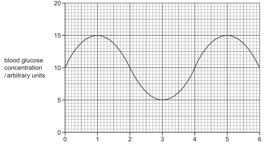
The graph shows blood glucose concentration over 6 hours in a day.
What is the set point for blood glucose concentration shown by this graph?
Options
A 5 arbitrary units
B 10 arbitrary units
C 15 arbitrary units
D 20 arbitrary units
Working
The set point is the normal, steady value that a homeostatic mechanism tries to maintain. In a graph showing homeostatic control, the variable oscillates around this set point. Looking at the graph, the blood glucose concentration oscillates between a maximum of 15 arbitrary units and a minimum of 5 arbitrary units. The midpoint, or set point, around which it oscillates is 10 arbitrary units. The curve starts at 10, crosses 10 at 2 hours, 4 hours, and returns to 10 at 6 hours.
Answer
B
B
Walkthrough
- Understand the concept of a set point: In homeostasis, the body maintains a variable (like blood glucose concentration) around a normal value called the set point. The body uses negative feedback to keep the concentration close to this value.
- Analyze the graph: The y-axis shows blood glucose concentration in arbitrary units (0 to 20). The x-axis shows time in hours (0 to 6).
- Observe the oscillation: The curve goes up to a peak of 15 arbitrary units at 1 hour and 5 hours, and down to a minimum of 5 arbitrary units at 3 hours.
- Find the midpoint: The set point is the central value around which the variable oscillates. It is the average of the maximum and minimum values in a symmetric oscillation: arbitrary units. You can also see that the curve starts at 10, crosses 10 at 2 hours, 4 hours, and returns to 10 at 6 hours.
- Conclude: The set point is 10 arbitrary units, which matches option B.
Key Takeaways
- Homeostasis involves maintaining a biological variable around a set point using negative feedback mechanisms.
- On a graph, the set point is the central value around which the variable oscillates over time.
- Reading the midpoint of a symmetric oscillating curve gives the set point.
Common Mistakes
- Choosing the maximum (15) or minimum (5) value on the graph as the set point instead of the average or central value.
- Misreading the y-axis scale and selecting an incorrect value.
- Confusing the set point with the range of variation or the amplitude of the oscillation.
Things to Be Careful About
- The set point is not the maximum or minimum value reached, but the normal baseline level being regulated by homeostasis.
- Ensure you read the correct axis: the y-axis is for blood glucose concentration, and the x-axis is for time.
- The oscillation is symmetric around 10, confirming it as the set point. The curve crosses this value multiple times as it oscillates between the peaks and troughs.
Which hormone can be used to treat Type 1 diabetes?
Options
A adrenaline
B oestrogen
C glucagon
D insulin
Working
Type 1 diabetes is caused by the failure of the pancreas to produce enough insulin, so blood glucose concentration stays high after meals. Insulin lowers blood glucose by promoting the uptake of glucose into cells and its storage as glycogen. Giving insulin by injection replaces the missing hormone, so it is the treatment for Type 1 diabetes.
- A adrenaline — released in response to stress; raises heart rate and prepares the body for action, not used to treat diabetes.
- B oestrogen — a female sex hormone; not involved in blood glucose control.
- C glucagon — raises blood glucose concentration, the opposite effect to what is needed.
Answer
D
D
Walkthrough
This question asks which hormone is used to treat Type 1 diabetes. You need to know what Type 1 diabetes is and which hormone corrects the problem.
In Type 1 diabetes, the pancreas does not make enough insulin. Insulin is the hormone that lowers blood glucose concentration after a meal: it makes cells take up glucose from the blood and encourages the liver and muscles to store glucose as glycogen. Without enough insulin, blood glucose stays too high. The treatment is to inject insulin, so the missing hormone is replaced.
Now check each option:
- Adrenaline is the 'fight or flight' hormone released in response to stress. It raises heart rate and increases blood glucose to provide energy, so it would make diabetes worse, not better.
- Oestrogen is a female sex hormone involved in the menstrual cycle and secondary sexual characteristics. It has nothing to do with blood glucose control.
- Glucagon is made by the pancreas and does the opposite of insulin — it raises blood glucose by converting glycogen back to glucose. This is the wrong direction for treating diabetes.
- Insulin is the correct answer because it lowers blood glucose and is the hormone that Type 1 diabetics lack.
Key Takeaways
- Type 1 diabetes results from the pancreas producing too little insulin, so blood glucose stays high.
- Insulin lowers blood glucose; it is given by injection to treat Type 1 diabetes.
- Glucagon raises blood glucose — it is the partner hormone with the opposite effect.
- Adrenaline and oestrogen are not involved in treating diabetes.
Common Mistakes
- Choosing glucagon because it is also a pancreatic hormone. Glucagon raises blood glucose, so it would make the problem worse, not better.
- Confusing Type 1 diabetes (insulin deficiency, treated with insulin injections) with Type 2 diabetes, which is managed differently.
- Thinking adrenaline is involved because it raises blood glucose — that effect is for emergency energy, not a treatment.
Things to Be Careful About
- Remember the exact function of insulin: it lowers blood glucose by increasing glucose uptake into cells and promoting glycogen storage.
- Know that the treatment for Type 1 diabetes is insulin injections, not glucagon.
- The mark scheme accepts only the single correct letter, D.
An athlete's resting body temperature is .
After vigorous exercise for 30 minutes, the athlete's body temperature rises to .
The athlete rests for 10 minutes.
After resting, the athlete's body temperature is measured again and is .
Which events take place in the athlete's body during the 10-minute rest to lower their body temperature?
Options
| arterioles near the surface of the skin | heat energy is lost through | |
|---|---|---|
| A | contract | evaporation of sweat |
| B | contract | shivering |
| C | dilate | evaporation of sweat |
| D | dilate | shivering |
Working
After vigorous exercise the body is too hot, so during the rest the body must lose heat.
- Arterioles near the surface of the skin dilate — this increases blood flow to the skin so heat energy is lost to the surroundings.
- Evaporation of sweat — sweat evaporating from the skin carries heat energy away, cooling the body.
- Shivering generates heat, so it would raise body temperature, not lower it.
- Arterioles contracting (vasoconstriction) reduce heat loss, so this would keep heat in the body.
Therefore, the correct events are: arterioles dilate and heat energy is lost through evaporation of sweat.
Answer
C
C
Walkthrough
Read the question carefully: the athlete's temperature rose from 37.0 °C to 38.6 °C during exercise, and during the 10-minute rest it came back down to 37.2 °C. The body needs to get rid of excess heat, so we are looking for mechanisms that remove heat, not ones that make or trap heat.
The body's temperature is controlled by a part of the brain that acts like a thermostat. When the blood temperature is too high, it sends nerve impulses to effectors that cool the body down. Two of the main cooling responses are:
- Vasodilation of skin arterioles — the arterioles near the skin surface widen, so more blood flows through the skin. Warm blood reaches the surface, and heat energy is lost to the cooler surroundings by radiation and convection.
- Sweating and evaporation — sweat glands release sweat onto the skin surface. For sweat to evaporate, it needs heat energy, which it takes from the skin. This removes heat and cools the body.
Now look at the options.
- Row A says arterioles contract and heat lost through evaporation. Contraction of arterioles (vasoconstriction) reduces blood flow to the skin and reduces heat loss — the opposite of what is needed for cooling.
- Row B says arterioles contract and heat lost through shivering. Shivering produces heat by rapid muscle contraction; it warms the body, which is for when you are too cold, not too hot.
- Row C says arterioles dilate and heat lost through evaporation of sweat. Both of these remove heat, so this is correct.
- Row D says arterioles dilate but heat lost through shivering. The dilation is right, but shivering generates heat, so this row is wrong.
So the answer is C.
Key Takeaways
- Body temperature is kept constant by negative feedback: the body detects a change and reverses it.
- When too hot: skin arterioles dilate (vasodilation), sweating increases, and heat is lost to the surroundings.
- When too cold: skin arterioles constrict (vasoconstriction), shivering occurs, and hairs stand up — all of these reduce heat loss or generate heat.
- Evaporation is a cooling process because it uses heat energy from the skin.
Common Mistakes
- Choosing contract (vasoconstriction): constricting skin arterioles reduces blood flow to the skin and slows heat loss. This is for keeping heat in, not for cooling down.
- Choosing shivering: shivering produces heat through muscle activity. It raises body temperature and would be used when the body is too cold, not too hot.
- Forgetting which direction the heat moves: the question asks about lowering temperature, so only heat-losing processes count.
Things to Be Careful About
- Read each row as a pair: for the answer to be correct, both parts of the row must fit the situation.
- The phrase "heat energy is lost through" tells you the mechanism must remove heat; only evaporation of sweat does that here.
- Do not confuse vasodilation (widening, increases heat loss) with vasoconstriction (narrowing, reduces heat loss).
Which endocrine gland produces follicle-stimulating hormone?
Options
A adrenal gland
B ovary
C pancreas
D pituitary gland
Working
FSH (follicle-stimulating hormone) is produced by the pituitary gland, the 'master' endocrine gland at the base of the brain. It travels in the blood to the ovary, where it stimulates follicle development.
- A adrenal gland — produces adrenaline; incorrect.
- B ovary — produces oestrogen and progesterone, not FSH; incorrect.
- C pancreas — produces insulin and glucagon; incorrect.
- D pituitary gland — produces FSH; correct.
Answer
D
D
Walkthrough
The question asks which endocrine gland produces follicle-stimulating hormone (FSH). FSH is one of several hormones released by the pituitary gland, a small gland hanging from the underside of the brain. In females, FSH travels in the blood to the ovaries and stimulates the growth of follicles, each of which contains an egg. In males, FSH stimulates sperm production in the testes. The crucial point is that the gland that PRODUCES FSH is the pituitary, even though FSH acts on the ovary. The ovary itself produces oestrogen and progesterone — a common confusion. The adrenal gland produces adrenaline, and the pancreas produces insulin and glucagon, so none of those can be correct.
Key Takeaways
- The pituitary gland is the 'master' endocrine gland: it produces several hormones that control other glands and organs.
- FSH is produced by the pituitary and acts on the ovary (and testes).
- The ovary produces oestrogen and progesterone, not FSH.
- Know the main endocrine glands and the hormones each one produces.
Common Mistakes
- Confusing the gland that produces a hormone with the organ it acts on. The ovary is the TARGET of FSH, not its source.
- Thinking the ovary produces all female reproductive hormones, including FSH.
- Mixing up the pituitary with the pancreas or adrenal gland.
Things to Be Careful About
- The mark scheme requires the exact term 'pituitary gland' — a vague answer like 'the brain' would not score.
- FSH is specifically a follicle-stimulating hormone; do not confuse it with LH (luteinising hormone), which the pituitary also produces and which triggers ovulation.
The diagram shows a plant grown on its side without light.
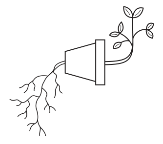
Which row explains the responses shown by the roots and the shoot?
Options
| roots | shoot | |
|---|---|---|
| A | positive gravitropism | positive phototropism |
| B | negative phototropism | positive phototropism |
| C | positive gravitropism | negative gravitropism |
| D | negative gravitropism | positive gravitropism |
Answer
C
C
Walkthrough
The plant is placed horizontally in the dark. Because there is no light, phototropism cannot be occurring. The only directional stimulus present is gravity. The roots are growing downwards, which is towards the pull of gravity. A growth response towards a stimulus is called positive tropism, so the roots are exhibiting positive gravitropism. The shoot is growing upwards, which is away from the pull of gravity. A growth response away from a stimulus is negative tropism, so the shoot is exhibiting negative gravitropism. Looking at the options, row C matches these two responses.
Key Takeaways
- Gravitropism (or geotropism) is the growth response of a plant to gravity.
- Positive gravitropism means growing towards gravity (roots).
- Negative gravitropism means growing away from gravity (shoots).
- Phototropism is the growth response to light. If a plant is in the dark, phototropism is not the cause of directional growth.
Common Mistakes
- Choosing an option with "phototropism" when the question states the plant is "without light".
- Confusing positive and negative tropisms: positive means towards the stimulus, negative means away from it.
- Thinking shoots grow up because of positive gravitropism; they grow up away from gravity, so it is negative gravitropism.
Things to Be Careful About
- Read the stem carefully: "without light" immediately rules out any phototropic responses.
- "Positive" and "negative" refer to the direction of growth relative to the stimulus (towards or away), not the physical direction (up or down) in space. Roots growing down is positive gravitropism; shoots growing up is negative gravitropism.
The tips of growing shoots of a plant are cut off and placed on small blocks of jelly which contain no auxin.
Small pieces of impermeable clear glass are placed in some of the jelly blocks, as shown in diagram X. Some of the shoot tips are divided by impermeable clear glass pieces, as shown in diagram Y.
The tips are left on the small blocks of jelly with light coming from one side only.
After 12 hours, the number of arbitrary units of auxin is measured in each of the jelly blocks and the means, shown in the diagrams, are calculated.
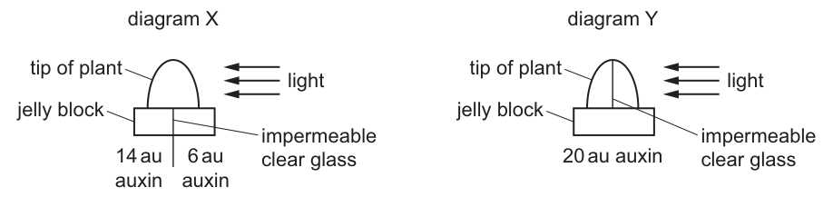
A student wrote down some conclusions from the results.
- Auxin moves downwards out of the shoot tip.
- The distribution of auxin in a shoot tip is affected by light intensity.
- Some auxin is destroyed by light.
- Auxin moves from areas of more light to areas of less light.
Which of the student's conclusions are correct?
Options
A 1, 2, 3 and 4
B 1, 2 and 4 only
C 1 and 3 only
D 2, 3 and 4 only
Working
Evaluate each conclusion against the data provided in the diagrams:
- Conclusion 1: In diagram X, auxin is found in the jelly blocks below the shoot tip (14 au and 6 au), which shows that auxin moves downwards out of the shoot tip. (Correct)
- Conclusion 2: Light is directed from one side only, creating a difference in light intensity across the shoot tip. The unequal distribution of auxin in diagram X (14 au on the shaded side vs 6 au on the lit side) shows that the distribution is affected by light intensity. (Correct)
- Conclusion 3: The total amount of auxin measured in diagram X is au. The total amount in diagram Y is also 20 au. Since the total amount of auxin is conserved, no auxin is destroyed by the light. (Incorrect)
- Conclusion 4: Light comes from the right side. In diagram X, the right side (more light) has 6 au of auxin and the left side (less light) has 14 au. This shows that auxin moves from areas of more light to areas of less light. (Correct)
Conclusions 1, 2, and 4 are correct.
Answer
B
B
Walkthrough
The question presents an experiment investigating how light affects the distribution of auxin in a plant shoot tip. We are given two diagrams, X and Y, and four conclusions written by a student. We must evaluate each conclusion against the data and biological principles.
Diagram X shows a shoot tip on a jelly block that is split into two halves, with an impermeable glass barrier only in the jelly below. Light shines from the right. After 12 hours, the left (shaded) half of the jelly has 14 arbitrary units (au) of auxin, and the right (lit) half has 6 au. The total auxin is au.
Diagram Y shows a shoot tip that is vertically bisected by an impermeable glass piece extending into the jelly block. This prevents any lateral movement of auxin within the tip itself. Light shines from the right. The total auxin measured in the jelly block is 20 au.
Now evaluate the student's conclusions:
- Auxin moves downwards out of the shoot tip. In diagram X, we measure auxin in the jelly blocks below the tip. This directly shows downward movement. (Correct)
- The distribution of auxin in a shoot tip is affected by light intensity. Light comes from one side only, meaning one side of the tip receives more light intensity than the other. The auxin distribution is unequal (14 au vs 6 au), demonstrating that the distribution is affected by this difference in light intensity. (Correct)
- Some auxin is destroyed by light. If light destroyed auxin, the total amount in diagram X would be less than the total in diagram Y (where the tip is blocked and perhaps less exposed, or simply as a baseline). However, the total in X is 20 au and the total in Y is 20 au. The amount is conserved, so no auxin is destroyed. (Incorrect)
- Auxin moves from areas of more light to areas of less light. In diagram X, the glass barrier is only in the jelly, so auxin can move laterally across the tip before moving down. Light is from the right. The right side (more light) has 6 au, and the left side (less light) has 14 au. This proves auxin moved from the lit side to the shaded side. (Correct)
Conclusions 1, 2, and 4 are correct, which matches option B.
Key Takeaways
- Auxin is produced in the shoot tip and moves downwards to promote cell elongation.
- Unilateral light causes auxin to move laterally from the lit side to the shaded side of the tip.
- The total amount of auxin remains constant; light does not destroy it, it only redistributes it.
- Impermeable barriers can be used in experiments to block lateral movement and prove that redistribution requires unobstructed tissue.
Common Mistakes
- Assuming light destroys auxin: Students often see less auxin on the lit side (6 au) and incorrectly conclude it was destroyed, forgetting to check the total amount (20 au) against the control/baseline (20 au in diagram Y).
- Confusing light direction with light intensity: While the experiment uses light from one side, the resulting gradient is a difference in light intensity across the tip. Concluding that distribution is affected by light direction is also true, but in the context of the options, the unequal distribution proves it is affected by the intensity gradient.
- Ignoring the total amount: Failing to add the values in diagram X () makes it impossible to correctly evaluate conclusion 3.
Things to Be Careful About
- Always calculate the total amount of the measured substance when evaluating claims about destruction or loss. Here, matches the 20 au in diagram Y, immediately falsifying the claim that auxin is destroyed.
- Pay close attention to where the impermeable barriers are placed. In diagram X, the barrier is only in the jelly, allowing lateral movement in the tip. In diagram Y, the barrier is in the tip, preventing lateral movement. This contrast is what proves auxin moves laterally from light to shade.
- Read the options carefully. Option B includes 1, 2, and 4, which are the three correct conclusions. Ensure you do not accidentally select an option that includes conclusion 3.
Which part of the flower develops into a fruit?
Options
A ovary
B ovule
C sepal
D stamen
Working
After fertilisation, the ovule develops into the seed, while the ovary wall develops into the fruit. The sepal and stamen do not develop into the fruit.
Answer
A
A
Walkthrough
This question asks you to recall what happens to the parts of a flower after pollination and fertilisation. The key change is:
- the ovary develops into the fruit;
- the ovule inside the ovary develops into the seed;
- the sepal usually withers or remains as a small structure at the base of the fruit;
- the stamen (anther and filament) usually withers and falls off after pollination.
So the correct answer is A, ovary. The other options are wrong because the ovule becomes the seed, not the fruit, and the sepal and stamen do not form the fruit.
Key Takeaways
- After fertilisation, the ovary becomes the fruit and the ovules inside it become the seeds.
- The ovary wall becomes the fruit wall, which protects the seeds and may help in dispersal.
- Knowing the fate of each floral part helps you understand how fruits and seeds form.
Common Mistakes
- Confusing the ovary with the ovule. The ovule becomes the seed, not the fruit.
- Choosing the stamen because it is a prominent part of the flower, but it does not develop into the fruit.
- Forgetting that the sepal is a non-reproductive part that usually does not form the fruit.
Things to Be Careful About
- Read the question carefully: it asks for the part that develops into a fruit, not a seed.
- Remember the exact terms: ovary → fruit, ovule → seed.
- In an exam, you only need to give the letter, so make sure you are confident about the biology before selecting it.
Chromosomes, DNA molecules and genes are pieces of inherited material with different sizes.
Which statement describes the relationship between these pieces of inherited material?
Options
A A chromosome is larger than a gene.
B A gene is larger than a chromosome.
C Multiple chromosomes make up a DNA molecule.
D Multiple DNA molecules make up a gene.
Working
A chromosome is made of a long DNA molecule, and a gene is a short section of a DNA molecule that codes for a protein. So the order of size is:
chromosome > DNA molecule > gene
Therefore a chromosome is larger than a gene. A is correct.
- B is wrong because it reverses the relationship.
- C is wrong — one DNA molecule makes up (part of) a chromosome, not the other way round.
- D is wrong — a gene is part of one DNA molecule, not several DNA molecules.
Answer
A
A
Walkthrough
This question tests the basic size relationship between three pieces of inherited material: chromosomes, DNA molecules and genes.
Start with the smallest unit. A gene is a short section of a DNA molecule that codes for a particular protein. It is only a small part of the whole molecule. Next, a single DNA molecule (along with proteins) makes up a chromosome. So a chromosome is a structure that contains a DNA molecule, and that DNA molecule contains many genes.
The correct order of size is therefore:
chromosome > DNA molecule > gene
So option A "A chromosome is larger than a gene" is correct.
The other options are wrong because:
- B says a gene is larger than a chromosome. This is the wrong way round — a gene is a tiny part of the DNA inside a chromosome.
- C says multiple chromosomes make up a DNA molecule. That is backward — the DNA molecule is part of the chromosome, not the other way round.
- D says multiple DNA molecules make up a gene. A gene is a section of a single DNA molecule, not a collection of DNA molecules.
Key Takeaways
- Know the size order: chromosome > DNA molecule > gene.
- A chromosome contains one DNA molecule. A DNA molecule contains many genes.
- Do not confuse the direction of the relationship: genes are inside DNA, and DNA is inside chromosomes.
Common Mistakes
- Reversing the order, as in option B: thinking a gene is larger than a chromosome.
- Confusing the direction of containment, as in options C and D: it is the chromosome that is built. from a DNA molecule, and the DNA molecule that contains many genes — not the other way round.
Things to Be Careful About
- Read each option as a precise biological statement. The mark is for the exact relationship, not just for the words "chromosome" and "gene" appearing together.
- a "gene" is a segment of a DNA molecule, so it is always smaller than the whole molecule.
- "DNA molecule" is the correct term for the thread that makes up a chromosome. A chromosome is the larger structure containing it.
During the division of a nucleus by meiosis, changes can happen that produce a gamete with an abnormal number of genes or an abnormal number of chromosomes.
Which change can result in a child having Down's syndrome?
Options
A a gamete with one extra chromosome
B a gamete with one less gene
C a gamete with one less chromosome
D a gamete with one extra gene
Working
Down's syndrome is caused by the presence of an extra copy of chromosome 21. This happens when a gamete formed by meiosis carries one extra chromosome, so the child inherits three copies of chromosome 21 instead of two.
- A is correct because an extra chromosome in a gamete can produce Down's syndrome.
- B and D are incorrect because a change in the number of individual genes does not cause Down's syndrome.
- C is incorrect because a gamete with one less chromosome would not produce the extra chromosome 21 that causes Down's syndrome.
Answer
A
A
Walkthrough
In humans, body cells have 23 pairs of chromosomes. Gametes are produced by meiosis, so each gamete normally has one copy of each chromosome. During meiosis, a pair of chromosomes can sometimes fail to separate properly. One gamete may then receive two copies of a chromosome, while another gamete receives none.
If a gamete with two copies of chromosome 21 is fertilised by a normal gamete with one copy, the resulting zygote has three copies of chromosome 21. This condition is called trisomy 21, and it leads to Down's syndrome.
The question asks which change can produce a child with Down's syndrome. The answer must therefore be a gamete with one extra chromosome. Options B and D talk about genes, not chromosomes. A single extra gene or a missing gene does not change the chromosome number and does not cause Down's syndrome. Option C talks about a missing chromosome, which would not give the extra chromosome 21 needed for Down's syndrome; a missing chromosome usually causes a much more severe, often fatal, outcome.
Key Takeaways
- Down's syndrome is caused by an extra chromosome 21, so the child's cells have 47 chromosomes instead of 46.
- This type of change is a chromosome number mutation, not a gene mutation.
- Meiosis is the cell division that produces gametes, and mistakes during meiosis can lead to gametes with the wrong number of chromosomes.
- A single gene being added or missing is not the cause of Down's syndrome.
Common Mistakes
- Choosing option C because the question mentions an "abnormal number of chromosomes". A missing chromosome does not cause Down's syndrome.
- Confusing "gene" with "chromosome". A chromosome carries many genes; an extra gene is not the same as an extra chromosome.
- Thinking Down's syndrome is a gene mutation inherited in a simple dominant or recessive pattern. It is caused by a change in chromosome number.
Things to Be Careful About
- Read the options carefully: B and D are about genes, while A and C are about chromosomes.
- Remember that "one extra chromosome" means an entire extra chromosome, not just an extra copy of one gene.
- Down's syndrome is specifically trisomy 21, so the gamete must carry an extra copy of chromosome 21.
- No calculation is needed here; this is a recall and discrimination question.
Which row shows possible effects of natural selection?
Options
| natural selection can cause mutations to occur | natural selection can lead to the extinction of a species | |
|---|---|---|
| A | no | no |
| B | no | yes |
| C | yes | no |
| D | yes | yes |
Working
Natural selection does not cause mutations. Mutations are random changes in DNA that provide variation; natural selection acts on the variation that is already present.
Natural selection can lead to the extinction of a species. If the environment changes and no individuals are adapted to survive the new conditions, the species may die out.
So the first statement is no and the second statement is yes.
Answer
B
B
Walkthrough
Look at the two statements in the table one at a time.
-
Can natural selection cause mutations to occur? No. Mutations are random changes in the DNA of an organism. They can happen because of mistakes during DNA copying or because of mutagens such as radiation or some chemicals. Natural selection does not make mutations happen. Instead, natural selection works on the variation that mutations have already produced. Without variation, there would be nothing for natural selection to act on.
-
Can natural selection lead to the extinction of a species? Yes. If the environment changes, such as a new predator arriving, the climate becoming warmer, or a food source disappearing, individuals that are not well adapted may die before they can reproduce. If no individuals in the species have the alleles needed to survive the new conditions, the whole species can die out. This is extinction.
The only row that matches “no” for the first statement and “yes” for the second statement is row B.
Key Takeaways
- Mutations are random changes in DNA and are a source of genetic variation.
- Natural selection does not cause mutations; it acts on the variation that already exists in a population.
- Natural selection can change the allele frequencies in a population over generations.
- If a species cannot adapt quickly enough to a changing environment, natural selection can lead to its extinction.
Common Mistakes
- Thinking that natural selection causes mutations because it causes evolution. Evolution happens because natural selection acts on mutations, not because natural selection creates them.
- Choosing A because the student thinks extinction is not an effect of natural selection. Extinction can happen when no individuals are adapted to survive new conditions.
- Choosing C or D because the student confuses mutation with variation. Natural selection does not make mutations occur.
Things to Be Careful About
- Read the table columns carefully: the first column asks whether natural selection can cause mutations, and the second asks whether it can lead to extinction.
- Use precise terms: a mutation is a change in DNA, while natural selection is the process by which better-adapted organisms survive and reproduce.
- Remember that natural selection can change allele frequencies, but it does not generate new alleles by itself. New alleles come from mutation.
All organisms in the same species show variation.
Some features of different types of variation are listed.
- an example is body mass
- has a limited number of possible forms with no intermediates
- has any value from a minimum to a maximum
- is unaffected by the environment
- is usually caused by genes only
Which statements refer to discontinuous variation?
Options
A 1, 3 and 5
B 1 and 3 only
C 2, 4 and 5
D 2 and 4 only
Working
Discontinuous variation has a limited number of distinct forms with no intermediates, is controlled mainly by genes, and is not affected by the environment. Statement 2 describes the limited distinct forms; statement 4 says it is unaffected by the environment; statement 5 says it is caused by genes only. Statements 1 and 3 describe continuous variation: body mass has any value from a minimum to a maximum.
Statements 2, 4 and 5 refer to discontinuous variation.
Answer
C
C
Walkthrough
This question lists five features and asks which ones describe discontinuous variation. Start with what discontinuous variation means: it produces a small number of distinct categories with no intermediates, such as being able or unable to roll the tongue, or having a particular blood group. It is usually controlled by genes only and is not affected much by the environment.
Now test each statement. Statement 1 says an example is body mass. Body mass can take any value between a minimum and a maximum, so it is an example of continuous variation, not discontinuous. Statement 2 says there is a limited number of possible forms with no intermediates — that is the defining feature of discontinuous variation. Statement 3 says the feature can have any value from a minimum to a maximum, which describes continuous variation. Statement 4 says the feature is unaffected by the environment, which is typical of discontinuous variation. Statement 5 says it is usually caused by genes only, which also matches discontinuous variation.
So the correct statements are 2, 4 and 5, which is option C.
Key Takeaways
- Continuous variation shows a range of values with no clear categories. Body mass, height and shoe size are examples.
- Discontinuous variation has a limited set of distinct categories with no intermediates, such as blood group and tongue-rolling.
- Continuous variation is often caused by both genes and the environment; discontinuous variation is usually controlled mainly by genes and is little affected by the environment.
Common Mistakes
- Choosing statement 1 (body mass) as an example of discontinuous variation — body mass varies gradually and has no discrete steps.
- Selecting option D, forgetting statement 5, which is also part of discontinuous variation because it is usually caused by genes only.
- Confusing the feature “unaffected by the environment” with continuous variation; in fact this is another mark of discontinuous variation.
Things to Be Careful About
The question asks only for statements that refer to discontinuous variation, so do not select features that describe continuous variation even if they seem familiar. Remember that statements 1 and 3 both describe continuous variation, so they should not be included.
Bacteria reproduce by splitting into two. This can happen every 20 minutes in ideal conditions of sufficient water, food and at a warm temperature. Small numbers of bacteria do not usually make people ill.
When a student takes her lunch out of the fridge, it contains 4 bacteria. After 2 hours at room temperature, it contains 256 bacteria.
From the time that the food is taken out of the fridge, how long will it take before there are 16 384 bacteria in her food?
Options
A 2 hours
B 3 hours
C 4 hours
D 5 hours
Working
Each division takes 20 minutes. From 4 bacteria to 256 bacteria is 6 divisions (), which takes 2 hours.
To reach 16 384 bacteria:
So 6 more divisions are needed, taking another 2 hours.
Total time hours.
Answer
C
C
Walkthrough
This question is about bacterial reproduction by splitting in two — a form of asexual reproduction called binary fission. Each split doubles the number of bacteria, and the question tells us one split happens every 20 minutes in ideal conditions.
We are given a check-point: starting from 4 bacteria, after 2 hours there are 256. Let's confirm that fits the rule. Two hours is 120 minutes, which is generations. Starting from 4 and doubling 6 times: . That matches, so the rule holds.
Now we need the time to reach 16 384 bacteria. The quickest route is to compare 16 384 with the 256 we already have at 2 hours:
So six more doublings are needed after the 2-hour point. Six doublings at 20 minutes each is minutes, which is 2 hours. Total time from the fridge is therefore hours.
Alternatively, work from the start: , so 12 doublings in total, and minutes hours. Both routes give option C.
Key Takeaways
- Bacteria reproduce asexually by splitting in two (binary fission), and each split doubles the population.
- The doubling time (here 20 minutes) lets you predict population size: after generations, the population is the starting number multiplied by .
- Converting between hours and 20-minute generations is the key step — 2 hours is 6 generations, not 2.
Common Mistakes
- Forgetting that 2 hours contains 6 generations of 20 minutes, not just 2. This leads to treating each hour as one doubling.
- Confusing the doubling count with the time: 12 doublings is 240 minutes, not 12 hours.
- Starting the count from the 2-hour point without adding the first 2 hours back on — this would give 2 hours (option A) instead of 4.
Things to Be Careful About
- The question asks for the time from when the food is taken out of the fridge, so the first 2 hours must be included in the total.
- Work in consistent units: convert hours to minutes or count generations, then convert back.
- The options are spaced 1 hour apart, so a single off-by-one-generation error lands on the wrong option — check that 16 384 is exactly times the starting 4.
What could be an advantage of genetically modifying crop plants?
Options
A increased biodiversity in an area
B increased chance that the inserted genes will transfer to wild species
C increased nutritional content of crop plants
D increased use of pesticides
Working
Genetic modification can add genes to crop plants to give useful traits, such as improved nutritional content. Option C is a genuine advantage. Option A is not a reliable advantage of GM crops, option B is a risk rather than an advantage, and option D is not an advantage because GM is often used to reduce pesticide use.
Answer
C
C
Walkthrough
Genetic modification means taking a gene from one organism and inserting it into the genome of another organism. In crop plants this is done to give the plant a new, useful characteristic, such as resistance to pests, tolerance of herbicides, or improved nutritional value.
The question asks for an advantage of genetically modifying crop plants, so we need a positive outcome.
- A increased biodiversity in an area — this is not a reliable advantage. Growing GM crops often involves large areas of a single crop variety, which can reduce biodiversity rather than increase it.
- B increased chance that the inserted genes will transfer to wild species — this is a risk or concern, not an advantage. Gene flow to wild relatives could have unpredictable effects.
- C increased nutritional content of crop plants — this is a real advantage. For example, golden rice has been genetically modified to contain beta-carotene, a source of vitamin A, to help prevent deficiency.
- D increased use of pesticides — this is not an advantage. Many GM crops are modified to be resistant to pests, which can reduce the need for pesticides.
So the correct answer is C.
Key Takeaways
- Genetic modification of crop plants can add desirable traits such as improved nutritional content, pest resistance, herbicide tolerance and better yield.
- Advantages are positive outcomes for farmers or consumers.
- Risks and concerns, such as genes spreading to wild species or reducing biodiversity, are not advantages.
- When answering multiple-choice questions, read the wording carefully: here the key word is "advantage".
Common Mistakes
- Choosing B because it is a possible effect of GM crops, without noticing that it is a risk, not an advantage.
- Choosing D because some GM crops are linked to herbicide use, without realising that increased pesticide use is not a benefit.
- Choosing A because biodiversity sounds positive, without checking whether GM crops actually increase biodiversity.
Things to Be Careful About
- The question asks for an advantage, so the answer must be a positive consequence.
- "Increased nutritional content" is a clear, well-known advantage of GM crops and is the correct option.
- Do not confuse possible risks of genetic modification with advantages.
An investigation is done to find out how effective different biological washing powders are at digesting protein.
Four equal-sized cubes of cooked egg white (protein) are cut and their masses measured.
The cubes of protein are then placed in test-tubes containing of solutions of four different biological washing powders of the same concentration. The cubes of protein are left in the solutions at room temperature.
After 24 hours, the cubes of protein are removed from the test-tubes and their masses remeasured.
| biological washing powder | mass of cube of protein / start of investigation | mass of cube of protein / end of investigation |
|---|---|---|
| 1 | 10.2 | 8.3 |
| 2 | 10.2 | 7.9 |
| 3 | 10.4 | 8.4 |
| 4 | 10.3 | 8.5 |
What is the percentage decrease in mass of the cube of protein in the most effective washing powder given to one decimal place?
Options
A 17.5%
B 19.2%
C 22.5%
D 29.1%
Working
The most effective powder gives the greatest percentage decrease in mass.
Powder 1:
Powder 2:
Powder 3:
Powder 4:
Powder 2 shows the greatest percentage decrease.
Answer
C
C
Walkthrough
Biological washing powders contain enzymes called proteases that break down protein molecules. The cooked egg white is protein, so a cube left in a powder that works well loses more mass as the protein is digested into smaller soluble products. The most effective powder is therefore the one that causes the greatest percentage decrease in mass.
Because the cubes do not all have exactly the same starting mass, compare percentage decreases rather than just grams lost. For each powder, subtract the end mass from the start mass, divide by the start mass, and multiply by 100.
Powder 1: decrease ; percentage .
Powder 2: decrease ; percentage .
Powder 3: decrease ; percentage .
Powder 4: decrease ; percentage .
Powder 2 has the largest percentage decrease, , so it is the most effective. This matches option C.
Key Takeaways
- Percentage decrease is calculated as .
- In this experiment, most effective means greatest percentage loss of protein mass.
- Biological washing powders contain enzymes that digest protein stains; egg white provides a convenient source of protein for testing.
- A fair test needs the same concentration, volume, cube size and temperature for each powder.
Common Mistakes
- Using the end mass as the denominator. For powder 2 this gives , which is option D. The original (start) mass must be used.
- Comparing only the absolute loss in grams. It happens to give the same answer here, but it is not a fair comparison when starting masses differ slightly.
- Forgetting to round to one decimal place.
- Misreading the table and using the wrong pair of masses.
Things to Be Careful About
- The formula uses the start mass as the denominator.
- Calculate all four values, or at least enough to identify the maximum; the options are the four possible answers.
- Give the answer to one decimal place: , not or .
- The correct option letter is C.
Which enzyme is added to fruit to aid juice production?
Options
A lactase
B lipase
C pectinase
D protease
Working
Pectinase is used in the food industry to break down pectin, the jelly-like substance that holds plant cells together. Adding pectinase to fruit pulp makes the cells separate, releasing more juice and making it clearer. Therefore, the enzyme added to fruit to aid juice production is pectinase.
- Lactase breaks down lactose in milk.
- Lipase breaks down lipids (fats and oils).
- Protease breaks down proteins.
Answer
C
C
Walkthrough
Start by recalling what each named enzyme acts on, because each enzyme is specific to one kind of substrate.
- Lactase breaks down lactose, the sugar found in milk. It is used to make lactose-free milk and other dairy products.
- Lipase breaks down lipids (fats and oils) into fatty acids and glycerol. It is part of the digestive process and also used in some industrial processes.
- Pectinase breaks down pectin, the sticky substance found in the cell walls and middle lamellae of plant cells. Pectin holds plant cells together, so when it is broken down, the cells separate and juice is released more easily. This is exactly what fruit-juice producers want.
- Protease breaks down proteins into amino acids. Proteases are used in biological washing powders and in digestion, not in fruit-juice making.
The question asks which enzyme is added to fruit to aid juice production. Only pectinase fits, because fruit and its juice are plant materials held together by pectin.
So the correct answer is C.
Key Takeaways
- Enzymes are specific: each works on one type of substrate.
- Pectinase is an industrial enzyme used to break down pectin in plant tissues, which releases juice from fruit and makes it clearer.
- Know the four common industrial enzymes in the 5090 syllabus: pectinase (juice production), lactase (lactose-free milk), proteases and lipases (biological washing powders), and pectinase also helps in juice extraction.
Common Mistakes
- Choosing protease because it sounds like something that breaks things down. In fruit juice, the targeted substance is pectin, not protein.
- Choosing lactase if the student confuses "fruit sugar" with the milk sugar lactose. Lactose is found in milk, not in fruit.
- Choosing lipase if the student vaguely remembers "lipids" — fruit juice extraction is not about fats.
Things to Be Careful About
- The question asks specifically for the enzyme that aids juice production from fruit. Pectinase is the only option that breaks down pectin, which is present in fruit pulp.
- Read the names carefully: "pectinase" looks similar to "petrol" in everyday words but it comes from the word pectin, which is the plant substance it breaks down.
- If the options included digestive enzymes, remember that industrial enzymes for fruit juice production are produced by microorganisms and are added directly to the fruit pulp; pectinase is the correct one.
Using the Mark Scheme
The mark scheme directly gives the correct answer as C. No working is required in the exam paper; the reasoning here is only to help you understand why C is correct. In the exam, answer C and move on.
The diagram shows a food web.
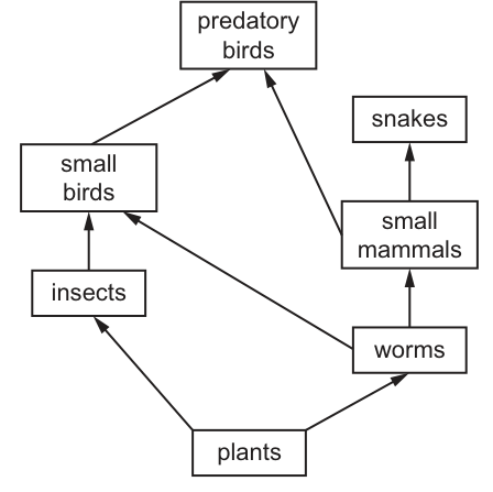
Which organisms in this food web are primary consumers?
Options
A insects
B plants
C small birds
D small mammals
Working
In a food web, arrows show the direction of energy flow, pointing from the organism being eaten to the organism that eats it.
- Producers are at the base of the food web. Here, plants are the producers.
- Primary consumers eat producers. The arrows from plants point to insects and worms, meaning insects and worms eat plants. Therefore, insects and worms are primary consumers.
- Secondary consumers eat primary consumers. Small birds eat insects and worms, and small mammals eat worms. These are secondary (or higher) consumers.
Looking at the options:
- A insects: primary consumer (eats plants).
- B plants: producer.
- C small birds: secondary/tertiary consumer.
- D small mammals: secondary consumer.
The only primary consumer listed in the options is insects.
Answer
A
A
Walkthrough
- Understand the arrows: In a food web diagram, arrows represent the flow of energy and matter. An arrow points from the organism being eaten to the organism that eats it. For example, an arrow from plants to insects means insects eat plants.
- Identify the producer: Producers are organisms that make their own food, usually through photosynthesis. They are at the base of the food web with no arrows pointing to them from other organisms. Here, plants are the producers.
- Identify primary consumers: Primary consumers are herbivores that eat producers. Follow the arrows originating from the plants. Arrows point from plants to insects and worms. This means both insects and worms are primary consumers.
- Identify higher consumers:
- Small mammals eat worms (arrow from worms to small mammals), so they are secondary consumers.
- Small birds eat insects and worms (arrows from insects and worms to small birds), so they are secondary and tertiary consumers.
- Snakes and predatory birds eat small mammals and small birds, making them tertiary or quaternary consumers.
- Evaluate the options:
- A (insects): Eats plants, so it is a primary consumer. This is correct.
- B (plants): These are producers, not consumers.
- C (small birds): Eats insects and worms (which are consumers), so small birds are secondary or tertiary consumers.
- D (small mammals): Eats worms (a primary consumer), so small mammals are secondary consumers.
Key Takeaways
- Arrows in a food web always point from the food source to the consumer (energy flow direction).
- Producers (usually plants) are at the start of food chains.
- Primary consumers are the first animals in a food chain; they eat producers (herbivores).
- Secondary consumers eat primary consumers (carnivores or omnivores).
Common Mistakes
- Reversing the arrow direction: A common error is thinking an arrow from A to B means A eats B. Remember, the arrow shows where the energy goes, so it points to the eater.
- Confusing trophic levels: Thinking that any animal is a primary consumer, or that the top predator is a primary consumer because it is at the top of the diagram.
- Ignoring the producer: Forgetting that primary consumers must eat producers directly. Small mammals eat worms, not plants, so they are not primary consumers.
Things to Be Careful About
- Always trace the arrows back to the producer to determine the trophic level.
- An organism can occupy multiple trophic levels if it eats from different levels (e.g., small birds eat both insects and worms, making them secondary and tertiary consumers). However, the question asks for primary consumers, which only have one trophic level.
- Read the options carefully; sometimes the correct organism is not listed, but here 'insects' is explicitly an option.
The processes listed occur in living organisms in a food chain.
- excretion
- photosynthesis
- respiration
Which processes would result in a loss of energy from a food chain?
Options
A 1 and 2
B 1 and 3
C 2 only
D 3 only
Working
Respiration releases energy from glucose and much of it is eventually lost as heat. Excretion removes waste substances that contain some energy. Photosynthesis is how energy enters the food chain in the first place, so it does not cause loss of energy.
Answer
B
B
Walkthrough
Energy enters a food chain when producers photosynthesise: sunlight energy is trapped and stored in the chemical bonds of glucose. Once this chemical energy passes along a food chain, it does not all move to the next organism. There are many points at which energy is lost.
- Respiration occurs in every living organism at every trophic level. During respiration, energy is released from glucose to power life processes, but not all of that energy is used. A large fraction is lost as heat to the surroundings. This heat energy cannot be passed on to the next trophic level, so respiration is a major loss of energy from a food chain.
- Excretion is the removal of the waste products of metabolism. These waste substances, such as urea, still contain some energy. Excreting them removes that energy from the organism and so from the food chain.
- Photosynthesis is the process by which light energy becomes chemical energy. It supplies energy to the chain, it does not remove it.
Statement 1 (excretion) and statement 3 (respiration) both result in loss of energy. Statement 2 (photosynthesis) does not. So the correct option is B.
Key Takeaways
- Energy enters a food chain only through photosynthesis in producers.
- Energy is measured at each step because organisms use it for respiration and lose some as heat.
- Excretion removes waste materials that still contain trapped energy, so it is another source of energy loss.
- A food chain cannot transmit all of the energy from one trophic level to the next; the loss limits the length of food chains.
Common Mistakes
- Choosing C (2 only) means thinking photosynthesis is a loss, when photosynthesis is actually the energy input.
- Choosing D (3 only) ignores that excretion also removes energy.
- Confusing "loss of energy from the food chain" with mechanisms of energy transfer. This is about loss, so respiration and excretion are the key processes.
- Saying respiration "releases energy" without mentioning that much is lost as heat. The heat is the reason it is a loss in the food chain.
Things to Be Careful About
- The question asks for "loss of energy", so think only of processes that remove energy from the chain. Do not include processes that transfer energy further.
- Excretion here means the removal of waste substances that still contain energy. In the food chain context, materials that are not fully eaten or absorbed also cause loss, but that is not asked for.
- Photosynthesis is the only process that adds energy to the food chain from outside.
Trees in a forest need a constant supply of nitrates to continue growing.
The diagram shows some of the stages of the nitrogen cycle that provide the necessary nitrates.
Which stage in this cycle is not brought about by living bacteria?
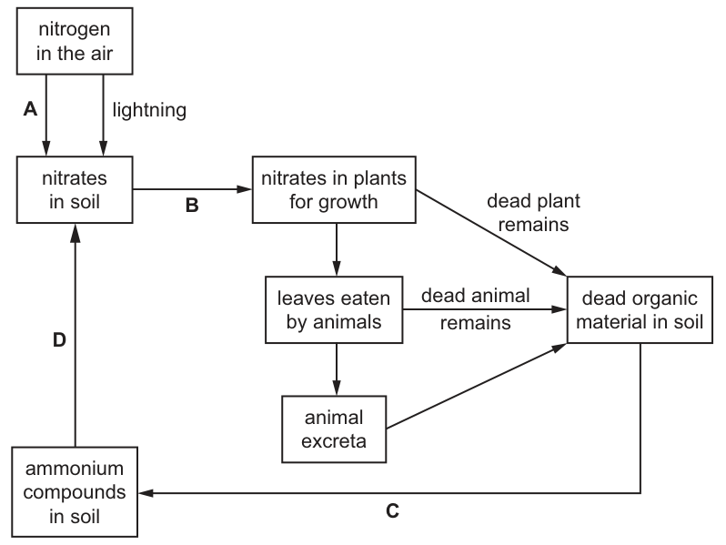
Options
A A
B B
C C
D D
Working
Analyze each stage in the nitrogen cycle diagram:
- Stage A (Nitrogen fixation): Atmospheric nitrogen is converted to nitrates in the soil. This is carried out by nitrogen-fixing bacteria (e.g., Rhizobium, Azotobacter), although lightning (an abiotic process) can also do this.
- Stage B (Absorption): Nitrates in the soil are absorbed by plant roots to form nitrates in plants for growth. This is an active transport process by the plant itself, not by bacteria.
- Stage C (Ammonification / Decomposition): Dead organic material is broken down into ammonium compounds. This is carried out by saprophytic bacteria and fungi.
- Stage D (Nitrification): Ammonium compounds are converted into nitrates. This is carried out by nitrifying bacteria (e.g., Nitrosomonas, Nitrobacter).
Stage B is the uptake of nitrates by plants, which is not brought about by living bacteria.
Answer
B
B
Walkthrough
The question asks to identify the stage in the nitrogen cycle that is not performed by living bacteria. We can analyze each labelled arrow in the diagram:
- Arrow A (Nitrogen fixation): Nitrogen gas from the air is converted into nitrates in the soil. While lightning (shown as a parallel arrow) can fix nitrogen abiotically, the biological pathway for this stage is carried out by nitrogen-fixing bacteria (such as Rhizobium in root nodules or free-living Azotobacter).
- Arrow B (Absorption): Nitrates in the soil are taken up by plant roots to form nitrates in plants for growth. Plants absorb mineral ions like nitrates from the soil solution through their root hair cells via active transport. This process is carried out by the plant itself, not by bacteria.
- Arrow C (Ammonification / Decomposition): Dead organic material (from dead plants, dead animals, and animal excreta) is broken down into ammonium compounds in the soil. This decomposition is carried out by saprophytic bacteria (and fungi) which secrete enzymes to break down proteins and nucleic acids into ammonia/ammonium.
- Arrow D (Nitrification): Ammonium compounds in the soil are converted into nitrates in the soil. This two-step oxidation process (ammonium to nitrite, then nitrite to nitrate) is carried out by nitrifying bacteria such as Nitrosomonas and Nitrobacter.
Therefore, stage B is the only stage not brought about by living bacteria; it is a physiological process of plant growth.
Key Takeaways
- The nitrogen cycle involves several distinct processes: nitrogen fixation, absorption, ammonification, and nitrification.
- Bacteria are central to the nitrogen cycle: nitrogen-fixing bacteria convert atmospheric to usable forms, saprophytic bacteria decompose organic matter into ammonium, and nitrifying bacteria convert ammonium to nitrates.
- Plants do not perform these bacterial conversions; they absorb pre-formed nitrates from the soil through their roots to synthesize their own amino acids and proteins.
Common Mistakes
- Confusing nitrogen fixation with absorption: Students might look at arrow A and see 'lightning' and think it's the only abiotic process, forgetting that bacteria also perform fixation. Conversely, they might think plants make nitrates from ammonium, confusing absorption with nitrification.
- Overlooking the role of plants: Assuming all movement of nitrogen in the soil-plant system is bacterial. Plants actively absorb nitrates (arrow B) using energy (ATP) from respiration; this is not a bacterial action.
- Misidentifying decomposition: Thinking arrow C is only fungi. While fungi are involved, saprophytic bacteria are the primary agents credited in the 5090 syllabus for ammonification.
Things to Be Careful About
- Read the diagram labels carefully: Arrow B goes from 'nitrates in soil' to 'nitrates in plants'. This is clearly uptake/absorption. Arrow A goes from 'nitrogen in the air' to 'nitrates in soil'. This is fixation.
- Remember the command 'not': The question asks for the stage not brought about by bacteria. Four options are given; three involve bacteria (A, C, D), one involves plants (B).
- Abiotic factors: Lightning is an abiotic factor that fixes nitrogen (part of arrow A's pathway), but the question asks about stages brought about by living bacteria. Since bacteria also do A, C, and D, B is the clear outlier as it is a plant process.
What is a method of conserving fish stocks?
Options
A fishing in protected areas
B fishing with nets with a small mesh size
C having closed fishing seasons
D only catching young, immature fish
Working
Closed fishing seasons stop fishing during the breeding period, so fish can reproduce and maintain the population.
- A is wrong: fishing in protected areas removes fish from exactly the places set aside to protect them.
- B is wrong: nets with a small mesh size catch young, small fish, reducing the breeding stock.
- C is correct: having closed fishing seasons protects fish while they are breeding.
- D is wrong: catching young, immature fish stops them from ever breeding.
Answer
C
C
Walkthrough
This question asks which of the four listed practices is a genuine method of conserving fish stocks. Conservation means managing a resource so that it is not used up and can continue to supply future generations. For fish, that means making sure enough fish survive to breed and replace the ones that are caught.
Look at each option in turn:
- A — fishing in protected areas. Protected areas (often called marine reserves or no-fishing zones) are places where fishing is banned so fish can breed safely. Fishing in them would defeat the whole purpose, so this is not a conservation method — it is the opposite.
- B — fishing with nets with a small mesh size. A small mesh catches everything, including very young, small fish. Removing young fish before they have bred reduces the number that will grow up to replace the stock. This damages the population, so it is not conservation.
- C — having closed fishing seasons. This is a real conservation method. During the breeding season, fishing is banned for a period, so adult fish can reproduce undisturbed. The young produced then restock the population. This is the correct answer.
- D — only catching young, immature fish. Immature fish have not yet bred. If they are all caught, they never get the chance to reproduce, and the population collapses. This is the opposite of conservation.
So the only option that actually conserves fish stocks is C.
Key Takeaways
- Conservation of fish stocks means managing fishing so that enough fish survive to breed and replace those removed.
- Common conservation methods include closed seasons (no fishing during breeding), size limits (only catching fish above a minimum size), quotas (limits on how many can be caught), and protected areas where fishing is banned.
- Practices that catch young, immature fish, or that catch fish during the breeding season, reduce the future population and are not sustainable.
Common Mistakes
- Choosing A — thinking that "protected areas" sounds positive. But the option says fishing in protected areas, which is the opposite of protection.
- Choosing B — thinking a small mesh catches more fish and is therefore efficient. The mark scheme rejects this because small mesh removes young fish that have not bred.
- Choosing D — thinking that leaving the large fish is good. In fact, catching immature fish prevents them from ever breeding, so the stock cannot be maintained.
Things to Be Careful About
- Read the exact wording of each option. "Fishing in protected areas" is not the same as "having protected areas".
- The key idea for conservation of fish is protecting the breeding stock — the fish that can reproduce. Any practice that removes breeding-age or immature fish is harmful.
- This question is one mark, so the answer is a single letter. Do not add extra explanation on the paper — just write C.
Your score so far
Answer a question to start scoring
Your marks add up here as you work through the paper.