Biology 5090/11 — May/June 2025
Cambridge O-Level · Multiple Choice · answer key with instant marking and worked solutions
Topics Coordination and Control · Cell Structure and Organisation · Transport in Humans · Enzymes · Disease and Immunity · Excretion · +11 more
Tap an option under each question to check it — your score builds as you go.
The diagram shows a plant cell.
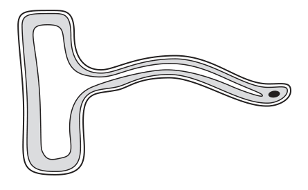
What is the function of this cell?
Options
A to carry mineral ions along stems
B to photosynthesise
C to take in carbon dioxide
D to take in water
Working
The diagram shows a plant cell with a long, thin projection extending from the main cell body, and a nucleus located near the tip of this projection. This is the characteristic shape of a root hair cell.
Root hair cells are specialised cells found in the roots of plants. Their elongated shape greatly increases the surface area in contact with the soil. Their primary function is to absorb water from the soil by osmosis, as well as mineral ions.
Evaluating the options:
- A: Carrying mineral ions along stems is the function of xylem vessels.
- B: Photosynthesis is the function of palisade mesophyll cells in the leaf.
- C: Taking in carbon dioxide is carried out by mesophyll cells for photosynthesis.
- D: Taking in water is the correct function for a root hair cell.
Answer
D
D
Walkthrough
- Identify the cell: The diagram shows a plant cell with a distinct long, narrow extension (projection) and a nucleus positioned near the tip of this extension. This morphology is the classic textbook representation of a root hair cell.
- Recall the function: Root hair cells are specialised cells located on the surface of plant roots. Their main role is to absorb water and mineral ions from the soil. The long projection increases the surface area, making absorption more efficient. Water is absorbed by osmosis down a water potential gradient.
- Evaluate the options:
- Option A: Transporting mineral ions along the stem is the function of xylem vessels, not root hair cells.
- Option B: Photosynthesis occurs in palisade mesophyll cells in the leaves, which are packed with chloroplasts. Root hair cells lack chloroplasts and are found underground in the dark.
- Option C: Taking in carbon dioxide is done by mesophyll cells in the leaf, with the gas entering through stomata.
- Option D: Taking in water is the primary function of root hair cells, matching the cell identified in the diagram.
Key Takeaways
- Root hair cells are specialised plant cells with a long, thin projection that increases surface area for absorption.
- Their main function is to absorb water (by osmosis) and mineral ions (by active transport) from the soil.
- Different specialised plant cells have different functions: xylem for transport, palisade mesophyll for photosynthesis, and root hair cells for absorption.
Common Mistakes
- Misidentifying the cell: Confusing root hair cells with other specialised cells like palisade mesophyll (which are rectangular and packed with chloroplasts) or xylem (which are hollow tubes).
- Forgetting the dual function: Root hair cells absorb both water and mineral ions. A candidate might hesitate if they only remember one, but in a multiple-choice format, you select the best available match.
- Ignoring the location: Root hair cells are found in the roots, underground. Any function related to gas exchange (carbon dioxide) or photosynthesis (which requires light) is immediately incorrect for a root cell.
Things to Be Careful About
- Look for the shape: The elongated projection is the key visual cue for a root hair cell. Always match the diagram's morphology to the correct specialised cell type.
- Read all options: Options A, B, and C describe valid biological processes, but they are the functions of other plant tissues (xylem, palisade mesophyll). Ensure the function matches the specific cell shown.
- Water vs. ions: While root hair cells absorb both, the question options separate them. Water uptake is the most fundamental and voluminous function of the root hair cell, making D the unambiguously correct choice here.
The diagram shows a structure that was observed using a microscope.
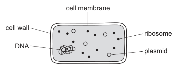
What does the diagram show?
Options
A an animal cell
B a bacterium
C a plant cell
D a virus
Working
The diagram shows a cell with a cell wall, a cell membrane, ribosomes, a single loop of DNA that is not enclosed in a nucleus, and plasmids. A nucleus, mitochondria, chloroplasts and a sap vacuole are absent, so it is not an animal or plant cell. It is far too large and complex to be a virus, which has no cell wall, membrane or ribosomes. These are the defining features of a bacterium.
Answer
B
B
Walkthrough
The figure labels four structures: a cell wall, a cell membrane, ribosomes, and DNA shown as a tangled loop, plus small rings labelled plasmids. Each label is a clue.
-
DNA as a free loop, with no nucleus. In animal and plant cells the DNA is enclosed inside a nucleus. Here the DNA floats freely in the cytoplasm as a single circular loop — the hallmark of a prokaryote. That alone rules out options A and C.
-
Plasmids. These small extra rings of DNA are found in bacteria and are a classic exam giveaway. Animal and plant cells do not have them.
-
Ribosomes but no other organelles. Bacteria do have ribosomes (they make proteins), but they lack mitochondria, chloroplasts and a sap vacuole. A plant cell would show chloroplasts and a vacuole; an animal cell would show a nucleus and no cell wall.
-
Not a virus. A virus is not a cell at all: it has no cell wall, no cell membrane, no cytoplasm and no ribosomes — just genetic material in a protein coat, far smaller than any cell. The labelled cell wall, membrane and ribosomes eliminate option D immediately.
So the structure is a bacterium — option B.
Key Takeaways
- The defining features of a bacterial cell: cell wall, cell membrane, cytoplasm, ribosomes, a single circular loop of DNA with no true nucleus, and often plasmids.
- Prokaryotes (bacteria) have no membrane-bound organelles — no nucleus, mitochondria or chloroplasts.
- A virus is not a cell: no membrane, no cytoplasm, no ribosomes.
Common Mistakes
- Choosing C (plant cell) because a cell wall is present — bacteria also have cell walls, but theirs surround a cell with no nucleus and no chloroplasts.
- Choosing A (animal cell) because no chloroplasts are shown — but the free DNA loop and plasmids are not animal-cell features.
- Choosing D (virus) because the structure is small and simple — but viruses have no cell wall, membrane or ribosomes at all.
- Thinking the labelled 'DNA' means there is a nucleus; the diagram shows the DNA loose in the cytoplasm, not enclosed.
Things to Be Careful About
- Check for a nucleus first: its absence with free circular DNA means prokaryote.
- Plasmids are the strongest single clue for a bacterium.
- Do not assume 'has a cell wall' means 'plant' — bacteria have cell walls too (not made of cellulose).
- In MCQs, eliminate options by finding one feature each option cannot have, rather than looking for a perfect match.
The diagram shows the structures found in cell X.
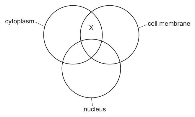
Which type of cell is X?
Options
A liver cell
B red blood cell
C root hair cell
D xylem vessel
Working
The Venn diagram shows that cell X is located in the intersection of the 'cytoplasm' and 'cell membrane' circles, but outside the 'nucleus' circle. This means cell X has a cell membrane and cytoplasm, but no nucleus.
- A liver cell is a typical animal cell and contains a nucleus.
- A root hair cell is a plant cell and contains a nucleus.
- A xylem vessel is dead at maturity and contains neither cytoplasm nor a nucleus (only a cell wall).
- A mature mammalian red blood cell lacks a nucleus to maximise space for haemoglobin, but it does contain a cell membrane and cytoplasm.
Therefore, cell X must be a red blood cell.
Answer
B
B
Walkthrough
The question presents a Venn diagram with three overlapping circles representing 'cytoplasm', 'cell membrane', and 'nucleus'. The region marked X is the overlap between 'cytoplasm' and 'cell membrane' only, meaning it excludes the 'nucleus'. We must identify which of the four given cell types has a cell membrane and cytoplasm but no nucleus.
- Liver cells (option A) are typical animal cells; they have a nucleus, cytoplasm, and cell membrane. This does not match region X.
- Red blood cells (option B), specifically mature mammalian red blood cells, lose their nucleus during maturation to make more room for haemoglobin. They retain a cell membrane and cytoplasm. This matches region X perfectly.
- Root hair cells (option C) are specialised plant cells for absorption; they have a nucleus, cytoplasm, cell membrane, cell wall, and a large vacuole. This does not match region X.
- Xylem vessels (option D) are dead at functional maturity; they have no cytoplasm and no nucleus, only a lignified cell wall. This does not match region X.
Key Takeaways
- Venn diagrams can be used to represent the presence or absence of cellular structures. Reading them requires identifying which regions are included and which are excluded.
- Specialised cells often have unique adaptations. Mature mammalian red blood cells lack a nucleus, while xylem vessels lack both nucleus and cytoplasm. Typical animal and plant cells retain a nucleus.
Common Mistakes
- Choosing xylem vessel (D) by forgetting that xylem vessels are dead and lack cytoplasm.
- Choosing liver cell (A) or root hair cell (C) by overlooking that both contain a nucleus, which is explicitly excluded from region X.
- Confusing red blood cells with other blood cells like white blood cells, which do have a nucleus.
Things to Be Careful About
- Read the Venn diagram carefully: X is in the overlap of cytoplasm and cell membrane, but outside the nucleus circle. This means the cell has cytoplasm and a cell membrane, but no nucleus.
- Remember that only mature mammalian red blood cells lack a nucleus; other red blood cells (e.g., in birds or amphibians) do have a nucleus. The 5090 syllabus focuses on mammalian blood cells in this context.
- Xylem vessels are dead at maturity, so they have no cytoplasm or nucleus, only a cell wall. Do not confuse them with living cells that simply lack a nucleus.
What is an example of an organ?
Options
A digestive system
B eye
C blood
D neurone
Working
An organ is made of different tissues working together to carry out a particular function.
The eye contains several tissues, such as muscle, nerve and blood, so it is an organ.
The digestive system is an organ system, blood is a tissue, and a neurone is a cell.
Answer
B
B
Walkthrough
The question asks you to identify which option is an organ. In the 5090 syllabus, the levels of organisation are: cell, tissue, organ, organ system and organism.
- A cell is the basic unit of life. A neurone is a single nerve cell, so option D is a cell, not an organ.
- A tissue is a group of similar cells working together. Blood is a tissue because it is made of red blood cells, white blood cells and platelets suspended in plasma. So option C is a tissue.
- An organ is made of different tissues working together to perform a specific function. The eye contains muscle tissue, nervous tissue and blood, all working together for vision, so option B is an organ.
- An organ system is a group of organs working together. The digestive system includes the stomach, intestines, liver and other organs, so option A is an organ system.
Therefore the correct answer is B.
Key Takeaways
- Know the order of biological organisation: cell → tissue → organ → organ system → organism.
- Be able to give examples of each level: e.g. neurone (cell), blood (tissue), eye (organ), digestive system (organ system).
- An organ is always made of more than one type of tissue.
Common Mistakes
- Choosing blood because it is a liquid: blood is a tissue, not an organ.
- Choosing the digestive system because it contains organs: it is an organ system, not a single organ.
- Choosing neurone because it has a specialised function: it is a single cell, not an organ.
Things to Be Careful About
- Read the options carefully and classify each one before deciding.
- The eye is a classic example of an organ in the syllabus.
- Do not confuse "organ" with "organ system"; the digestive system is a group of organs, not one organ.
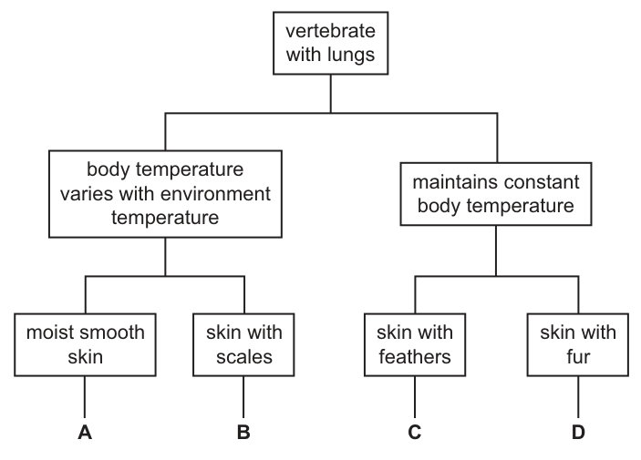
A new type of small vertebrate with lungs is discovered. This is identified as a reptile.
Which vertebrate in the key is a reptile?
Options
A A
B B
C C
D D
Answer
B
A reptile is an ectothermic (cold-blooded) vertebrate with lungs and skin covered in dry scales. Following the key:
- Start at vertebrate with lungs.
- Reptiles do not maintain a constant body temperature, so follow the branch body temperature varies with environment temperature.
- Reptiles have dry skin with scales, so follow the branch skin with scales.
- This leads to B.
The other options represent:
- A: Amphibian (moist smooth skin, variable body temperature).
- C: Bird (feathers, constant body temperature).
- D: Mammal (fur, constant body temperature).
B
Walkthrough
The question asks us to identify a reptile using a dichotomous key. A dichotomous key is a tool that splits organisms into two groups at each step based on a contrasting pair of features. We start at the top and follow the branches that match the features of our target organism — a reptile.
- Start at the top: The key begins with vertebrate with lungs. All four options (A, B, C, D) are vertebrates with lungs, so we proceed to the next split.
- Body temperature: The key splits into body temperature varies with environment temperature (ectothermic / cold-blooded) and maintains constant body temperature (endothermic / warm-blooded). Reptiles are ectothermic; they rely on the environment to regulate their body heat. Birds and mammals are endothermic. So we follow the left branch: body temperature varies with environment temperature.
- Skin covering: This branch splits into moist smooth skin and skin with scales. Amphibians (like frogs) have moist, smooth, permeable skin used for gas exchange. Reptiles have dry skin covered in keratinised scales to prevent water loss. So we follow the branch skin with scales.
- Result: This leads directly to B.
For completeness, let us identify the other options:
- A (moist smooth skin, variable temperature) describes an amphibian.
- C (skin with feathers, constant temperature) describes a bird.
- D (skin with fur, constant temperature) describes a mammal.
Key Takeaways
- A dichotomous key works by making a series of binary choices based on observable features. Always start at the top and follow the path that matches the organism.
- The four main groups of lung-bearing vertebrates (tetrapods) can be distinguished by two key features: thermoregulation (constant vs. variable body temperature) and skin covering (feathers, fur, scales, or moist smooth skin).
- Reptiles are ectothermic vertebrates with dry, scaly skin.
Common Mistakes
- Following the wrong branch at the first split: Some candidates see "lungs" and immediately think of mammals or birds, forgetting that reptiles and amphibians also have lungs. They must use the next feature (body temperature) to split the group.
- Confusing reptiles with amphibians: Choosing A because both are ectothermic vertebrates. The deciding feature is the skin: amphibians have moist, smooth, permeable skin, while reptiles have dry, scaly skin.
- Misreading the key: Reading "skin with scales" as applying to the constant-temperature branch. Always trace the lines carefully from the box you are in.
Things to Be Careful About
- Follow the key strictly: Do not skip steps or jump to the answer based on prior knowledge without verifying through the key's stated features. The key is the authority for the question.
- Read feature descriptions precisely: "Moist smooth skin" and "skin with scales" are mutually exclusive in this key. Ensure you match the exact wording.
- Know the basic vertebrate groups: At O Level, you must be able to recall the defining features of amphibians, reptiles, birds, and mammals. If you do not know whether a reptile is warm-blooded or cold-blooded, you cannot use the key correctly.
Molecules and ions can be moved into and out of cells by active transport.
Which statement about active transport is not correct?
Options
A Active transport takes place only in plants.
B Active transport moves molecules and ions against a concentration gradient.
C Active transport uses energy to move molecules and ions.
D Molecules and ions are moved across partially permeable membranes by active transport.
Working
Active transport is the movement of molecules and ions across a partially permeable membrane against a concentration gradient, using energy released by respiration. It occurs in both plants (e.g. root hair cells taking up mineral ions) and animals (e.g. glucose absorption in the villi), so statement A is not correct.
- A — Incorrect: active transport occurs in animals as well as plants.
- B — Correct: it moves substances against the concentration gradient.
- C — Correct: it requires energy.
- D — Correct: it occurs across partially permeable membranes.
Answer
A
A
Walkthrough
The question lists four statements about active transport and asks which one is not correct. The best approach is to recall the exact definition of active transport from the 5090 syllabus: movement of molecules and ions across a partially permeable membrane against a concentration gradient, using energy released by respiration.
Check each statement against that definition:
- A says active transport takes place only in plants. This is false. Active transport happens in animal cells too — for example, the villi of the small intestine use active transport to absorb glucose, and the kidney tubules use it to reabsorb useful substances. So this is the incorrect statement.
- B says active transport moves substances against a concentration gradient. This is exactly the definition, so it is correct.
- C says active transport uses energy. Correct — this energy comes from respiration.
- D says substances are moved across partially permeable membranes. Correct — the cell surface membrane is partially permeable, and active transport moves substances across it.
Since only A is wrong, A is the answer.
Key Takeaways
- Active transport moves substances against the concentration gradient, from a lower to a higher concentration.
- It requires energy released by respiration.
- It happens across partially permeable membranes.
- Active transport occurs in both plants and animals — root hair cells in plants take up mineral ions this way, and the villi in humans absorb glucose this way.
- This is the opposite of diffusion, which is passive and moves substances down the concentration gradient.
Common Mistakes
- Choosing B, C or D because they are all true statements — the question asks for the statement that is not correct, so you must identify the false one.
- Thinking active transport only happens in plants because the syllabus's main example is root hair cells. Remember the villi example from Human Nutrition.
- Confusing active transport with osmosis or diffusion — those are passive processes that do not use energy.
Things to Be Careful About
- Read the question stem carefully: it asks for the statement that is not correct, not the correct one.
- The mark scheme requires the letter A — the answer is the option letter, not a rewritten statement.
- Remember the exact wording: "against a concentration gradient" and "partially permeable membrane" are the precise terms the syllabus uses; vague phrases like "against the gradient" or "through a membrane" would not be credited in a written answer.
Frogs are amphibians and so have a partially permeable skin. They live in and near water such as lakes and marshes, where the water contains a low concentration of mineral ions. They absorb any water they need through their skin.
Biologists are concerned that frogs living near coasts will be endangered if global warming leads to a rise in sea level. Sea water contains a high concentration of mineral ions.
What would happen to the frogs if their habitat became flooded with sea water?
Options
A Water would move into the frog by osmosis.
B Mineral ions would move out of the frog by diffusion.
C Water would move into the frog by diffusion.
D Water would move out of the frog by osmosis.
Working
Sea water has a high concentration of mineral ions, so its water potential is lower than the water potential of the frog's body fluids. Water moves by osmosis from the region of higher water potential (inside the frog) to the region of lower water potential (the sea water).
Therefore water would move out of the frog by osmosis.
Answer
D
D
Walkthrough
The frog's skin is partially permeable, which means water can pass through it but dissolved substances (like mineral ions) cannot pass through freely. This is exactly the condition needed for osmosis to occur.
Osmosis is the net movement of water molecules from a region of higher water potential to a region of lower water potential, through a partially permeable membrane.
The key is to work out which side has the higher water potential.
- Fresh water (the frog's normal habitat) contains a low concentration of mineral ions, so it has a high water potential.
- Sea water contains a high concentration of mineral ions, so it has a low water potential.
- The frog's body fluids contain some mineral ions, so their water potential is somewhere in between — higher than sea water but lower than fresh water.
If the habitat becomes flooded with sea water, the frog's body fluids now have a HIGHER water potential than the surrounding sea water. Water therefore moves by osmosis out of the frog, across its partially permeable skin, into the sea water. The frog would lose water and could become dehydrated — this is why the biologists are concerned.
So the correct answer is D.
Key Takeaways
- Osmosis is the movement of water, not of dissolved substances, through a partially permeable membrane.
- Water always moves from a region of higher water potential to a region of lower water potential.
- A high concentration of dissolved ions means a LOW water potential; a low concentration of dissolved ions means a HIGH water potential.
- An animal with a partially permeable skin in a more concentrated solution loses water by osmosis.
Common Mistakes
- Choosing A (water moves into the frog by osmosis): this confuses the direction. Water moves INTO the frog only in fresh water, where the surrounding water has a higher water potential than the frog's body. In sea water the gradient is reversed.
- Choosing C (water moves into the frog by diffusion): water does not move by diffusion — the movement of water through a partially permeable membrane is osmosis. Diffusion is the movement of other substances (like dissolved gases or ions).
- Choosing B (mineral ions move out of the frog by diffusion): the concentration of mineral ions is higher in sea water than in the frog, so if anything, ions would tend to diffuse INTO the frog, not out. Also, the question is about what happens to the water, not the ions.
- Thinking that "high ion concentration" means "lots of water" — it means the opposite: less water available, so a lower water potential.
Things to Be Careful About
- The mark scheme requires the precise term osmosis, not "diffusion" or "water moving out".
- State the direction correctly: water moves OUT of the frog because the sea water has a lower water potential than the frog's body fluids.
- Remember that a partially permeable membrane lets water through but not dissolved ions — this is why the ions themselves do not equalise the concentrations.
- On an O Level paper, "high concentration of mineral ions" is a direct clue for "low water potential" — connect the two ideas explicitly in your reasoning.
The diagram shows an enzyme molecule.
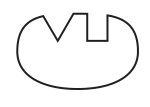
Which diagram shows the substrate molecule for this enzyme?
Options
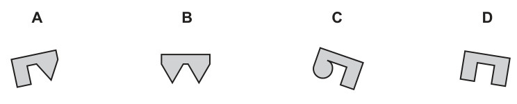
Answer
A
A
Walkthrough
The lock-and-key hypothesis explains enzyme specificity: the substrate molecule must have a shape that is exactly complementary to the active site of the enzyme. The active site is the region on the enzyme where the substrate binds. In Fig. 5, the enzyme's active site has two distinct notches: one is triangular (V-shaped) and the other is square (rectangular).
To find the correct substrate, we look for a molecule with protrusions that match these notches perfectly. A triangular protrusion will fit into the V-shaped notch, and a rectangular protrusion will fit into the square notch.
- Option A has a rectangular protrusion and a triangular/angled protrusion that are complementary to the enzyme's active site.
- Option B has two triangular protrusions, which would not fill the square notch.
- Option C has a circular and a rectangular protrusion, which do not match the triangular notch.
- Option D has two rectangular protrusions, which would not fill the triangular notch.
Therefore, molecule A is the correct substrate.
Key Takeaways
- Enzyme-substrate reactions are highly specific because the substrate must fit the active site.
- The lock-and-key model requires the substrate to have a shape complementary to the active site (protrusions on the substrate match notches on the enzyme).
Common Mistakes
- Choosing an option that matches only one of the notches but not the other (e.g., picking a shape with only triangles or only rectangles).
- Selecting a shape that is identical to the active site rather than complementary (the substrate needs bumps where the enzyme has holes).
Things to Be Careful About
- Always check all features of the active site; a partial match is not enough to score the mark.
- Remember that the substrate and active site are complementary, not identical in shape.
The enzyme amylase catalyses the breakdown of starch into maltose.
The effect of different pHs on the time it took for this breakdown to occur when all other variables were controlled was investigated.
Which graph shows the results of this investigation?
Options
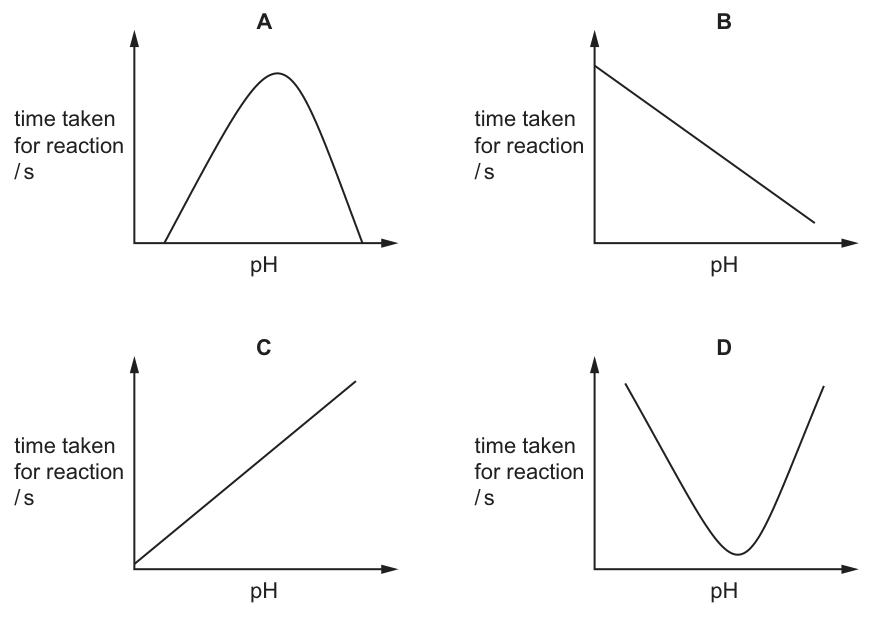
Answer
D
D
Walkthrough
The investigation measures the time taken for the enzyme amylase to break down starch at different pH values. Enzymes have an optimum pH at which their activity (rate of reaction) is highest. At the optimum pH, the reaction is completed as quickly as possible, so the time taken is at its minimum. As the pH moves away from the optimum in either direction, the enzyme's active site changes shape (denaturation) or substrate binding is less effective, so the rate of reaction decreases and the time taken increases. This produces a U-shaped curve with a minimum at the optimum pH. Graph D shows exactly this. Graph A shows a peak at the optimum pH, which would be correct if the y-axis were 'rate of reaction', but since the y-axis is 'time taken', A is the inverse of the correct answer. Graphs B and C show linear relationships, which do not reflect enzyme behaviour.
Key Takeaways
- Enzymes have an optimum pH where the rate of reaction is maximum.
- The time taken for a reaction is inversely related to the rate of reaction: highest rate = shortest time.
- Graphs of 'time taken' against a factor like pH or temperature are U-shaped (minimum at optimum), whereas graphs of 'rate of reaction' are bell-shaped (maximum at optimum).
Common Mistakes
- Choosing Graph A because it is the familiar bell-shaped curve for enzyme activity. This happens when the candidate forgets that the y-axis is 'time taken' rather than 'rate of reaction'.
- Assuming a linear relationship between pH and enzyme activity.
Things to Be Careful About
- Always check the label on the y-axis. 'Time taken' and 'rate of reaction' are inversely proportional. A peak in rate is a trough in time.
- The optimum pH for amylase is around 7 (neutral), but the exact value does not matter for identifying the shape of the curve; only the fact that there is a minimum time at the optimum pH is needed.
What are features of enzymes?
- They act on specific substrates.
- They are carbohydrates.
- They are catalysts.
- They are involved in some, but not all, metabolic reactions.
Options
A 1, 2 and 3
B 1 and 3 only
C 1 and 4
D 2, 3 and 4
Working
Statement 1: true — each enzyme acts on a specific substrate because its active site has a particular shape (lock-and-key).
Statement 2: false — enzymes are proteins, not carbohydrates.
Statement 3: true — enzymes are biological catalysts that speed up reactions without being used up.
Statement 4: false — enzymes are involved in essentially all metabolic reactions, not just some.
The true statements are 1 and 3 only.
Answer
B
B
Walkthrough
This question lists four statements about enzymes and asks which of them are true. The cleanest way to answer is to judge each statement one at a time, then match the true ones to the options.
Statement 1 — "They act on specific substrates." This is true. Each enzyme has an active site with a particular shape that only its specific substrate can fit into — the lock-and-key model. For example, amylase only breaks down starch, not proteins or fats.
Statement 2 — "They are carbohydrates." This is false. Enzymes are proteins. Being a protein is a defining feature of an enzyme, so any statement that calls an enzyme a carbohydrate is wrong.
Statement 3 — "They are catalysts." This is true. Enzymes are biological catalysts: they speed up chemical reactions without being used up or permanently changed themselves.
Statement 4 — "They are involved in some, but not all, metabolic reactions." This is false. Enzymes are involved in essentially all the metabolic reactions that happen in living organisms. Every metabolic reaction is catalysed by an enzyme, so "some, but not all" is incorrect.
The two true statements are 1 and 3, which matches option B.
Key Takeaways
- Enzymes are protein catalysts — never carbohydrates.
- Each enzyme is specific to one substrate because of the shape of its active site (lock-and-key).
- Enzymes catalyse all metabolic reactions, not just a selection of them.
- When a multiple-choice question lists numbered statements, judge each statement independently before matching to the options.
Common Mistakes
- Choosing statement 2 because of confusion between carbohydrates and proteins — enzymes are always proteins.
- Choosing statement 4 by thinking that some reactions do not need enzymes — in fact every metabolic reaction is enzyme-catalysed.
- Picking option C (1 and 4) by accepting statement 4 without checking it carefully.
Things to Be Careful About
- Read each numbered statement on its own merits before looking at the options.
- Statement 4 is the trap: the phrase "some, but not all" is wrong because enzymes catalyse all metabolic reactions.
- The mark scheme gives only the single letter B — there is no partial credit for getting two statements right but choosing the wrong option.
The diagram shows a transverse section through a leaf.
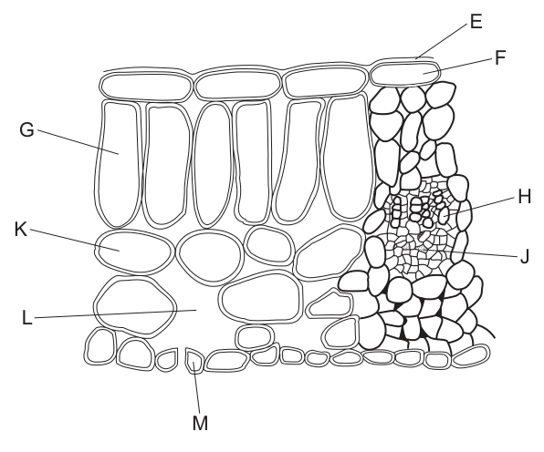
In which parts of the leaf can photosynthesis take place?
Options
A K and L
B E and F
C G and M
D H and J
Working
Photosynthesis takes place in cells that contain chloroplasts. In a transverse section of a leaf:
- G (palisade mesophyll cells) contain many chloroplasts.
- M (guard cells) contain chloroplasts.
- K (spongy mesophyll cells) contain chloroplasts, but L is an air space and contains no cells.
- E (cuticle), F (upper epidermis), H (xylem), and J (phloem) do not contain chloroplasts.
Evaluating the options:
- A: K and L — L is an air space, so no photosynthesis.
- B: E and F — neither contains chloroplasts.
- C: G and M — both contain chloroplasts and can carry out photosynthesis.
- D: H and J — vascular tissue, no chloroplasts.
Answer
C
C
Walkthrough
The question asks to identify which labelled parts of the leaf cross-section can carry out photosynthesis. Photosynthesis requires the pigment chlorophyll, which is located inside chloroplasts. Therefore, we must identify which of the labelled structures are living cells that contain chloroplasts.
Let us examine each label based on the diagram of a typical dicotyledonous leaf:
- E is the waxy cuticle, a non-living layer that prevents water loss. No photosynthesis.
- F is the upper epidermis. Epidermal cells are transparent and lack chloroplasts to allow light to reach the mesophyll below. No photosynthesis.
- G points to the palisade mesophyll cells. These are elongated cells packed tightly together just below the upper epidermis. They contain the highest concentration of chloroplasts in the leaf and are the main site of photosynthesis.
- H is the xylem within the vascular bundle. Xylem vessels are dead, hollow tubes for water transport and contain no chloroplasts.
- J is the phloem. Phloem sieve tubes lack most organelles, including chloroplasts, for sugar transport.
- K is the spongy mesophyll cell. These cells do contain chloroplasts and can photosynthesise, but they are loosely arranged with large air spaces between them.
- L points to an air space within the spongy mesophyll. An air space is a gap filled with gas, not a cell, so it cannot carry out photosynthesis.
- M points to a guard cell surrounding the stoma. Unlike ordinary epidermal cells, guard cells contain chloroplasts and can carry out photosynthesis to help regulate the opening of the stoma.
Now we evaluate the given options:
- A (K and L): K is a spongy mesophyll cell (can photosynthesise), but L is an air space (cannot). Incorrect.
- B (E and F): Neither the cuticle nor the upper epidermis contains chloroplasts. Incorrect.
- C (G and M): Both the palisade mesophyll cell (G) and the guard cell (M) contain chloroplasts. Correct.
- D (H and J): Xylem and phloem are vascular tissues that do not contain chloroplasts. Incorrect.
Key Takeaways
- Photosynthesis only occurs in cells containing chloroplasts.
- In a leaf, the palisade mesophyll, spongy mesophyll, and guard cells contain chloroplasts.
- Epidermal cells, the cuticle, air spaces, and vascular tissues (xylem and phloem) do not contain chloroplasts.
Common Mistakes
- Confusing spongy mesophyll with air spaces: Candidates often see K (spongy mesophyll) and L (air space) and assume both are cells. Remember that L is a gap filled with gas, not a cell.
- Forgetting that guard cells have chloroplasts: Many students assume only the mesophyll cells have chloroplasts and that guard cells are like ordinary epidermal cells. Guard cells are unique among epidermal cells because they do contain chloroplasts.
- Misidentifying vascular tissue: Xylem and phloem are transport tissues and do not carry out photosynthesis.
Things to Be Careful About
- Always read the diagram labels carefully. L points to the empty space between spongy mesophyll cells, not to a cell itself.
- Remember that the question asks where photosynthesis can take place, not just where the majority of it occurs. Both palisade and guard cells are capable of photosynthesis.
- In multiple-choice questions with combinations of labels, eliminate options containing any structure that cannot perform the process. Here, eliminating L, E, F, H, and J leaves only option C.
Which transverse section photomicrograph shows the position of the xylem and phloem in a non-woody dicotyledonous stem?
Options
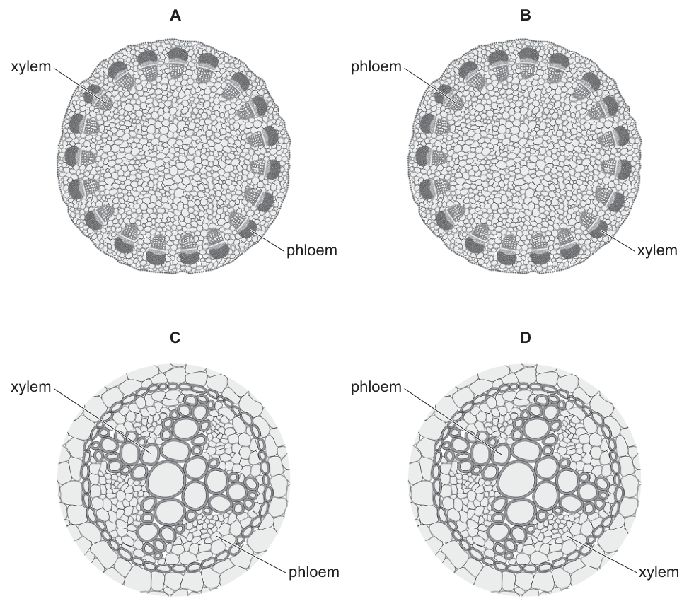
Working
The question asks for a non-woody dicotyledonous stem.
- In a dicot stem, vascular bundles are arranged in a ring around the periphery. This matches diagrams A and B. Diagrams C and D show a central vascular cylinder, which is characteristic of a root, so they are eliminated.
- Within each vascular bundle in a dicot stem, the xylem is positioned towards the inside (centre) and the phloem is positioned towards the outside (periphery).
- Diagram A correctly labels the inner tissue as xylem and the outer tissue as phloem. Diagram B has the labels reversed.
Answer
A
A
Walkthrough
- Identify the organ: The question specifies a "non-woody dicotyledonous stem". Look at the arrangements. Diagrams A and B show vascular bundles arranged in a distinct ring around the edge of the cross-section. This is the classic arrangement for a dicot stem. Diagrams C and D show a central vascular cylinder (stele) with xylem forming a central star or cross shape. This is characteristic of a dicot root, not a stem. So we can eliminate C and D.
- Identify the tissue positions: In a dicot stem vascular bundle, the xylem (which transports water and minerals upwards) is located towards the inside (centripetal side) of the bundle, closer to the pith. The phloem (which transports sugars) is located towards the outside (centrifugal side), closer to the epidermis. Diagram A has these labels correct; diagram B has them swapped.
- Conclusion: Diagram A is the only correct representation of a dicot stem cross-section.
Key Takeaways
- Dicot stem cross-sections show vascular bundles arranged in a ring; dicot root cross-sections show a central vascular cylinder.
- Within a dicot stem vascular bundle, xylem is always towards the centre and phloem is towards the periphery.
Common Mistakes
- Confusing stem and root cross-sections: roots have a central vascular cylinder (often with a star-shaped xylem core), whereas stems have a ring of discrete vascular bundles.
- Reversing xylem and phloem positions: in stems, xylem is always towards the centre and phloem towards the periphery. Students may incorrectly assume phloem is inner because it is mentioned first in some textbooks, but the positional rule is strict.
Things to Be Careful About
- Read the question carefully: it asks for a "stem", not a "root". C and D are roots.
- Pay attention to the labels on the diagrams. A and B have the correct anatomical arrangement but B has the wrong labels.
- Distinguish between monocot and dicot stems if asked (monocots have scattered bundles, dicots have a ring). Here, the ring arrangement confirms dicot.
Students set up apparatus to measure the uptake of water by a leafy shoot.
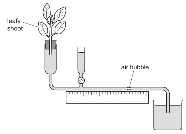
They recorded the position of the air bubble at the start. They observed the apparatus for 10 minutes, and recorded the distance travelled by the air bubble at one-minute intervals.
They then switched on a fan pointed towards the leafy shoot and recorded the distance travelled by the air bubble from the start, each minute, for another 10 minutes.
Which graph would they draw from their data?
Options
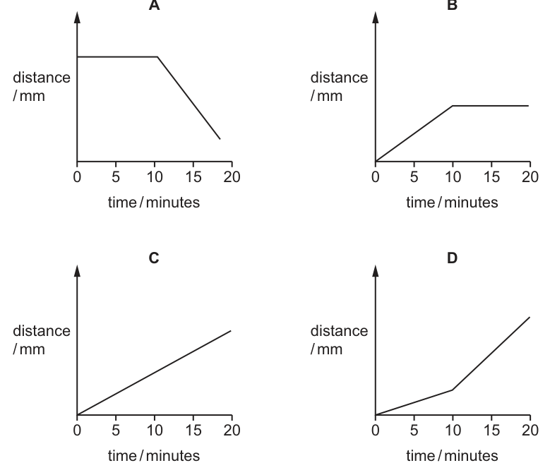
Working
A potometer measures the rate of water uptake by a leafy shoot, which is approximately equal to the rate of transpiration. As the plant loses water vapour through the leaves, it takes up water from the apparatus, moving the air bubble. The data recorded is the "distance travelled ... from the start", which is a cumulative distance. Therefore, the graph of distance against time will always go upwards (positive slope), and the gradient (slope) of the line represents the rate of water uptake.
- First 10 minutes (0–10 min): The shoot transpires at a normal rate. Water is taken up steadily, so the air bubble moves a constant distance each minute. The graph is a straight line with a constant positive slope.
- Next 10 minutes (10–20 min): A fan is switched on. Air movement over the leaves removes water vapour more quickly, maintaining a steeper water potential gradient between the leaf interior and the surrounding air. This increases the rate of transpiration. Consequently, the plant takes up water faster, and the air bubble moves further each minute. The rate is higher, so the slope of the distance-time graph must be steeper.
Comparing the options:
- A: Shows distance decreasing; incorrect as uptake is cumulative and always increases.
- B: Shows uptake stopping (flat line); incorrect as the fan increases uptake.
- C: Shows a constant rate throughout; incorrect as the fan changes the rate.
- D: Shows a moderate slope initially (0–10 min) and a steeper slope afterwards (10–20 min), representing an increased rate of uptake. This matches the expected results.
Answer
D
D
Walkthrough
The question asks to predict the shape of a graph based on an experiment using a potometer. We need to break this down into three parts: what the apparatus measures, what the fan does, and how to read the graph.
1. What the apparatus measures:
The image shows a potometer. A leafy shoot is sealed into the apparatus. As the leaves transpire (lose water vapour), the plant pulls water up the xylem to replace it. This water comes from the reservoir through the capillary tube. As water moves into the plant, the air bubble in the capillary tube moves towards the plant. The question states they record the "distance travelled by the air bubble from the start". This is a cumulative distance. If the bubble moves 2 mm in minute 1 and 2 mm in minute 2, the recorded distance at minute 2 is 4 mm. Therefore, the y-axis (distance) must always increase. The plant is taking up water, so the bubble never moves back towards the reservoir (unless there's a leak or pressure change, but we assume ideal conditions). This eliminates graph A, which shows distance decreasing.
2. The effect of the fan:
Transpiration is driven by the water potential gradient between the inside of the leaf (high water potential, moist air) and the outside air (lower water potential). A fan blows air across the leaves. This removes the layer of humid air that builds up around the stomata and replaces it with drier air. This steepens the water potential gradient, causing water to diffuse out of the stomata faster. Therefore, the rate of transpiration increases.
Since water uptake is driven by transpiration (the transpiration pull), the rate of water uptake also increases.
3. Interpreting the graph:
The graph plots cumulative distance (y-axis) against time (x-axis). In a distance-time graph, the gradient (slope) represents speed or rate.
- 0 to 10 minutes: Normal conditions. Steady transpiration rate. The graph is a straight line with a constant slope.
- 10 to 20 minutes: Fan is on. Transpiration rate is higher. The bubble moves further each minute. The rate is faster, so the line must be steeper.
Graph D shows a line starting with a moderate slope (0-10 min) and then bending upwards to a steeper slope (10-20 min). This correctly represents an increase in rate. Graph C shows a constant rate (incorrect). Graph B shows the rate dropping to zero (incorrect).
Key Takeaways
- A potometer measures water uptake, which is a proxy for transpiration rate.
- The data recorded is usually cumulative distance, so the graph of distance vs time always has a positive slope.
- The slope of a distance-time graph represents the rate. A steeper slope means a faster rate.
- Air movement (wind/fan) increases the rate of transpiration by removing water vapour and maintaining a steep water potential gradient.
Common Mistakes
- Confusing rate with cumulative distance: Students might look for a graph that shows "rate" (which would be a flat line at a low value, then a flat line at a higher value). But the axis is "distance", so the line must keep going up, just faster.
- Thinking the bubble moves back: Graph A shows distance decreasing. This would happen if water was being pushed out, but here the plant is pulling water in.
- Thinking transpiration stops: Graph B shows the line going flat. This would happen if the leaves were removed or the stomata closed, but a fan increases transpiration.
- Ignoring the fan's effect: Graph C assumes the rate stays constant. Students must remember that wind is a factor affecting transpiration rate.
Things to Be Careful About
- Axis labels: Always check what is on the y-axis. Here it is "distance / mm" (cumulative), not "rate". If it were rate, the graph would look like steps (flat, then higher flat). Since it's distance, it's a slope that changes.
- Cumulative vs instantaneous: The text says "recorded the distance travelled by the air bubble from the start". This confirms it is cumulative. If it said "distance travelled in that minute", the graph would be different (roughly constant then higher constant).
- Slope interpretation: Remember that in a distance-time graph, a curve upwards means acceleration (increasing rate). Here, the change is step-wise (fan switched on), so it's two straight lines with different slopes. Graph D correctly shows two linear segments with the second being steeper.
The percentages of each of the gases in inspired and expired air are measured when a student is resting and when a student is exercising.
The results are shown.
| gas | percentage in inspired air | percentage in expired air when resting | percentage in expired air when exercising |
|---|---|---|---|
| nitrogen | 78 | 78 | 78 |
| oxygen | 21 | 16 | 14.5 |
| carbon dioxide | 0.04 | 5.04 | 6.54 |
From the data, which statement is not correct?
Options
A The difference between the percentages of carbon dioxide in expired air when exercising and in expired air when resting is 1.5.
B When resting, the difference between the percentage of oxygen in inspired and expired air is due to oxygen being used by respiration in the body.
C Inspired air contains 52.5 times more oxygen than carbon dioxide.
D The percentage of oxygen taken from inspired air is the same as the percentage of carbon dioxide added to expired air when exercising.
Working
Check each statement against the data.
A: — correct.
B: ; oxygen is used by respiration — correct.
C: , not — not correct.
D: ; — correct.
Answer
C
C
Walkthrough
This question asks you to check each statement against the data in the table and find the one that is not correct. Work through each option one at a time.
Option A: Subtract the resting expired carbon dioxide from the exercising expired carbon dioxide: . This matches the statement exactly, so A is correct.
Option B: When resting, inspired oxygen is 21% and expired oxygen is 16%, a difference of 5%. The body uses oxygen in respiration to release energy, and carbon dioxide is produced as a waste product. This statement is correct.
Option C: The statement claims inspired air contains 52.5 times more oxygen than carbon dioxide. Inspired oxygen is 21% and carbon dioxide is 0.04%. The ratio is times, not 52.5. This is a factor-of-ten error, so C is not correct — this is the answer.
Option D: When exercising, oxygen taken from inspired air is %. Carbon dioxide added to expired air is %. These are equal, so D is correct.
Therefore the incorrect statement is C.
Key Takeaways
- During gas exchange, oxygen is taken up by the body and carbon dioxide is produced during respiration.
- When exercising, the body needs more energy, so more oxygen is used and more carbon dioxide is produced — expired air during exercise has less oxygen and more carbon dioxide than at rest.
- Nitrogen is not used or produced by the body, so its percentage stays at 78% in inspired and expired air.
- Always check calculations carefully when a statement gives a specific number, especially ratios.
Common Mistakes
- Miscalculating the ratio: , not 52.5. A factor-of-ten slip makes option C look correct.
- Misreading the question: it asks for the statement that is not correct, so the answer is the one that fails the check, not the one that passes.
- Assuming option A is wrong without doing the subtraction — exactly.
- Ignoring the tiny carbon dioxide value in inspired air (0.04%) when comparing ratios — this is what makes the ratio so large.
Things to Be Careful About
- Read the command carefully: "which statement is not correct" — the answer is the false statement.
- Do the arithmetic precisely: .
- In option D, both calculations give 6.5%, so the statement is correct even though the numbers look different at first glance.
- Note the units are percentages throughout — no conversion needed.
Which balanced chemical equation shows aerobic respiration?
Options
A
B
C
D
Working
Aerobic respiration uses glucose and oxygen and produces carbon dioxide and water:
- A is the reverse reaction — this is photosynthesis.
- B shows glucose breaking down to lactic acid — anaerobic respiration in humans.
- C shows glucose breaking down to ethanol and carbon dioxide — anaerobic respiration in yeast.
- D matches the balanced equation for aerobic respiration.
Answer
D
D
Walkthrough
Aerobic respiration is the release of energy from glucose using oxygen. The reactants are glucose and oxygen; the products are carbon dioxide and water. Option D shows exactly this:
The equation balances: there are 6 carbon atoms, 12 hydrogen atoms and 18 oxygen atoms on each side.
Option A is the reverse — it is the summary equation for photosynthesis, where carbon dioxide and water are used with light energy to make glucose and oxygen. The arrow points the wrong way for respiration.
Option B shows glucose breaking down to lactic acid (). This is anaerobic respiration in humans, which happens when oxygen is in short supply, for example during vigorous exercise.
Option C shows glucose breaking down to ethanol and carbon dioxide:
This is anaerobic respiration in yeast, used in brewing and bread-making.
So only D is aerobic respiration.
Key Takeaways
- Aerobic respiration: glucose + oxygen → carbon dioxide + water.
- The equation must balance — count the atoms on both sides.
- Anaerobic respiration in humans: glucose → lactic acid.
- Anaerobic respiration in yeast: glucose → ethanol + carbon dioxide.
- Photosynthesis is the reverse of aerobic respiration.
Common Mistakes
- Choosing A: that is photosynthesis, not respiration — the arrow points the wrong way.
- Confusing B and C: both are anaerobic respiration, but in different organisms. B is humans (lactic acid), C is yeast (ethanol).
- Forgetting that aerobic respiration requires oxygen as a reactant, so oxygen must appear on the left-hand side of the equation.
Things to Be Careful About
- The syllabus expects both the word equation and the balanced symbol equation for aerobic respiration; memorise both.
- Check the direction of the arrow — respiration consumes glucose and oxygen, it does not make them.
Blood is made up from a number of different components.
The pie chart shows the percentage volume of each of the components of blood.
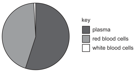
What is the percentage volume of blood for the component that has the function of transporting oxygen?
Options
A 1%
B 30%
C 44%
D 55%
Answer
Oxygen is transported by red blood cells.
Looking at the pie chart (Fig. 8) and its key:
- Plasma (dark grey) is approximately 55%.
- Red blood cells (light grey) are approximately 44%.
- White blood cells (white) are approximately 1%.
The percentage volume for the component transporting oxygen (red blood cells) is 44%.
Answer
C
C
Walkthrough
- Identify the component: The question asks for the blood component that transports oxygen. Recall that red blood cells (erythrocytes) contain the pigment haemoglobin, which binds to oxygen in the lungs and transports it to the body's tissues. Plasma transports dissolved substances (like glucose, amino acids, carbon dioxide), and white blood cells are involved in immunity.
- Read the chart: Look at Fig. 8. The key indicates:
- Dark grey = plasma.
- Light grey = red blood cells.
- White = white blood cells.
The light grey section (red blood cells) occupies roughly 44% of the circle (almost half, slightly less than the dark grey plasma section which is about 55%).
- Match to options: The value 44% corresponds to option C.
Key Takeaways
- Blood is composed mainly of plasma (~55%), red blood cells (~44%), and a small layer of white blood cells and platelets (~1%).
- The primary function of red blood cells is oxygen transport due to their haemoglobin content.
- Pie charts represent parts of a whole as percentages adding up to 100%.
Common Mistakes
- Choosing plasma (55%): A candidate might choose the largest percentage (55%) without checking which component it represents, or might mistakenly think plasma carries the oxygen (it carries dissolved carbon dioxide and some oxygen, but the main transport is via red blood cells).
- Choosing white blood cells (1%): Confusing the function of white blood cells (fighting infection) with oxygen transport.
- Misreading the chart: Assuming the light grey section is 30% (option B) without estimating the angle or area correctly. 44% is close to half (50%), and the light grey section is clearly almost half the circle.
Things to Be Careful About
- Function recall: Ensure you know that red blood cells, not plasma, are responsible for the bulk of oxygen transport.
- Chart reading: The chart is not to scale with exact numbers, but the description and visual estimation confirm red blood cells are around 44%. The options are 1%, 30%, 44%, 55%. 55% is plasma. 1% is white blood cells. 44% is red blood cells. 30% is a distractor not matching any major component in this simplified model.
- Option B (30%): This is a distractor, possibly for someone misreading the light grey section or thinking of a different composition.
What is the total number of atria and ventricles that the blood passes through during two complete circuits of the human body?
Options
A 2
B 4
C 8
D 16
Working
In one complete circuit of the human body, blood passes through the heart twice: once through the right side (to the lungs) and once through the left side (to the body). Each time it passes through one atrium and one ventricle.
So in one complete circuit, blood passes through:
- right atrium
- right ventricle
- left atrium
- left ventricle
That is 4 chambers in one circuit.
In two complete circuits:
Answer
C
C
Walkthrough
The human circulatory system is a double circulation: blood passes through the heart twice for every complete journey around the body.
Let's trace one complete circuit. Blood leaves the left ventricle through the aorta, travels around the body, and returns to the right atrium. It then goes to the right ventricle, is pumped to the lungs, returns to the left atrium, and finally enters the left ventricle again before leaving through the aorta. So in one complete circuit, blood passes through all four chambers: right atrium, right ventricle, left atrium, left ventricle.
The question asks for the total number of atria and ventricles the blood passes through during two complete circuits. Since one circuit involves 4 chambers, two circuits involve chambers.
Therefore the correct option is C.
Key Takeaways
- The heart has four chambers: two atria and two ventricles.
- In a double circulation, blood passes through the heart twice per complete circuit of the body.
- Each passage through the heart involves one atrium and one ventricle.
- To count chambers, multiply the number of circuits by 4.
Common Mistakes
- Counting only one side of the heart, giving 2 or 4 instead of 8.
- Thinking one complete circuit means passing through the heart only once.
- Confusing the number of times blood enters the heart with the number of chambers it enters.
- Choosing 16 by counting each chamber twice per circuit incorrectly.
Things to Be Careful About
- The question asks for the number of atria and ventricles, not the number of times blood enters the heart.
- Remember that a complete circuit includes both the pulmonary and systemic circulations.
- Read the options carefully: 8 is the correct total for two complete circuits.
The table shows the range of percentages of different types of tissue found in the walls of the aorta of people of different ages.
Collagen tissue is present in the walls of blood vessels to provide strength and support.
| type of tissue | percentage (%) young adult | percentage (%) old adult |
|---|---|---|
| elastic | 38–46 | 28–40 |
| muscle | 14–21 | 9–37 |
| collagen | 13–36 | 24–51 |
Which conclusion about the range of percentages is correct?
Options
A The range of collagen tissue found in people of both age groups is 39.
B The range of each type of tissue is smaller in old adults than in young adults.
C The range of muscle tissue in old adults is four times greater than in young adults.
D The range of elastic tissue in people of both age groups is the greatest of all three types of tissue.
Working
Range = highest value − lowest value.
Elastic: young ; old .
Muscle: young ; old .
Collagen: young ; old .
Check each option:
- A: collagen combined range , not 39. Wrong.
- B: elastic range is larger in old adults (12) than in young adults (8). Wrong.
- C: muscle old range 28 is young range 7. Correct.
- D: elastic combined range , smaller than collagen (38) and muscle (28). Wrong.
Answer
C
C
Walkthrough
This question tests whether you can work out the range of a set of data and then compare ranges. The range is the difference between the highest and lowest values. For each tissue type, the table gives a range of percentages for young adults and for old adults.
First, work out the range for each tissue in each age group:
- Elastic: young ; old .
- Muscle: young ; old .
- Collagen: young ; old .
Now check each conclusion:
- A says the range of collagen in both age groups combined is 39. The lowest collagen value is 13 (young) and the highest is 51 (old), so the combined range is , not 39. A is wrong.
- B says the range of each tissue is smaller in old adults. But elastic is 8 in young adults and 12 in old adults — it is larger in old adults. B is wrong.
- C says the range of muscle in old adults is four times greater than in young adults. Young muscle range is 7, old muscle range is 28, and . C is correct.
- D says elastic has the greatest combined range of all three tissues. Elastic combined range is , which is less than collagen (38) and muscle (28). D is wrong.
The aorta is an artery, and its wall contains elastic tissue (to stretch and recoil as blood is pumped out of the heart), muscle (to control the diameter of the vessel) and collagen (to provide strength and support). As people age, the proportions of these tissues change, which is why the ranges differ between young and old adults.
Key Takeaways
- The range of a data set is the difference between the largest and smallest values.
- When a table shows a range such as 38–46, the range as a single number is .
- To compare combined ranges across groups, use the overall lowest and highest values across both columns.
- Read each conclusion carefully and check it against your calculated values before choosing.
Common Mistakes
- Confusing the range written in the table (e.g. 38–46) with the range as a single number ().
- For option A, forgetting to combine both age groups: the lowest collagen value is in young adults (13) and the highest is in old adults (51), giving 38, not 39.
- For option B, assuming all ranges shrink with age without checking each tissue — elastic actually increases from 8 to 12.
- Arithmetic errors in subtraction, especially with the larger numbers such as .
Things to Be Careful About
- The range is highest minus lowest, not the midpoint of the two values.
- When the question says "in people of both age groups", you must use the overall minimum and maximum across both columns.
- Check every option, not just the one that looks plausible — the correct answer is the only one that is arithmetically true.
The table shows information about three different blood vessels, P, Q and R.
One of the vessels is an artery, one is a capillary and one is a vein.
| blood vessel | P | Q | R |
|---|---|---|---|
| thickness of wall / | 500 | <1 | 1000 |
| diameter of lumen / | 5 | 0.008 | 4 |
Using the information from the table, what is the cross-sectional area of the lumen of the vein?
(Assume )
Options
A
B
C
D
Working
The capillary is Q (wall < 1 , lumen 0.008 mm).
Between P and R, the artery has the thicker wall and the smaller lumen, so R (wall 1000 , lumen 4 mm) is the artery and P (wall 500 , lumen 5 mm) is the vein.
Vein lumen diameter = 5 mm, so radius = 2.5 mm.
Answer
B
B
Walkthrough
First, identify which vessel is which. The capillary has the thinnest wall and the smallest lumen, so Q (wall < 1 , lumen 0.008 mm) is clearly the capillary.
That leaves P and R. An artery has a thicker wall and a smaller lumen than a vein. R has a wall of 1000 and a lumen of 4 mm; P has a wall of 500 and a lumen of 5 mm. So R is the artery and P is the vein. This makes sense biologically: veins carry blood at lower pressure, so their walls are thinner, and they have a wider lumen to hold a greater volume of blood.
The vein's lumen diameter is 5 mm. The cross-sectional area of a circular lumen is given by , where is the radius. The radius is half the diameter:
Rounded to one decimal place, this is 19.6 , which is option B.
Key Takeaways
- Arteries have thick, muscular, elastic walls and a relatively small lumen; veins have thinner walls and a larger lumen; capillaries have walls only one cell thick and a tiny lumen.
- The cross-sectional area of a circular lumen is , and the radius is always half the diameter.
- When a table gives structural data, use the distinguishing features (wall thickness and lumen size) to identify each vessel before doing any calculation.
Common Mistakes
- Confusing the artery and the vein. The artery has the thicker wall (1000 ) and the smaller lumen (4 mm), so R is the artery. Choosing R as the vein gives option A (12.6 ).
- Using the diameter instead of the radius. (option D) is the trap for forgetting to halve the diameter.
- Using the artery's lumen. (option A) uses the artery's radius; (option C) uses the artery's diameter as if it were the radius.
Things to Be Careful About
- Read the question carefully: it asks for the vein's lumen area, so identify the vein first.
- Always halve the diameter to get the radius before squaring it in the area formula.
- Use the value of given in the question (3.14), not 3.14159.
- The final value 19.625 rounds to 19.6 , matching option B exactly.
The table shows the number of deaths estimated to be due to smoking in England each year from 2009 to 2019.
| year | number of deaths estimated to be due to smoking |
|---|---|
| 2009 | 82 000 |
| 2010 | 80 300 |
| 2011 | 78 600 |
| 2012 | 78 700 |
| 2013 | 78 200 |
| 2014 | 77 800 |
| 2015 | 79 100 |
| 2016 | 77 900 |
| 2017 | 77 800 |
| 2018 | 77 000 |
| 2019 | 74 600 |
Using the information in the table, which statement is correct?
Options
A The number of deaths decreased every year from 2009 to 2019.
B The number of deaths decreased by 9% between 2009 and 2019.
C The lowest number of deaths in one year occurred in 2018.
D The greatest decrease in the number of deaths in one year occurred between 2017 and 2018.
Working
The number of deaths fell from 82 000 in 2009 to 74 600 in 2019, a decrease of 7 400.
So statement B is correct.
Checking the other statements:
- A is false: deaths increased from 78 600 (2011) to 78 700 (2012).
- C is false: the lowest number (74 600) occurred in 2019, not 2018.
- D is false: the greatest one-year decrease was 2 400 (2018 to 2019), not the 200 between 2017 and 2018.
Answer
B
B
Walkthrough
This question tests your ability to read a data table and test statements against it. The table lists estimated deaths due to smoking in England each year. Work through each statement one at a time.
Statement A says deaths decreased every year from 2009 to 2019. Check each consecutive pair. From 2011 (78 600) to 2012 (78 700) the number went UP, not down. Since one increase is enough to break "every year", A is false.
Statement B says deaths decreased by 9% between 2009 and 2019. Calculate the percentage change using the original (2009) value as the denominator. The decrease is 82 000 − 74 600 = 7 400. As a fraction of the original: 7 400 ÷ 82 000 = 0.0902, which is 9.02%, about 9%. B is correct.
Statement C says the lowest number of deaths in one year occurred in 2018. The lowest value in the table is 74 600, which is in 2019, not 2018. C is false.
Statement D says the greatest decrease in one year occurred between 2017 and 2018. Compare every year-on-year change. The largest drop is 2 400, between 2018 and 2019, not the 200 between 2017 and 2018. D is false.
Only B is correct.
Key Takeaways
- Percentage change is always calculated against the ORIGINAL value, not the final value.
- A statement claiming "every year" must be checked for all consecutive pairs — a single counterexample disproves it.
- "Lowest number" means the smallest value in the table, wherever it occurs.
- "Greatest decrease" means the largest difference between two consecutive years, which requires comparing all the year-on-year changes.
Common Mistakes
- Assuming deaths fell every year without checking each pair — the rise from 2011 to 2012 disproves statement A.
- Using the final value (74 600) as the denominator in the percentage calculation instead of the original (82 000); that would give about 9.9%, which is wrong.
- Choosing 2018 for the lowest value because it looks low, without noticing that 2019 (74 600) is lower than 2018 (77 000).
- For statement D, guessing the largest drop without computing the differences; the drop from 2018 to 2019 (2 400) is the largest.
Things to Be Careful About
- Write the percentage calculation clearly: decrease ÷ original × 100, and round only at the end (9.02% rounds to 9%).
- Read the table carefully — values are given in thousands, but the arithmetic works the same whether you use 82 000 or 82.
- Check every statement independently against the data rather than assuming the first true-looking one is the answer.
What is a method used to control the malarial vector?
Options
A antibiotics
B antibodies
C insecticide
D herbicide
Working
Malaria is spread by the female Anopheles mosquito, which is the vector. Controlling the vector means reducing the mosquito population, for example by killing the mosquitoes with an insecticide.
- A antibiotics — kill bacteria, not mosquitoes; they treat bacterial infections, not malaria.
- B antibodies — proteins made by the immune system; they are not used to control the mosquito population.
- C insecticide — a chemical that kills insects, so it can kill the mosquito vector. Correct.
- D herbicide — a chemical that kills plants, not insects.
Answer
C
C
Walkthrough
This question tests your knowledge of how malaria is controlled. Malaria is caused by a parasite called Plasmodium, which is carried from one person to another by the female Anopheles mosquito. The mosquito is called the vector — the organism that transmits the pathogen without itself being the cause of the disease.
To control malaria, we can do two broad things: kill the parasite inside people (using antimalarial drugs) or stop the mosquito from biting people and breeding. Controlling the vector means attacking the mosquito. The most direct way is to spray insecticide to kill the mosquitoes, especially in and around houses and in their breeding sites.
Now look at each option:
- A antibiotics — these kill bacteria. Malaria is caused by a protozoan parasite, not a bacterium, so antibiotics do not work against malaria, and they certainly do not kill mosquitoes.
- B antibodies — these are proteins produced by lymphocytes to neutralise pathogens. They are part of the body's immune response, not something you spray to control mosquitoes.
- C insecticide — a chemical designed to kill insects. The mosquito is an insect, so insecticide kills the vector. This is the correct answer.
- D herbicide — a chemical designed to kill plants (weeds). It has no effect on mosquitoes.
Key Takeaways
- A vector is an organism that transmits a pathogen from one host to another without being the cause of the disease itself.
- For malaria, the vector is the female Anopheles mosquito.
- Vector control methods include using insecticides to kill mosquitoes, removing standing water where they breed, and using mosquito nets to prevent bites.
- Know the difference between killing the pathogen (drugs), killing the vector (insecticide), and the body's own defences (antibodies).
Common Mistakes
- Choosing A antibiotics — antibiotics act on bacteria, and malaria is caused by a protozoan parasite, so they are useless here.
- Choosing B antibodies — antibodies are made by the body to fight infection; they are not a method of vector control.
- Confusing herbicide with insecticide — remember the prefixes: "herb-" refers to plants, "insect-" refers to insects.
Things to Be Careful About
- The question specifically asks for control of the vector (the mosquito), not treatment of the disease. Insecticide targets the mosquito, so it is the vector-control method.
- Learn the precise definition of a vector and the named example (Anopheles mosquito for malaria) — 5090 often asks for these exact terms.
Which disease can be treated by using antibiotics?
Options
A cholera
B HIV
C malaria
D scurvy
Working
Cholera is caused by a bacterium, and antibiotics kill bacteria. HIV is caused by a virus, malaria by a protozoan, and scurvy by a lack of vitamin C, so none of these is treated with antibiotics.
Answer
A
A
Walkthrough
The question asks which disease can be treated with antibiotics. Antibiotics are drugs that kill or stop the growth of bacteria, so the correct answer must be a disease caused by a bacterium.
Cholera is caused by the bacterium Vibrio cholerae, which is usually spread through contaminated water or food. Because it is a bacterial infection, antibiotics can be used to treat it, alongside rehydration therapy.
HIV is caused by a virus. Antibiotics do not kill viruses, so they are not used to treat HIV.
Malaria is caused by a protozoan parasite called Plasmodium, which is transmitted by mosquitoes. Antibiotics do not work against protozoa; malaria is treated with antimalarial drugs.
Scurvy is not an infection at all. It is a deficiency disease caused by a lack of vitamin C in the diet. Antibiotics cannot cure a vitamin deficiency; scurvy is treated by eating foods containing vitamin C.
Therefore the only disease that can be treated with antibiotics is cholera.
Key Takeaways
Antibiotics are only effective against bacteria. To answer this type of question, identify the causative agent of each disease: bacterium, virus, protozoan, fungus, or a nutritional deficiency. Cholera is bacterial, HIV is viral, malaria is protozoan, and scurvy is a deficiency disease.
Common Mistakes
Choosing malaria because it is an infectious disease: malaria is caused by a protozoan, not a bacterium, so antibiotics do not treat it.
Choosing HIV because it is a serious infectious disease: HIV is a virus, and antibiotics have no effect on viruses.
Choosing scurvy because it is a disease: scurvy is not caused by a pathogen at all, so antibiotics cannot help.
Things to Be Careful About
Remember that antibiotics target bacteria only. Overusing antibiotics for viral infections does not cure the infection and contributes to antibiotic resistance. In this question, the key is to know the cause of each disease: cholera is the only bacterial disease listed.
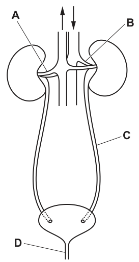
Which structure is the ureter?
Options
A A
B B
C C
D D
Answer
The ureter is the tube that carries urine from the kidney to the urinary bladder. Examining the diagram:
- A points to a renal blood vessel (renal vein or artery).
- B points to the kidney.
- C points to the ureter.
- D points to the urethra (the tube leaving the bladder).
Therefore, structure C is the ureter.
Answer
C
C
Walkthrough
The question asks to identify the ureter from a labelled diagram of the human urinary system. We can do this by tracing the pathway of urine from its formation to its exit from the body:
- Kidney (Label B): Urine is produced in the kidneys. Label B points to the bean-shaped organ, the kidney.
- Ureter (Label C): Urine leaves the kidney via a narrow tube called the ureter. Label C points to this tube, which carries urine down to the urinary bladder.
- Urinary bladder: Urine is stored in the bladder.
- Urethra (Label D): Urine leaves the body through the urethra. Label D points to this final tube exiting the bottom of the bladder.
- Renal vessels (Label A): Blood enters and leaves the kidney via the renal artery and renal vein. Label A points to one of these blood vessels attached to the kidney.
Since the ureter is the tube connecting the kidney to the bladder, label C is the correct answer.
Key Takeaways
- The human urinary system consists of two kidneys, two ureters, a urinary bladder, and a urethra.
- The kidney produces urine.
- The ureter transports urine from the kidney to the bladder.
- The bladder stores urine.
- The urethra expels urine from the body.
- Blood vessels (renal artery and vein) carry blood to and from the kidney; they are not part of the urine pathway.
Common Mistakes
- Confusing the ureter and the urethra: These names are similar. Remember: the ureter has an 'e' and connects the kidney to the bladder (the 'e' could stand for 'entering the bladder'). The urethra has an 'a' and is the exit tube from the bladder.
- Identifying blood vessels as ureters: Label A points to the renal blood vessels. These carry blood, not urine, and are much larger and positioned differently than the ureters.
- Naming the kidney instead of the ureter: Label B is the kidney itself, not the tube carrying urine away from it.
Things to Be Careful About
- Always read the diagram labels carefully. In this diagram, C is clearly the tube running from the kidney to the bladder, which is the definition of the ureter.
- Do not confuse the ureter (C) with the urethra (D). The urethra is the short tube at the very bottom leaving the bladder.
- This is a straightforward identification question; no complex reasoning is needed, just accurate recall of the anatomy of the excretory system.
The processes of filtration and reabsorption take place in the kidneys.
The daily amounts of some of the substances entering the kidneys in the blood and of these substances leaving the kidneys in the urine are shown.
| substance | amount entering the kidneys in the blood | amount leaving the kidneys in the urine |
|---|---|---|
| water | ||
| glucose | ||
| urea |
Which percentage of each substance is reabsorbed by the kidneys during a day?
Options
| % of substance reabsorbed by kidneys: water | % of substance reabsorbed by kidneys: glucose | % of substance reabsorbed by kidneys: urea | |
|---|---|---|---|
| A | 99 | 100 | 40 |
| B | 148.5 | 150 | 20 |
| C | 0.99 | 1.0 | 0.4 |
| D | 1.0 | 0.0 | 60 |
Working
For each substance, the amount reabsorbed is the amount entering minus the amount leaving, and the percentage reabsorbed is:
Water:
Glucose:
Urea:
These match option A.
Answer
A
A
Walkthrough
The kidney filters blood at the glomerulus, producing filtrate. Most of this filtrate is reabsorbed back into the blood as it passes along the nephron — this is selective reabsorption. The table gives the amount of each substance entering the kidney in the blood (which is what gets filtered) and the amount leaving in the urine. The difference between these is what has been reabsorbed.
For water: 150 dm³ enters, 1.5 dm³ leaves in urine, so 148.5 dm³ is reabsorbed. As a percentage: 148.5 / 150 × 100 = 99%. This makes sense — the kidney reabsorbs almost all the water, which is why urine is so concentrated.
For glucose: 150 g enters, 0 g leaves in urine. All 150 g is reabsorbed, so 100%. This is the key point of selective reabsorption — glucose is actively transported back into the blood in the proximal convoluted tubule, so none is lost in urine in a healthy person.
For urea: 50 g enters, 30 g leaves. So 20 g is reabsorbed, which is 20 / 50 × 100 = 40%. Urea is a waste product, so it is only partially reabsorbed (it diffuses back to some extent) and most is excreted.
The correct option is A.
Key Takeaways
- Reabsorption in the kidney: what enters minus what leaves in urine equals what is reabsorbed.
- Percentage reabsorbed = (amount entering − amount in urine) / amount entering × 100.
- Glucose is fully reabsorbed (100%) in a healthy kidney — its presence in urine indicates diabetes.
- Water is mostly reabsorbed (about 99%).
- Urea, a waste product, is only partially reabsorbed (here 40%).
Common Mistakes
- Confusing the amount reabsorbed with the amount in urine. The urine value is what is NOT reabsorbed.
- Option B gives the actual amounts reabsorbed (148.5, 150, 20) rather than percentages — a common trap.
- Option C divides the urine amount by the entering amount instead of using the reabsorbed amount.
- Option D gives the percentage of the entering amount that is NOT reabsorbed (water: 1.5/150 = 1%; glucose: 0%; urea: 30/50 = 60%).
Things to Be Careful About
- The question asks for percentage reabsorbed, so you must subtract the urine value from the entering value first.
- Work each substance separately and check all three values against the options.
- Units (dm³ and g) cancel in the percentage calculation, so they do not affect the answer.
Where are the receptors for the pupil reflex located?
Options
A brain
B iris
C pupil
D retina
Working
The pupil reflex is a reflex action: light enters the eye and is detected by light receptors, which send impulses along a sensory neurone to the brain, which then sends impulses along a motor neurone to the muscles of the iris to change the size of the pupil.
The receptors that detect the light are therefore in the retina, the light-sensitive layer at the back of the eye.
- A brain — the brain coordinates the reflex but is not where the receptors are.
- B iris — the iris contains the muscles that change pupil size, not the light receptors.
- C pupil — the pupil is just the opening through which light enters; it contains no receptors.
- D retina — correct, this is where the light receptors are located.
Answer
D
D
Walkthrough
The pupil reflex controls how much light enters the eye. When light is bright, the circular muscles of the iris contract and the pupil gets smaller (so less light enters); when light is dim, the radial muscles contract and the pupil gets larger (so more light enters).
For this reflex to work, something must first detect the light. That job belongs to the light-sensitive cells (photoreceptors) in the retina, the layer at the back of the eye. The retina detects the light and sends a nerve impulse along a sensory neurone to the brain. The brain then sends an impulse along a motor neurone to the muscles of the iris, which respond by changing the size of the pupil.
So the question asks where the receptors are. Receptors are the structures that detect a stimulus — here, light. They are in the retina (option D).
The other options are wrong for clear reasons:
- The brain is the coordinator of the reflex, not the receptor. It receives the impulse and sends out the response.
- The iris contains the muscles that carry out the response (the effectors), not the receptors.
- The pupil is just the hole in the middle of the iris through which light passes — it has no receptors at all.
Key Takeaways
- A reflex arc always has the same sequence: stimulus → receptor → sensory neurone → coordinator (brain or spinal cord) → motor neurone → effector → response.
- In the pupil reflex, the stimulus is light, the receptor is in the retina, the coordinator is the brain, the effectors are the muscles of the iris, and the response is a change in pupil size.
- Receptors detect stimuli; effectors carry out the response. Do not confuse the two.
Common Mistakes
- Choosing iris — the iris is the effector (it contains the muscles that change pupil size), not the receptor. This is the most tempting wrong answer.
- Choosing brain — the brain coordinates the reflex but does not detect the light.
- Choosing pupil — the pupil is just an opening; it has no sensory function.
Things to Be Careful About
- The question asks specifically for the receptors, so answer with the structure that detects the stimulus, not the structure that responds.
- Remember the retina is the light-sensitive layer at the back of the eye — it contains the photoreceptors (rods and cones).
The internal body temperature of some animals, including humans, remains constant even when the external temperature changes.
The graph shows how the oxygen used by one of these animals is affected by change in the external temperature.
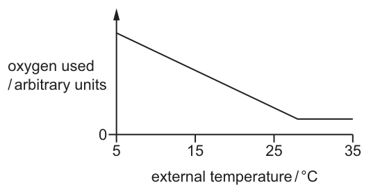
Which conclusion can be made from this graph?
Options
A The animal stops respiring when the external temperature is greater than .
B The animal’s respiration rate increases as the external temperature decreases from .
C The internal temperature of the animal decreases as the external temperature increases.
D The animal’s respiration rate increases as the external temperature increases.
Working
The question states the animal maintains a constant internal body temperature, meaning it is an endotherm. Endotherms use energy from aerobic respiration to generate heat and maintain this temperature. Oxygen is consumed during aerobic respiration, so the amount of oxygen used is a direct measure of the respiration rate (metabolic rate).
Reading the graph:
- The x-axis is external temperature, and the y-axis is oxygen used.
- As the external temperature drops from 28 °C down to 5 °C (moving left along the x-axis), the line slopes upwards, meaning more oxygen is used.
- More oxygen used means a higher respiration rate. The animal must respire faster to produce more heat to maintain its constant internal temperature in a cold environment.
- As the external temperature rises from 5 °C to 28 °C, less oxygen is used, so the respiration rate decreases.
- Above 28 °C, the line is horizontal at a positive value, meaning oxygen is still being used and respiration does not stop.
Evaluating the options:
- A is false: the graph levels off at a value above zero, meaning oxygen is still being used and respiration continues.
- B is true: as external temperature decreases from 28 °C (moving left), the graph shows oxygen used increases, which means the respiration rate increases.
- C is false: the stem explicitly states the internal temperature remains constant.
- D is false: the graph shows oxygen used (respiration rate) decreases as external temperature increases from 5 °C to 28 °C.
Answer
B
B
Walkthrough
- Understand the premise: The stem tells us the animal keeps a constant internal temperature. This is the definition of an endotherm (warm-blooded animal). To keep warm in a cold environment, it must produce more heat.
- Link oxygen to respiration: Animals produce heat through aerobic respiration, which uses oxygen. Therefore, the amount of oxygen used is a direct indicator of the respiration rate. Higher oxygen use = higher respiration rate.
- Interpret the graph: Look at the line from 28 °C down to 5 °C (moving left along the x-axis). The line goes up, meaning oxygen used increases. This confirms that as it gets colder externally, the animal respires faster to generate more heat. From 5 °C to 28 °C (moving right), oxygen used decreases. Above 28 °C, the line is flat, meaning oxygen use is constant and not zero.
- Evaluate options:
- A claims respiration stops above 28 °C. The graph does not touch the x-axis (y=0) above 28 °C; it stays at a positive constant value. Respiration continues.
- B claims respiration rate increases as temperature decreases from 28 °C. This matches our interpretation of the upward slope when moving left from 28 °C.
- C claims internal temperature decreases. This directly contradicts the first sentence of the stem.
- D claims respiration rate increases as external temperature increases. This contradicts the downward slope from 5 °C to 28 °C.
Key Takeaways
- Oxygen consumption is a reliable proxy for respiration rate in whole-organism experiments.
- Endotherms increase their metabolic (respiration) rate in cold environments to produce enough heat to maintain a constant internal body temperature.
- Graph interpretation requires careful attention to axis directions; a downward slope on a left-to-right graph means an increase in the y-variable as the x-variable decreases.
Common Mistakes
- Misreading the graph direction: Students often look at the downward slope from left to right and conclude 'as temperature increases, respiration increases' (Option D) without checking the axes properly or reading the graph from right to left for Option B.
- Assuming respiration stops: Option A is a trap for students who see the line level off and assume it means 'zero'. The line is clearly above the x-axis (y=0), meaning oxygen is still being used.
- Ignoring the stem: Option C is a trap for students who do not read the first sentence. The stem explicitly states the internal temperature remains constant, making C impossible.
Things to Be Careful About
- Axis labels and directions: Always read the axes carefully. The x-axis is 'external temperature' and the y-axis is 'oxygen used'. Remember that oxygen used is a measure of respiration rate.
- Positive vs. zero values: When a graph levels off, check if it levels off at zero or at a positive value. Here, it levels off at a positive value, meaning the process (respiration) continues at a steady rate.
- Stem information: The first sentence of the stem provides critical context ('internal body temperature... remains constant') that immediately eliminates Option C. Always read the entire stem before evaluating options.
Which statements about synapses are correct?
- Synapses are junctions between neurones.
- Synapses ensure impulses travel in only one direction.
- Neurotransmitter chemicals diffuse across synaptic gaps.
Options
A 1, 2 and 3
B 1 and 2 only
C 1 and 3 only
D 2 and 3 only
Working
All three statements are correct:
- Statement 1: a synapse is the junction between two neurones.
- Statement 2: neurotransmitter is released only from the presynaptic neurone, so the impulse can only pass in one direction.
- Statement 3: neurotransmitter molecules diffuse across the synaptic gap to the next neurone.
Since 1, 2 and 3 are all correct, the answer is A.
Answer
A
A
Walkthrough
This question asks you to judge three statements about synapses and then choose the option that lists the correct ones.
Statement 1 says synapses are junctions between neurones. This is correct. A synapse is the tiny gap between the end of one neurone and the next neurone (or between a neurone and a muscle or gland).
Statement 2 says synapses ensure impulses travel in only one direction. This is also correct. The neurotransmitter is released only from the presynaptic neurone, and the receptors for it are only on the postsynaptic neurone. Because the chemicals cannot be released from the receiving side, the impulse cannot go backwards.
Statement 3 says neurotransmitter chemicals diffuse across synaptic gaps. This is correct too. After the impulse arrives at the end of the first neurone, tiny vesicles release neurotransmitter molecules into the synaptic gap, and these molecules diffuse across to bind to receptors on the next neurone.
Because all three statements are correct, the correct option is A: 1, 2 and 3.
Key Takeaways
- A synapse is a junction between neurones.
- Transmission across a synapse involves the release of a neurotransmitter, its diffusion across the synaptic gap, and its binding to receptors on the next neurone.
- Synapses make impulse transmission one-way because only the presynaptic neurone releases the neurotransmitter.
Common Mistakes
- Choosing an option that omits statement 2. Some students think impulses can pass both ways at a synapse, but the arrangement of release and receptors makes transmission one-way.
- Confusing the synapse with the reflex arc. The synapse is a junction between neurones; the reflex arc is the whole pathway of a reflex action.
- Thinking that the impulse itself jumps across the gap. It does not — the chemical neurotransmitter diffuses across.
Things to Be Careful About
- Read each numbered statement separately before looking at the options.
- Use the exact term "neurotransmitter" in explanations; the mark scheme for this type of question expects the biological vocabulary.
- In an MCQ, once you have decided that all three statements are correct, select the option that includes 1, 2 and 3 — here option A.
Which process is not part of a homeostatic mechanism in mammals?
Options
A change in flow of blood in vessels of the skin
B growth of longer hairs from the skin
C increased rate of sweat production
D contraction of hair erector muscles
Working
Homeostasis maintains a constant internal environment using rapid, reversible responses. A, C and D are all immediate temperature-control responses: changing skin blood flow, increasing sweat production, and contracting hair erector muscles. Growing longer hairs is a slow, long-term change, not a homeostatic mechanism.
Answer
B
B
Walkthrough
Homeostasis is the maintenance of a constant internal environment, despite changes inside and outside the body. It works mainly through negative feedback: when a condition such as body temperature moves away from the normal level, the body makes rapid, reversible changes to bring it back.
Look at each option:
- A – change in flow of blood in vessels of the skin. This is vasodilation or vasoconstriction. When the body is too hot, skin blood vessels widen to lose heat; when too cold, they narrow to reduce heat loss. This is a homeostatic mechanism.
- C – increased rate of sweat production. Sweating cools the body as the water evaporates from the skin surface. This is another homeostatic response to a rise in body temperature.
- D – contraction of hair erector muscles. When it is cold, these tiny muscles contract and make hairs stand up. In mammals this traps a layer of insulating air near the skin, reducing heat loss. This is also a homeostatic mechanism.
- B – growth of longer hairs from the skin. This is a slow, long-term change, such as growing a thicker winter coat. It is not an immediate response to a change in the internal environment and is not controlled by the rapid negative-feedback mechanisms of homeostasis.
Therefore the process that is not part of a homeostatic mechanism is B.
Key Takeaways
- Homeostatic mechanisms are rapid, reversible responses that keep internal conditions constant.
- Body temperature is controlled by several effectors: skin blood vessels, sweat glands, and hair erector muscles.
- Long-term changes such as hair growth are not homeostatic mechanisms.
Common Mistakes
- Thinking that contraction of hair erector muscles is not homeostatic — it is a genuine temperature-control response.
- Confusing "growth of longer hairs" with "contraction of hair erector muscles". The first is slow and long-term; the second is immediate and reversible.
- Choosing A because the candidate does not realise that changing blood flow in skin vessels is a key part of thermoregulation.
Things to Be Careful About
- The question asks for the process that is not part of a homeostatic mechanism, so look for the odd one out.
- Remember the 5090 definition of homeostasis: maintenance of a constant internal environment.
- Distinguish between short-term physiological control and long-term growth or development.
A nurse tests a sample of urine from a patient using Benedict’s reagent. The sample tests positive.
What is a possible explanation for this result?
Options
A The patient’s liver is not deaminating protein efficiently.
B The patient’s pancreas is not producing sufficient insulin.
C The patient has eaten too much glucose.
D The patient’s concentration of plasma proteins is too high.
Working
Benedict's reagent tests for reducing sugars. A positive result means glucose is present in the urine. Glucose is normally filtered out of the blood in the kidney and then completely reabsorbed back into the blood. Glucose appears in the urine only when the blood glucose concentration is so high that the kidney cannot reabsorb it all. This happens in diabetes mellitus, when the pancreas does not produce enough insulin to move glucose into cells.
- A is wrong: deamination of protein in the liver produces urea, not glucose in the urine.
- B is correct: insufficient insulin raises blood glucose above the kidney's reabsorption limit, so glucose spills into the urine.
- C is wrong: eating too much glucose does not normally cause glucose in the urine, because insulin stores the excess as glycogen.
- D is wrong: a high concentration of plasma proteins does not put glucose into the urine.
Answer
B
B
Walkthrough
Benedict's reagent is the food test for reducing sugars. It is blue when no reducing sugar is present, and turns green, yellow, orange or brick-red when a reducing sugar such as glucose is present. A positive result on a urine sample therefore means the patient has glucose in their urine.
Normally there is no glucose in urine. In the kidney, blood is filtered in the glomerulus, and the filtrate contains glucose. As the filtrate flows along the nephron, all the glucose is reabsorbed by active transport back into the blood. So a healthy person's urine contains no glucose.
Glucose appears in urine when the blood glucose concentration becomes so high that the kidney's reabsorption mechanism cannot keep up — the reabsorption limit is exceeded and glucose 'spills over' into the urine. The most common cause is diabetes mellitus, in which the pancreas does not produce enough insulin. Insulin normally lowers blood glucose by helping glucose enter cells and by promoting its storage as glycogen. Without enough insulin, blood glucose stays high, and glucose leaks into the urine.
Now check the options:
- A — The liver deaminates excess amino acids, producing urea, which is excreted in urine. If the liver were not deaminating efficiently, the problem would be with urea, not glucose. This does not explain glucose in the urine.
- B — Correct. Insufficient insulin means blood glucose stays high, exceeding the kidney's reabsorption capacity, so glucose appears in the urine.
- C — Eating too much glucose does not normally cause glucose in the urine. A healthy pancreas releases extra insulin to store the excess glucose as glycogen, keeping blood glucose within the normal range.
- D — Plasma proteins are too large to be filtered into the glomerular filtrate, so their concentration in the blood does not affect glucose in the urine.
Key Takeaways
- Benedict's reagent tests for reducing sugars; a colour change from blue to green/yellow/orange/brick-red is a positive result.
- Glucose is filtered out of the blood in the kidney and then completely reabsorbed by active transport in the nephron, so normal urine contains no glucose.
- Glucose appears in urine when blood glucose is too high for the kidney to reabsorb — this happens in diabetes mellitus when the pancreas produces insufficient insulin.
- Insulin lowers blood glucose; a lack of insulin leads to high blood glucose and glucose in the urine.
Common Mistakes
- Choosing C ('ate too much glucose') — this confuses a temporary dietary excess with a failure of blood glucose control. A healthy pancreas handles excess glucose by storing it as glycogen, so glucose does not appear in the urine.
- Choosing A — confusing the products of deamination (urea) with glucose. Deamination is about amino acids and urea, not sugar.
- Thinking that any glucose in the blood automatically appears in urine — glucose only appears in urine when its concentration exceeds the kidney's reabsorption capacity.
Things to Be Careful About
- The mark scheme requires the precise link: insufficient insulin → high blood glucose → glucose exceeds the kidney's reabsorption limit → glucose in urine. The answer must name insulin, not just 'diabetes'.
- Benedict's reagent detects reducing sugars, not just glucose — but in a urine test the relevant reducing sugar is glucose.
- Remember that glucose reabsorption in the nephron is by active transport, and it is normally complete; it is only when the filtered load exceeds the transport capacity that glucose appears in urine.
Why do plant shoots grow towards light?
Options
A Auxin inhibits cell elongation on one side.
B They grow more rapidly on the lighter side.
C More auxin accumulates on the darker side.
D Auxin stimulates cell division.
Working
Phototropism: auxin moves sideways from the light side to the shaded side of the shoot tip. The higher auxin concentration on the darker side causes cells there to elongate more rapidly. The darker side grows longer, so the shoot bends towards the light.
- A is wrong: auxin promotes, not inhibits, cell elongation.
- B is wrong: growth is faster on the darker side, not the lighter side.
- D is wrong: auxin stimulates cell elongation, not cell division.
Answer
C
C
Walkthrough
A shoot grows towards light because of the plant hormone auxin. Auxin is produced at the shoot tip and moves sideways, away from the light, so more auxin accumulates on the darker (shaded) side. Auxin causes cells in the shoot to elongate, and because there is more auxin on the darker side, those cells elongate more. This makes the darker side grow longer than the lighter side, so the shoot bends towards the light.
Now check each option:
- A says auxin inhibits cell elongation on one side. This is wrong because auxin promotes cell elongation in shoots, and it accumulates on the darker side, not the side facing the light.
- B says shoots grow more rapidly on the lighter side. This is the opposite of what happens: the darker side grows faster.
- C says more auxin accumulates on the darker side. This is correct and explains the bending.
- D says auxin stimulates cell division. Auxin mainly stimulates cell elongation in this response, not cell division.
Key Takeaways
- Phototropism is the growth of a plant shoot towards light.
- Auxin moves sideways in the shoot tip, away from light, so it accumulates on the darker side.
- Auxin promotes cell elongation, not cell division.
- The side with more auxin elongates more, causing the shoot to bend towards the light.
Common Mistakes
- Saying the shoot grows more on the lighter side: the darker side grows faster.
- Saying auxin inhibits cell elongation: auxin promotes elongation in shoots.
- Saying auxin causes cell division: the key effect here is cell elongation.
- Confusing the direction of bending: the shoot bends towards the light because the darker side grows longer.
Things to Be Careful About
- Use the precise term "cell elongation" rather than just "growth".
- Remember the auxin accumulates on the darker side, not the lighter side.
- The question asks "why", so the answer must give the mechanism, not just state the outcome.
Which statement about nuclear division is correct?
Options
A Meiosis results in the production of cells for the growth and repair of organisms.
B Meiosis results in genetically different daughter cells.
C Mitosis gives rise to cells where the chromosome number is halved.
D Mitosis results in the production of four daughter cells.
Working
Meiosis produces four genetically different daughter cells, each with half the chromosome number of the parent cell. Mitosis produces two genetically identical daughter cells with the same chromosome number as the parent cell, and is used for growth and repair.
Option B is the only correct statement.
Answer
B
B
Walkthrough
This question asks you to recall the key differences between the two types of nuclear division: mitosis and meiosis.
Mitosis is the type of division used for growth, repair and asexual reproduction. It produces two daughter cells that are genetically identical to the parent cell and to each other. The chromosome number stays the same, so if the parent cell is diploid, the daughter cells are also diploid.
Meiosis is the type of division used to produce gametes. It produces four daughter cells, and each one has half the chromosome number of the parent cell. The daughter cells are genetically different from each other and from the parent cell because of the way chromosomes are separated and exchanged during meiosis.
Now look at each option:
- A says meiosis produces cells for growth and repair. This is wrong because growth and repair use mitosis, not meiosis.
- B says meiosis results in genetically different daughter cells. This is correct.
- C says mitosis gives rise to cells where the chromosome number is halved. This is wrong: mitosis keeps the chromosome number the same; meiosis halves it.
- D says mitosis results in the production of four daughter cells. This is wrong: mitosis produces two daughter cells; meiosis produces four.
So the correct answer is B.
Key Takeaways
- Mitosis: two genetically identical daughter cells, same chromosome number, used for growth, repair and asexual reproduction.
- Meiosis: four genetically different daughter cells, half the chromosome number, used to produce gametes.
- The chromosome number is halved only in meiosis, not in mitosis.
Common Mistakes
- Confusing the number of daughter cells: mitosis gives two, meiosis gives four.
- Confusing the purpose: growth and repair use mitosis, while gamete production uses meiosis.
- Thinking mitosis halves the chromosome number. It does not; meiosis does.
- Thinking meiosis produces identical cells. It produces genetically different cells.
Things to Be Careful About
- Read each statement carefully and match it to the correct type of division.
- The word "genetically different" is a key phrase that points to meiosis.
- Remember that "halved chromosome number" is a meiosis feature, not a mitosis feature.
- Do not choose an option just because it mentions a familiar term; check all parts of the statement.
Stem cells are found in human embryos.
Which statements about stem cells are correct?
- Stem cells are unspecialised cells.
- Stem cells can divide by mitosis.
- Stem cells are haploid.
Options
A 1, 2 and 3
B 1 and 2 only
C 1 and 3 only
D 2 and 3 only
Working
Statement 1: Stem cells are unspecialised (undifferentiated) cells — correct.
Statement 2: Stem cells divide by mitosis to produce more cells — correct.
Statement 3: Stem cells are diploid, not haploid; only gametes are haploid — incorrect.
Statements 1 and 2 are correct, so option B is the answer.
Answer
B
B
Walkthrough
This question tests your knowledge of stem cells and the difference between haploid and diploid cells.
Statement 1 — Stem cells are unspecialised cells. This is correct. A stem cell is an undifferentiated cell: it has not yet become a specialised cell such as a muscle cell or a nerve cell. Its defining feature is that it can divide and then differentiate into different cell types.
Statement 2 — Stem cells can divide by mitosis. This is correct. Stem cells divide by mitosis to make more cells. Some of the new cells remain as stem cells (self-renewal) and others go on to specialise. Mitosis produces genetically identical diploid cells, which is exactly what is needed for growth and repair.
Statement 3 — Stem cells are haploid. This is incorrect. Stem cells are diploid — they contain two sets of chromosomes (46 in humans), one set from each parent. Haploid cells contain only one set of chromosomes, and in humans these are only the gametes — sperm and egg cells, each with 23 chromosomes.
So statements 1 and 2 are correct and statement 3 is wrong. The option that matches "1 and 2 only" is B.
Key Takeaways
- A stem cell is an unspecialised cell that can divide by mitosis and differentiate into specialised cells.
- Stem cells are diploid, like all body (somatic) cells.
- Haploid cells in humans are only the gametes (sperm and egg), produced by meiosis.
- This is the numbered-statements format: judge each statement true or false, then read off the option that matches.
Common Mistakes
- Thinking stem cells are haploid — they are diploid. Only gametes are haploid. This is the trap behind option C (1 and 3 only) and option A (1, 2 and 3).
- Confusing mitosis with meiosis: stem cells divide by mitosis for growth and repair, not by meiosis.
- Assuming that because stem cells can become many cell types they must be haploid — differentiation has nothing to do with chromosome number.
Things to Be Careful About
- Read each numbered statement independently and mark it true or false before looking at the options.
- Statement 3 is the discriminating one — it is false, which eliminates options A and C immediately.
- Remember that "haploid" means one set of chromosomes (n) and "diploid" means two sets (2n). Human stem cells are 2n = 46.
In the placenta, which substance passes from the blood of the embryo to the blood of the mother down a concentration gradient?
Options
A amino acids
B carbon dioxide
C glucose
D oxygen
Working
The placenta exchanges substances between the embryo's blood and the mother's blood. Oxygen, glucose and amino acids all move from the mother's blood into the embryo's blood. Carbon dioxide is a waste product of the embryo's respiration, so its concentration is higher in the embryo's blood and it diffuses down its concentration gradient into the mother's blood.
Answer
B
B
Walkthrough
The placenta is the organ where the embryo's blood and the mother's blood come very close together, separated only by thin membranes, so that substances can be exchanged by diffusion. The embryo gets what it needs from the mother and gets rid of its waste products into the mother's blood.
Look at each option:
- Amino acids (A) — these are building blocks the embryo needs to make proteins. They move from the mother's blood into the embryo's blood, not the other way round.
- Carbon dioxide (B) — this is a waste product made by the embryo's cells during respiration. The embryo's blood has a higher concentration of carbon dioxide than the mother's blood, so carbon dioxide diffuses down its concentration gradient from the embryo to the mother. This is the correct answer.
- Glucose (C) — the embryo needs glucose for energy (respiration), so glucose moves from the mother's blood into the embryo's blood.
- Oxygen (D) — the embryo needs oxygen for respiration, so oxygen moves from the mother's blood into the embryo's blood.
So the only substance in the list that passes from the embryo to the mother is carbon dioxide.
Key Takeaways
- The placenta is the exchange surface between the embryo and the mother.
- Substances the embryo needs (oxygen, glucose, amino acids) move from mother to embryo.
- Waste products of the embryo's metabolism (carbon dioxide, urea) move from embryo to mother.
- Diffusion always happens down a concentration gradient — from where a substance is more concentrated to where it is less concentrated.
Common Mistakes
- Choosing oxygen or glucose because they are the most familiar substances exchanged across the placenta — but they move in the opposite direction, from mother to embryo.
- Forgetting that carbon dioxide is a waste product produced by the embryo's own respiration, so it must leave the embryo.
- Confusing the direction of movement: the question asks for the substance passing from embryo to mother, not from mother to embryo.
Things to Be Careful About
- Read the direction carefully: "from the blood of the embryo to the blood of the mother".
- The mark scheme requires the single correct option letter, B.
- Remember that "down a concentration gradient" simply means by diffusion — the substance moves from where it is more concentrated to where it is less concentrated.
What is the main function of the prostate gland?
Options
A producing adrenaline
B producing testosterone
C producing a liquid in which the sperm can swim
D storing mature sperm cells
Working
The prostate gland is part of the male reproductive system. Its main function is to produce a liquid (seminal fluid) that mixes with the sperm to form semen, providing a medium in which the sperm can swim.
- A is wrong: adrenaline is produced by the adrenal glands, not the prostate.
- B is wrong: testosterone is produced by the testes.
- C is correct: the prostate produces a liquid in which the sperm can swim.
- D is wrong: mature sperm cells are stored in the epididymis, not the prostate.
Answer
C
C
Walkthrough
The question asks for the main function of the prostate gland, a structure in the male reproductive system. This is a recall question: you need to know what each part of the male reproductive system does.
The prostate gland surrounds the urethra just below the bladder. It produces a milky liquid called seminal fluid. During ejaculation, this fluid mixes with sperm from the testes to form semen. The liquid provides a medium in which the sperm can swim, and it also helps to nourish and protect them.
Now check each option:
- A — producing adrenaline. Adrenaline is a hormone made by the adrenal glands, which sit on top of the kidneys. It prepares the body for stress (the fight-or-flight response). The prostate has nothing to do with adrenaline.
- B — producing testosterone. Testosterone is the main male sex hormone. It is produced by the testes, not the prostate. The testes are the primary male reproductive organs.
- C — producing a liquid in which the sperm can swim. This is exactly what the prostate does. The seminal fluid it produces is the liquid part of semen.
- D — storing mature sperm cells. Sperm are made in the testes and then mature and are stored in the epididymis, a coiled tube on the back of each testis. The prostate does not store sperm.
So C is the only correct statement.
Key Takeaways
- The prostate gland produces seminal fluid, the liquid part of semen, in which sperm can swim.
- The testes produce both sperm and the hormone testosterone.
- The epididymis stores mature sperm cells.
- The adrenal glands produce adrenaline.
- Knowing which gland or organ produces which hormone and which structure stores which cell type lets you eliminate wrong options quickly.
Common Mistakes
- Confusing the prostate with the testes: the testes produce testosterone, the prostate does not.
- Thinking the prostate stores sperm — that is the job of the epididymis.
- Mixing up the prostate with the adrenal glands, which produce adrenaline.
- A common general error in this topic is confusing the seminal vesicle with the prostate; both add fluid to semen, but the question here is specifically about the prostate.
Things to Be Careful About
- The question asks for the "main" function — answer with the primary role (producing the liquid for sperm to swim in), not a secondary detail.
- Match each hormone to its exact source gland: testosterone → testes, adrenaline → adrenal glands.
- Remember that sperm storage happens in the epididymis, which is part of the testes region, not the prostate.
The graph shows the changes in levels of four hormones during the menstrual cycle.
Which line represents progesterone?
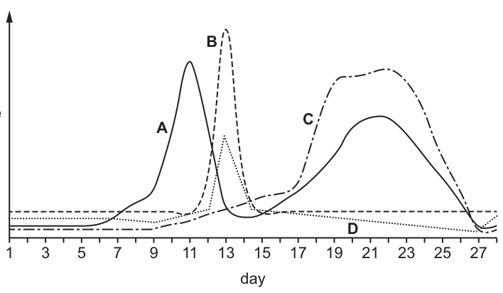
Options
A A
B B
C C
D D
Working
On the graph of the menstrual cycle:
- Line A peaks before day 14, representing oestrogen.
- Line B peaks sharply at day 14, representing luteinising hormone (LH).
- Line C rises after ovulation (day 14) and peaks around day 21, representing progesterone secreted by the corpus luteum.
- Line D remains low throughout.
Progesterone is the hormone that rises after ovulation to maintain the uterine lining, which matches line C.
Answer
C
C
Walkthrough
The menstrual cycle is controlled by four main hormones: follicle-stimulating hormone (FSH), oestrogen, luteinising hormone (LH), and progesterone. Their levels change predictably over the 28-day cycle.
- FSH (not clearly shown as a major peak here, but line D could represent a low-level hormone like FSH or inhibin) stimulates the development of the follicle.
- Oestrogen (line A) is secreted by the developing follicle and rises to a peak just before ovulation (around day 11–12).
- LH (line B) is released from the pituitary gland in a sharp surge at around day 13–14, which triggers ovulation (the release of the egg).
- Progesterone (line C) is secreted by the corpus luteum (the remains of the follicle after ovulation). Its level is low before ovulation, rises after day 14, and peaks around day 21 to maintain the uterine lining for a potential pregnancy.
By matching these known patterns to the graph, line C is the only curve that rises after ovulation and peaks in the second half of the cycle, identifying it as progesterone.
Key Takeaways
- Oestrogen peaks before ovulation; progesterone peaks after ovulation.
- LH has a sharp, brief peak that triggers ovulation.
- The corpus luteum produces progesterone in the luteal phase (days 15–28).
Common Mistakes
- Confusing oestrogen and progesterone: Oestrogen (line A) peaks before day 14, while progesterone (line C) peaks after day 14. Students often pick the highest peak or the first peak without considering the timing relative to ovulation.
- Confusing LH and progesterone: LH (line B) has a sharp spike at day 14, but it falls immediately after. Progesterone rises more gradually and stays elevated for about a week.
- Naming the hormone instead of the letter: The question asks 'Which line represents progesterone?', so the answer must be the letter C, not the word 'progesterone'.
Things to Be Careful About
- Read the x-axis carefully: ovulation occurs at day 14. Progesterone must be low before day 14 and high after day 14.
- Ensure you select the correct option letter (A, B, C, or D) that corresponds to the line on the graph.
- Remember that the corpus luteum forms from the ruptured follicle after ovulation, which is why progesterone production only begins in earnest after day 14.
Which row identifies the factors causing continuous and discontinuous variation?
Options
| continuous variation | discontinuous variation | |
|---|---|---|
| A | both the environment and the genes | both the environment and the genes |
| B | the environment only | both the environment and the genes |
| C | the genes only | the environment only |
| D | both the environment and the genes | the genes only |
Working
Continuous variation is influenced by both the genes and the environment (for example, height depends on inherited alleles and on nutrition). Discontinuous variation is controlled by genes alone and is not affected by the environment (for example, blood group or eye colour).
- Option A is wrong because discontinuous variation is not caused by the environment.
- Option B is wrong because continuous variation is not caused by the environment alone.
- Option C is wrong because continuous variation is not caused by genes alone, and discontinuous variation is not caused by the environment.
- Option D is correct.
Answer
D
D
Walkthrough
This question tests the difference between the two types of variation and what causes each.
Continuous variation is variation where there is a complete range of values between two extremes — for example, height, mass or shoe size. There are no distinct categories; individuals fall anywhere along a scale. This type of variation is caused by both the genes and the environment. Your genes set the range of heights you could reach, but whether you actually reach the top of that range depends on your diet and health.
Discontinuous variation is variation where individuals fall into distinct, separate categories with no intermediates — for example, blood group, sex, or whether you can roll your tongue. This type of variation is caused by the genes only. The environment does not change your blood group or your sex.
Looking at the rows:
- Row A says both types are caused by both genes and environment — wrong, because discontinuous variation is not affected by the environment.
- Row B says continuous variation is caused by the environment only — wrong, because genes also matter.
- Row C says continuous variation is caused by genes only and discontinuous by environment only — both halves are wrong.
- Row D says continuous variation is caused by both genes and environment, and discontinuous by genes only — this matches the correct biology.
So the answer is D.
Key Takeaways
- Continuous variation shows a smooth range of values (height, mass) and is caused by both genes and environment.
- Discontinuous variation shows distinct categories (blood group, tongue rolling) and is caused by genes only.
- A quick way to remember: if the environment can influence the trait, the variation is continuous; if it cannot, the variation is discontinuous.
Common Mistakes
- Choosing A because both types involve genes — forgetting that discontinuous variation is not influenced by the environment.
- Choosing B because continuous traits like height seem to depend on diet — forgetting that genes also set the limits.
- Confusing the two: thinking discontinuous variation (like height ranges) has many intermediates, when in fact it is the one with clear separate categories.
Things to Be Careful About
- Read the table carefully: the left column is continuous variation and the right column is discontinuous variation; check both before selecting a row.
- The mark scheme accepts only D — there is no partial credit for getting one column right.
- Remember the classic examples: height and mass are continuous; blood group and sex are discontinuous.
Which row shows what happens in the eye when focusing on a nearby object?
Options
| ciliary muscle | shape of lens | |
|---|---|---|
| A | relaxes | becomes thinner |
| B | relaxes | becomes rounder |
| C | contracts | becomes thinner |
| D | contracts | becomes rounder |
Working
When focusing on a nearby object, the ciliary muscles contract. This slackens the suspensory ligaments, allowing the lens to become rounder (more convex) so it can refract light more strongly.
Answer
D
D
Walkthrough
The eye must change the shape of its lens to focus light from objects at different distances. This process is called accommodation.
For a nearby object, the ciliary muscles contract. When they contract, the suspensory ligaments attached to the lens become slack. This allows the elastic lens to spring back into a rounder, more convex shape. A rounder lens refracts light more strongly, which is needed to focus light from a near object onto the retina.
Looking at the table:
- The ciliary muscle should be contracts, not relaxes. This removes options A and B.
- The lens should become rounder, not thinner. This removes option C.
So the correct row is D: ciliary muscle contracts and lens becomes rounder.
Key Takeaways
- Accommodation is the change in lens shape that allows the eye to focus on near and distant objects.
- For near objects: ciliary muscles contract, suspensory ligaments slacken, lens becomes rounder/more convex.
- For distant objects: ciliary muscles relax, suspensory ligaments become taut, lens becomes thinner/flatter.
Common Mistakes
- Choosing A or B: thinking the ciliary muscle relaxes when focusing on a near object. This is the opposite of what happens.
- Choosing C: knowing the ciliary muscle contracts but thinking the lens becomes thinner. A thinner lens is for distant objects, not near objects.
- Confusing the lens becoming "rounder" with "thinner". The lens must become more curved to increase refraction for near vision.
Things to Be Careful About
- The mark scheme requires the exact idea: ciliary muscle contracts and lens becomes rounder.
- Do not mix up near and distant accommodation. For a distant object, the ciliary muscle relaxes and the lens becomes thinner.
- This is a one-mark question, so the answer is simply the correct option letter, D.
What is the principal source of energy in most biological systems?
Options
A bacteria
B photosynthesis
C respiration
D sunlight
Working
The Sun is the principal (original) source of energy for almost all biological systems. Light energy from the Sun is trapped by chlorophyll during photosynthesis and converted into chemical energy in food. Photosynthesis and respiration are processes that transfer or release energy — they are not the source of the energy itself. Bacteria are organisms, not an energy source.
Answer
D
D
Walkthrough
The question asks for the principal source of energy in most biological systems — the original place the energy comes from, not a process that uses or releases it.
- Option D, sunlight, is correct. The Sun is the ultimate energy source for nearly all life on Earth. Plants and algae trap light energy from the Sun using chlorophyll during photosynthesis and store it as chemical energy in carbohydrates. When other organisms eat plants (or eat organisms that ate plants), that energy passes along food chains. So every food chain ultimately starts with sunlight.
- Option B, photosynthesis, is wrong because photosynthesis is the process that converts light energy into chemical energy — it transfers the Sun's energy but does not create it.
- Option C, respiration, is wrong because respiration is the process that releases energy from glucose to power life processes. It uses energy that was already captured, so it cannot be the source.
- Option A, bacteria, is wrong because bacteria are organisms. Some bacteria are decomposers or producers, but they are not the original source of energy for biological systems.
The key distinction is between the source of energy (the Sun) and the processes that transfer it (photosynthesis) or release it (respiration).
Key Takeaways
- The Sun is the principal energy source for almost all biological systems.
- Photosynthesis transfers light energy into chemical energy in food; respiration releases that chemical energy to power life processes.
- Energy flows from the Sun through producers to consumers along food chains.
Common Mistakes
- Choosing photosynthesis or respiration because they are familiar energy-related processes — but the question asks for the source, not the process.
- Confusing 'source of energy' with 'transfer of energy'. Photosynthesis does not make energy; it converts light energy into chemical energy.
Things to Be Careful About
- Read the word principal — it means 'main' or 'original'. The Sun is the ultimate source even though the energy a plant uses day-to-day is in the form of glucose.
- This is a one-mark recall question — no calculation or long explanation is needed. The answer is simply the Sun (sunlight).
Untreated sewage contains nitrates and phosphates.
What will happen in the lake if untreated sewage is added?
Options
A increased biodiversity
B increased eutrophication
C increased fish populations
D increased light penetration
Working
Untreated sewage adds nitrates and phosphates to the lake. These nutrients cause rapid growth of algae, forming an algal bloom. When the algae die, bacteria decompose them and use up oxygen, so the water becomes depleted of oxygen and many organisms die. This is eutrophication.
- A is wrong: oxygen depletion reduces biodiversity.
- C is wrong: fish die from lack of oxygen.
- D is wrong: the algal bloom reduces light penetration.
Answer
B
B
Walkthrough
The question asks what will happen in a lake if untreated sewage is added. Untreated sewage contains nitrates and phosphates, which are plant nutrients. When these enter a lake, algae grow very quickly and cover the surface in an algal bloom. The algae block light from reaching plants below, and when the algae die, bacteria decompose them. These bacteria respire and use up oxygen dissolved in the water. The drop in oxygen kills fish and other aquatic organisms, so biodiversity falls. This whole process is called eutrophication, so B is correct.
Option A is incorrect because biodiversity decreases, not increases, when oxygen runs out. Option C is incorrect because fish populations fall when oxygen is used up by the decomposing bacteria. Option D is incorrect because the algal bloom makes the water cloudy and reduces light penetration, not increases it.
Key Takeaways
- Nitrates and phosphates are nutrients that cause rapid algal growth when they enter water.
- Eutrophication is the sequence: nutrient enrichment → algal bloom → death of algae → decomposition by bacteria → oxygen depletion → death of fish and other organisms.
- Adding untreated sewage to water is a form of water pollution that harms the ecosystem.
Common Mistakes
- Choosing A: students think more nutrients mean more life, but the oxygen depletion that follows kills organisms and lowers biodiversity.
- Choosing D: students forget that an algal bloom blocks light, so light penetration decreases.
- Writing only "algae grow" without mentioning the later oxygen depletion: the full sequence is needed to explain eutrophication.
Things to Be Careful About
- The mark scheme requires the precise term "eutrophication", so use that word in the answer.
- Link both nitrates and phosphates to the algal bloom, not just one nutrient.
- Remember that the final effect on the lake is harmful: fish die and biodiversity falls.
DDT is an insecticide that is harmful to living organisms. It builds up in the cells of living organisms as it is not excreted.
The diagram shows the path taken by DDT when it enters a lake.

Which organisms would the DDT harm the most?
Options
A aquatic plants
B carnivorous fish
C herbivorous fish
D fish-eating birds
Working
DDT is not excreted, so it accumulates in the tissues of every organism that absorbs or eats it. As it passes along the food chain — aquatic plants → herbivorous fish → carnivorous fish → fish-eating birds — each organism takes in the DDT already accumulated by everything it ate, so the concentration builds up with each trophic level. The organism at the end of the chain, the fish-eating birds, therefore carries the highest concentration.
Answer
D — fish-eating birds
D
Walkthrough
The diagram gives you a five-step food chain: DDT dissolved in the lake water is absorbed by aquatic plants, which are eaten by herbivorous fish, which are eaten by carnivorous fish, which are eaten by fish-eating birds. The key fact in the stem is that DDT is not excreted — once it enters an organism's cells it stays there for life. This is bioaccumulation.
Trace the concentration up the chain. Each aquatic plant absorbs a little DDT from the water. A herbivorous fish eats many plants over its lifetime, so it collects all the DDT from every one of them, plus more from the water. A carnivorous fish eats many herbivorous fish, gathering the DDT stored in each of them. By the top of the chain, the fish-eating bird has eaten many carnivorous fish, so it receives the combined DDT load of a large number of organisms from every level below it. Because it never excretes the insecticide, its body concentration is the highest of any organism in the chain.
This is why the question asks which organisms are harmed the most — not which are exposed first. The plants are exposed first but carry the least DDT per organism; the birds are last in the chain but carry the most. This same principle explains why top predators such as birds of prey suffered shell-thinning when DDT was widely used.
Key Takeaways
- Bioaccumulation: a non-excreted, non-biodegradable substance becomes more concentrated at each successive trophic level of a food chain.
- Top predators are always the worst affected by persistent pollutants such as DDT, even though they are furthest from the source.
- A food chain shows the direction of energy (and here, pollutant) flow: producer → primary consumer → secondary consumer → tertiary consumer.
Common Mistakes
- Choosing A (aquatic plants) because they are the first to absorb the DDT — being exposed first does not mean accumulating the most.
- Choosing C (carnivorous fish) because they are 'high up' the chain — but the fish-eating birds are one level higher still, and the question asks for the maximum harm.
- Thinking the DDT dilutes as it spreads through the lake — the stem states it is not excreted, so it only accumulates.
- Confusing bioaccumulation (build-up within individuals up the chain) with simple presence of a pollutant in the water.
Things to Be Careful About
- Read the stem carefully: the phrase 'not excreted' is the whole basis of the answer — without it there would be no build-up.
- 'Harm the most' means the greatest concentration in the body, which is always the final consumer in the chain.
- Count the arrows: the fish-eating birds are the fourth organism in the chain, one step beyond the carnivorous fish, so do not stop one level early.
Your score so far
Answer a question to start scoring
Your marks add up here as you work through the paper.