Biology 5090/12 — October/November 2024
Cambridge O-Level · Multiple Choice · answer key with instant marking and worked solutions
Topics Transport in Humans · Coordination and Control · Organisms and Their Environment · Movement Into and Out of Cells · Disease and Immunity · Inheritance · +15 more
Tap an option under each question to check it — your score builds as you go.
The diagram shows three different types of microscopic structure.
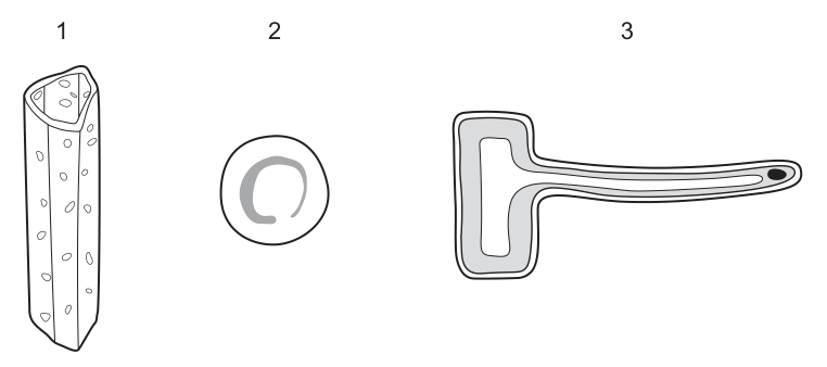
What are these structures?
Options
| 1 | 2 | 3 | |
|---|---|---|---|
| A | xylem vessel | root hair cell | red blood cell |
| B | red blood cell | xylem vessel | root hair cell |
| C | root hair cell | red blood cell | xylem vessel |
| D | xylem vessel | red blood cell | root hair cell |
Answer
D
D
Walkthrough
The question presents three diagrams of microscopic structures and asks for their identification. We evaluate each structure based on its visible features and match them to known specialised cells and tissues.
- Structure 1 is a long, cylindrical tube with pitted walls and no end walls (open ends). At maturity, these cells lose their cytoplasm and nucleus to form a continuous hollow tube. This is the characteristic structure of a xylem vessel, which transports water and mineral ions up the plant. The pitted walls allow water to move sideways between adjacent vessels.
- Structure 2 is a biconcave disc shape with no visible nucleus. This is the classic appearance of a red blood cell (erythrocyte). The biconcave shape maximises the surface area to volume ratio for efficient gas exchange, and the lack of a nucleus leaves more space for haemoglobin to bind oxygen.
- Structure 3 is an elongated cell with a long, thin hair-like extension and a nucleus located at the base of the extension. This is a root hair cell. The long extension greatly increases the surface area for absorbing water and mineral ions from the soil. The nucleus is retained to control the cell's metabolic activities, including active transport of ions.
Matching these identifications to the columns: 1 = xylem vessel, 2 = red blood cell, 3 = root hair cell. This corresponds exactly to option D.
Key Takeaways
- Xylem vessels are dead, hollow tubes with pitted walls and no end walls, adapted for water transport.
- Red blood cells are biconcave discs without a nucleus, adapted for oxygen transport.
- Root hair cells have a long cytoplasmic extension with a nucleus, adapted to increase surface area for water and ion uptake.
- Identifying specialised cells relies on recognising these key structural adaptations that match their specific functions.
Common Mistakes
- Confusing xylem with phloem: Phloem sieve tubes have end walls with sieve plates and are accompanied by companion cells. Xylem vessels have no end walls and no companion cells.
- Confusing root hair cells with nerve cells: Both have long extensions. However, root hair cells have a cell wall and a large central vacuole (often visible), and the extension is a simple cytoplasmic projection without a myelin sheath or dendrites. Nerve cells have a distinct cell body with dendrites and an axon.
- Forgetting that mature xylem vessels and red blood cells lack a nucleus: If a student assumes all cells have a nucleus, they might misidentify structure 1 or 2.
Things to Be Careful About
- Read the diagram carefully: Structure 1 has open ends (no end walls), which is the definitive feature of a xylem vessel at this level.
- Structure 2 is a simple biconcave disc; do not overcomplicate it by thinking of white blood cells, which have a visible nucleus and irregular shape.
- Structure 3 has a nucleus at the base of the hair; root hair cells do not have a nucleus in the hair extension itself.
- Option D is the only sequence that correctly matches all three structures. Ensure you read across the row for the correct letter, not down the columns.
Species of organisms have scientific names made up of two parts.
What is the system used to name species?
Options
A the classification system
B the genus system
C the binomial system
D the dichotomous key system
Working
The scientific name of a species has two parts, for example Homo sapiens. The system that gives each species a two-part name is called the binomial system.
- A is too general: the classification system is the overall way organisms are grouped, not the naming system.
- B is only part of the name: the genus is the first word, but the full two-part name is not called the genus system.
- D is an identification tool, not a naming system.
Answer
C
C
Walkthrough
This question asks for the name of the system used to give species their scientific names. A scientific name such as Homo sapiens is made up of two parts: the genus name (Homo) and the species name (sapiens). The system that uses two names is called the binomial system.
Look at each option:
- A, the classification system — this is the whole way of arranging organisms into groups such as kingdom, phylum, class, order, family, genus and species. It is not specifically about giving an organism a two-part name.
- B, the genus system — the genus is only the first part of a scientific name. The full name needs both parts, so this is not the correct name for the system.
- C, the binomial system — "bi-" means two and "nomial" means name. This is exactly the system that gives each species a two-part scientific name.
- D, the dichotomous key system — a dichotomous key is a tool used to identify an unknown organism by answering a series of yes/no questions. It does not name species.
Therefore the correct answer is C.
Key Takeaways
- Every species has a scientific name made of two parts: the genus name followed by the species name.
- This two-part naming system is called the binomial system.
- The binomial system is part of classification, but it is specifically the naming system, not the whole process of grouping organisms.
- A dichotomous key is used for identification, not for naming.
Common Mistakes
- Choosing A because classification and naming sound similar. Classification is grouping; the binomial system is naming.
- Choosing B because the genus is part of the scientific name. The system is named for both parts, not just the genus.
- Choosing D because keys are used in classification work. Keys identify organisms; they do not give them scientific names.
Things to Be Careful About
- Remember the exact phrase binomial system — it is the precise term needed.
- The scientific name is written in italics with the genus capitalised and the species name in lower case, e.g. Homo sapiens.
- This is a one-mark recall question, so no extra reasoning is needed in the answer.
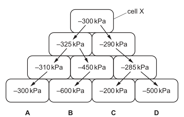
The diagram shows the water potentials measured in units of in a group of plant cells. A larger negative number indicates a lower water potential.
Which sequence of arrows shows the net movement of water from cell X?
Answer
Water moves by osmosis from a region of higher water potential (less negative) to a region of lower water potential (more negative).
Tracing the pathways from cell X ():
- Path A: . The water potential increases from to , so there is no net movement down the gradient.
- Path B: . The water potential continuously decreases (becomes more negative), representing a continuous net movement down the gradient.
- Path C: . The water potential continuously increases, so water would move in the opposite direction.
- Path D: . The water potential increases from to , breaking the downward gradient.
Only path B shows a continuous net movement of water from cell X.
Answer
B
B
Walkthrough
The question asks for the sequence of arrows showing the net movement of water from cell X. Water moves by osmosis down a water potential gradient, meaning it moves from a region of higher water potential (less negative) to a region of lower water potential (more negative). Cell X has a water potential of . We must trace a path from X to one of the options where every step goes to a more negative value.
- Path A goes through , then , then . The step from to is an increase in water potential (less negative), so net water movement would be backwards along that segment.
- Path B goes through , then , then . Every step is to a more negative value (), so this is a valid continuous path down the gradient.
- Path C goes through , then , then . Every step is to a less negative value, meaning water would move in the opposite direction (up the gradient).
- Path D goes through , then , then . The step from to is an increase in water potential, breaking the downward gradient from cell X.
Only path B represents a continuous net movement of water from cell X to the final cell.
Key Takeaways
- Water potential is measured in negative units (); a higher water potential is a less negative number (e.g., is higher than ).
- Net movement of water by osmosis is always from higher water potential to lower water potential (less negative to more negative).
- When tracing pathways, every single step must follow the gradient; a single reversal invalidates the entire path.
Common Mistakes
- Confusing the sign convention: thinking that is lower than because . Remember that is mathematically higher (less negative) than .
- Selecting a path that ends at a very low water potential (like ) without checking if the intermediate steps actually follow the gradient.
- Assuming water moves towards the cell with the most negative value regardless of the direct pathway.
Things to Be Careful About
- Always read the water potential values as negative numbers. is higher than .
- Check every segment of the path. A path like D looks like it goes to a low value (), but the segment from to goes up the gradient, so water cannot flow that way from cell X.
Which statements about active transport are correct?
- Ions move from a region of high concentration to a region of low concentration.
- Ions move across the cell membrane.
- Energy released during respiration is used to move ions into or out of a cell.
Options
A 1, 2 and 3
B 1 and 2 only
C 1 and 3 only
D 2 and 3 only
Working
Statement 1 is incorrect: active transport moves ions against the concentration gradient, from a low to a high concentration. Moving from high to low concentration describes diffusion.
Statement 2 is correct: active transport moves ions across the cell membrane.
Statement 3 is correct: active transport uses energy released during respiration.
So only statements 2 and 3 are correct.
Answer
D
D
Walkthrough
Read each numbered statement against the definition of active transport.
-
Ions move from a region of high concentration to a region of low concentration. This is the opposite of active transport. Active transport moves substances against the concentration gradient, from a region of lower concentration to a region of higher concentration. Moving from high to low concentration is diffusion, not active transport. So statement 1 is false.
-
Ions move across the cell membrane. Yes. Active transport happens across cell membranes, for example when root hair cells take up mineral ions from the soil. The ions are moved through carrier proteins in the membrane. So statement 2 is true.
-
Energy released during respiration is used to move ions into or out of a cell. Yes. Moving ions against the concentration gradient needs energy. This energy comes from respiration. So statement 3 is true.
Only statements 2 and 3 are correct, so the answer is D.
Key Takeaways
- Active transport is the movement of particles against a concentration gradient, from a lower to a higher concentration.
- Active transport requires energy, which is released during respiration.
- Active transport takes place across cell membranes, often using carrier proteins.
- Diffusion is different: it is the movement of particles down a concentration gradient, from high to low, and it does not require energy.
Common Mistakes
- Choosing A or C because statement 1 is wrongly thought to describe active transport. Statement 1 describes diffusion, not active transport.
- Forgetting that active transport goes against the concentration gradient. This is the key difference from diffusion.
- Thinking that active transport does not need energy. It always needs energy from respiration.
- Confusing the direction of movement: active transport is from low to high concentration, not high to low.
Things to Be Careful About
- The phrase "against the concentration gradient" is essential. If a question asks for the definition of active transport, you must include this idea.
- Statement 2 is true, but it is not enough on its own to identify active transport. The question asks which statements are correct, not which statement is unique to active transport.
- In multiple-choice questions with numbered statements, check each statement separately before choosing the option.
- Remember the example of root hair cells: they use active transport to take up mineral ions from the soil, even when the soil concentration is lower than inside the root.
Which biological molecule contains the chemical elements carbon, hydrogen, nitrogen and phosphorus?
Options
A DNA
B cellulose
C lipid
D starch
Working
DNA contains the elements carbon, hydrogen, oxygen, nitrogen and phosphorus. Cellulose, starch and lipids contain only carbon, hydrogen and oxygen (lipids may also contain small amounts of other elements, but not phosphorus as a defining feature).
Answer
A
A
Walkthrough
The question asks which biological molecule contains carbon, hydrogen, nitrogen and phosphorus. Recall the elements found in each option:
- DNA is a nucleic acid. Its nucleotides contain a sugar, a phosphate group and a nitrogen-containing base. Therefore DNA contains carbon, hydrogen, oxygen, nitrogen and phosphorus.
- Cellulose is a carbohydrate made of glucose units. Carbohydrates contain only carbon, hydrogen and oxygen.
- Lipids are made of fatty acids and glycerol. They contain carbon, hydrogen and oxygen (and sometimes phosphorus in phospholipids, but a general lipid does not contain nitrogen).
- Starch is also a carbohydrate, so it contains only carbon, hydrogen and oxygen.
Only DNA contains all four elements listed, so the correct option is A.
Key Takeaways
- Carbohydrates (starch, cellulose, sugars) contain carbon, hydrogen and oxygen.
- Lipids contain carbon, hydrogen and oxygen.
- Proteins contain carbon, hydrogen, oxygen, nitrogen and usually sulfur.
- DNA contains carbon, hydrogen, oxygen, nitrogen and phosphorus.
- The presence of nitrogen and phosphorus together points strongly to DNA.
Common Mistakes
- Choosing lipid because some lipids contain phosphorus. The question asks for nitrogen AND phosphorus together, and a general lipid does not contain nitrogen.
- Choosing cellulose or starch because they are common biological molecules, without recalling their elemental composition.
- Confusing DNA with protein: proteins contain nitrogen but do not contain phosphorus as a standard element.
Things to Be Careful About
- Read the question carefully: it requires both nitrogen and phosphorus.
- Remember that carbohydrates contain only C, H and O.
- The mark scheme gives the answer as A; no working is needed on the exam paper.
Catalase is an enzyme found in potato tissue. It catalyses the breakdown of hydrogen peroxide into water and oxygen.
The apparatus shown was used to investigate the activity of catalase.
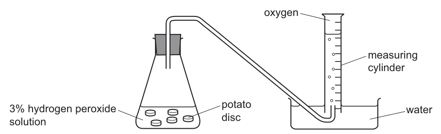
Five identical potato discs were dropped into of a hydrogen peroxide solution at a temperature of . The time taken for of oxygen to be produced was recorded.
This procedure was repeated at each of the following temperatures: , , , and .
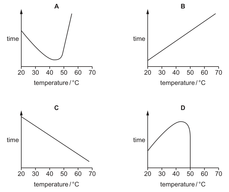
Which graph shows the results of the investigation?
Options
Working
The investigation measures the time taken for a fixed volume of oxygen () to be produced. Time is inversely related to reaction rate: a faster reaction means less time is taken.
- As temperature increases from to the optimum (), kinetic energy increases, enzyme activity rises, and the reaction is faster. Therefore, the time taken decreases.
- At the optimum temperature, activity is at its maximum and time is at its minimum.
- Above the optimum temperature, the enzyme (catalase) denatures, activity drops rapidly, the reaction slows down, and the time taken increases sharply.
The graph of time against temperature must therefore be U-shaped, with a minimum around . Graph A shows this U-shaped curve. Graph D is an inverted U-shape (bell curve), which would be correct if the y-axis represented rate or volume of oxygen, but not time.
Answer
A
A
Walkthrough
- Identify what is being measured: The y-axis on all four graphs is time (taken to produce of oxygen). Time is inversely proportional to the rate of reaction. If the reaction is fast, the time is short. If the reaction is slow, the time is long.
- Recall the effect of temperature on enzyme activity: As temperature rises from , the kinetic energy of the molecules increases. This leads to more frequent and more energetic collisions between the catalase enzyme and the hydrogen peroxide substrate. Enzyme activity increases, the reaction speeds up, and the time taken decreases. This continues until the optimum temperature is reached (around for catalase), where the time taken is at its minimum.
- Consider temperatures above the optimum: Beyond the optimum, the increased thermal energy breaks the bonds maintaining the enzyme's tertiary structure. The active site changes shape (denaturation), and the enzyme can no longer bind the substrate. Activity drops rapidly, the reaction slows down dramatically, and the time taken increases sharply.
- Match the prediction to the graphs: Because time decreases then increases, the graph must be U-shaped. Graph A matches this perfectly, with a minimum around . Graph D is an inverted U-shape (bell curve), which is the classic graph for enzyme rate against temperature, but it is incorrect here because the y-axis is time, not rate.
- Conclude: Graph A is the correct representation of the results.
Key Takeaways
- Time vs. rate: When plotting an enzyme investigation, always check the y-axis. If it is rate (or volume of product per unit time), the graph is a bell curve (inverted U). If it is time (to produce a fixed amount of product), the graph is a U-shape.
- Enzyme kinetics: Enzyme activity increases with temperature up to an optimum due to increased kinetic energy, then decreases sharply due to denaturation of the active site.
Common Mistakes
- Choosing Graph D: This is the most common error. Students memorise the "bell curve" for enzyme activity and select Graph D without noticing that the y-axis is time, not rate. A bell curve means activity peaks and then falls; a U-curve means time is shortest at the peak and then rises.
- Assuming a linear relationship: Choosing Graph B or C ignores the denaturation phase. Enzyme activity does not increase or decrease linearly with temperature across this entire range; it has a distinct optimum and a sharp drop-off due to denaturation.
- Misidentifying the optimum: The minimum time on Graph A occurs around , which is consistent with the known optimum temperature for catalase. Graphs B and C have no minimum, and Graph D has a maximum time, both of which contradict enzyme behaviour.
Things to Be Careful About
- Always read the axis labels carefully. The distinction between "time taken" and "rate of reaction" is one of the most frequently tested traps in 5090 enzyme questions.
- Unit consistency and scale: Ensure the temperature range on the x-axis matches the investigation () and that the curve reflects the sharp drop in activity above the optimum (denaturation), rather than a gradual decline.
- Recall the exact mechanism: Denaturation is the loss of the active site's shape due to the breaking of hydrogen and other bonds; it is not simply "the enzyme melting" or "the enzyme dying". Use precise terminology like denatured and active site if asked to explain the curve.
The graphs show factors affecting the rate of photosynthesis.
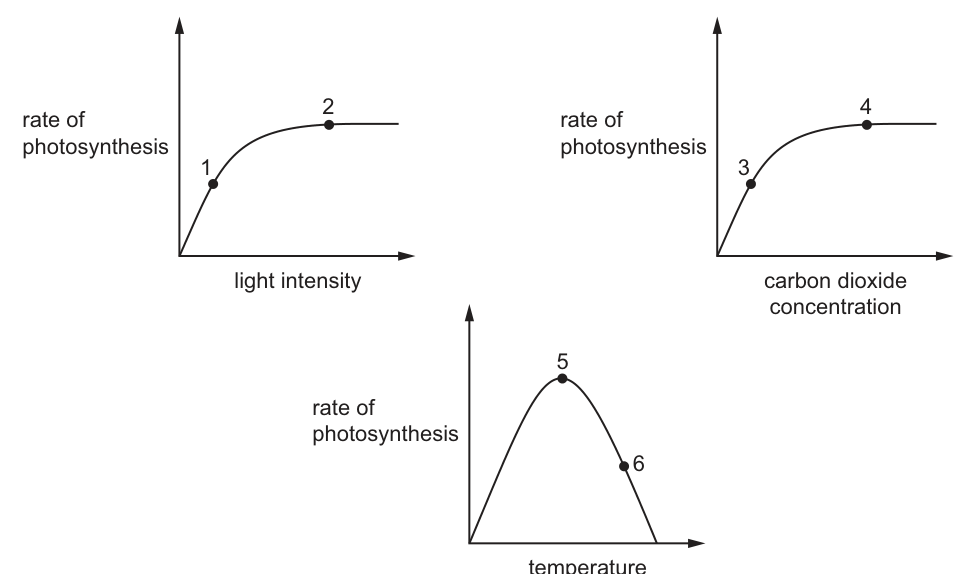
At which points on the graphs could the rate of photosynthesis be limited by the carbon dioxide concentration?
Options
A 1, 3 and 5
B 1, 4 and 6
C 2, 3 and 5
D 2, 4 and 6
Working
To determine where the rate of photosynthesis is limited by carbon dioxide concentration, we must look for points where increasing carbon dioxide would increase the rate. This happens when the current limiting factor is NOT carbon dioxide (i.e., we are on a plateau for the graph's x-axis variable, or the x-axis variable is at an optimum).
- Graph 1 (light intensity): At point 1, the rate is rising, so it is limited by light intensity. At point 2, the rate has plateaued; light is no longer limiting, so the rate is limited by another factor such as carbon dioxide concentration or temperature. Thus, point 2 could be limited by carbon dioxide.
- Graph 2 (carbon dioxide concentration): At point 3, the rate is rising with carbon dioxide, so carbon dioxide is the limiting factor. At point 4, the rate has plateaued; carbon dioxide is no longer limiting, so it is limited by light or temperature. Thus, point 3 is limited by carbon dioxide, but point 4 is not.
- Graph 3 (temperature): At point 5, the temperature is at the optimum; increasing temperature does not increase the rate, so the rate is limited by another factor such as light or carbon dioxide. Thus, point 5 could be limited by carbon dioxide. At point 6, the temperature is above the optimum and enzymes are denaturing; the rate is limited by temperature, not carbon dioxide.
The points where carbon dioxide could be the limiting factor are 2, 3, and 5.
Answer
C
C
Walkthrough
- Understand limiting factors: A limiting factor is any variable that, when increased, causes the rate of photosynthesis to increase. If a factor is NOT limiting, increasing it will not change the rate. We are looking for points where carbon dioxide concentration is the limiting factor, meaning that if we added more carbon dioxide, the rate would go up.
- Analyze Graph 1 (light intensity vs. rate): Point 1 is on the rising slope, meaning light intensity is the limiting factor. Point 2 is on the plateau, meaning light is no longer limiting. The rate must now be limited by another factor, such as carbon dioxide concentration or temperature. Therefore, carbon dioxide could be limiting at point 2.
- Analyze Graph 2 (carbon dioxide concentration vs. rate): Point 3 is on the rising slope, meaning carbon dioxide is directly limiting the rate. Point 4 is on the plateau, meaning carbon dioxide is no longer limiting; the rate is now limited by light or temperature. Therefore, carbon dioxide is limiting at point 3, but not at point 4.
- Analyze Graph 3 (temperature vs. rate): Point 5 is at the optimum temperature. The rate is at its maximum for temperature, so temperature is not the limiting factor. The rate is limited by another factor, such as carbon dioxide. Point 6 is on the descending slope; the high temperature is causing enzymes to denature, so temperature (specifically enzyme denaturation) is the limiting factor, not carbon dioxide.
- Combine the findings: Points 2, 3, and 5 are the locations where carbon dioxide concentration could be the limiting factor. This matches option C.
Key Takeaways
- A limiting factor is one that, if increased, would increase the rate of reaction.
- On a graph of rate vs. a specific factor, the rising portion indicates that factor is limiting.
- The plateau or peak indicates that factor is no longer limiting, and another factor (like light, temperature, or carbon dioxide) has become limiting.
- On the descending side of a temperature graph, enzyme denaturation is the limiting factor, not the other reactants.
Common Mistakes
- Choosing point 1 or 4: At point 1, light is limiting, not carbon dioxide. At point 4, carbon dioxide is on the plateau, so it is NOT limiting; the rate is limited by light or temperature.
- Choosing point 6: At point 6, the temperature is too high and enzymes are denaturing. The rate is limited by temperature (enzyme denaturation), not carbon dioxide.
- Assuming the plateau on the carbon dioxide graph means carbon dioxide is limiting: The plateau on the carbon dioxide graph (point 4) means carbon dioxide is in excess and no longer limiting.
Things to Be Careful About
- Always check the x-axis variable for each graph. If you are on the rising part of the curve, the x-axis variable is the limiting factor. If you are on the plateau or peak, the x-axis variable is NOT the limiting factor.
- Remember that at point 5 (optimum temperature), the rate is limited by the next limiting factor in the sequence, which is often carbon dioxide concentration or light intensity, not temperature itself.
- On the falling side of the temperature graph (point 6), the limiting factor is enzyme denaturation due to high temperature, not a lack of reactants like carbon dioxide.
The diagram shows a section through a leaf.
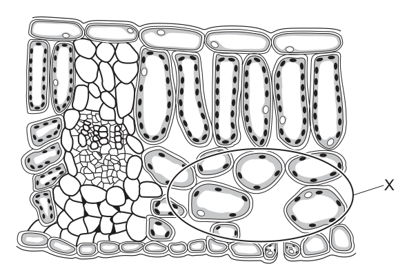
What is the main function of the region labelled X?
Options
A conduction and support
B gaseous exchange
C photosynthesis
D prevention of water loss
Working
The diagram shows a transverse section through a leaf. Region X encloses the spongy mesophyll layer, which is characterised by loosely arranged cells and large intercellular air spaces. These air spaces allow gases (carbon dioxide and oxygen) to diffuse freely throughout the leaf tissue, making gaseous exchange their main function.
- Option A (conduction and support) refers to the vascular bundle (xylem and phloem).
- Option C (photosynthesis) is the main function of the palisade mesophyll, which is tightly packed with chloroplasts.
- Option D (prevention of water loss) is the function of the waxy cuticle.
Thus, X relates to gaseous exchange.
Answer
B
B
Walkthrough
The question asks for the main function of the region labelled X in a leaf cross-section. We identify X by its position and cellular arrangement. X is located below the vascular bundle and above the lower epidermis, and it contains loosely arranged cells with large gaps between them. This is the spongy mesophyll layer. The large intercellular air spaces in the spongy mesophyll are an adaptation for gaseous exchange: they allow carbon dioxide to diffuse to the photosynthesising cells and oxygen (a product of photosynthesis) to diffuse out to the stomata. We then evaluate the other options to confirm:
- Option A (conduction and support) is the role of the vascular bundle (xylem and phloem).
- Option C (photosynthesis) is the primary role of the palisade mesophyll, which is tightly packed with chloroplasts.
- Option D (prevention of water loss) is the function of the waxy cuticle and epidermis.
Therefore, region X is adapted for gaseous exchange, making B the correct answer.
Key Takeaways
- Leaf cross-sections can be interpreted by matching cellular arrangement to tissue type: palisade mesophyll (tightly packed, columnar cells) versus spongy mesophyll (loosely arranged, large air spaces).
- Structure and function must be linked: air spaces in the spongy mesophyll facilitate gaseous exchange, which is the distinguishing function of this layer compared to the palisade layer.
Common Mistakes
- Confusing the spongy mesophyll with the palisade mesophyll. Candidates might choose "photosynthesis" because spongy mesophyll cells do contain chloroplasts, but the question asks for the main function of the region (especially the air spaces), which is gaseous exchange.
- Misidentifying the vascular bundle's function as gaseous exchange.
- Choosing "prevention of water loss" by looking at the wrong layer, such as the cuticle or epidermis.
Things to Be Careful About
- Read the diagram carefully: X is circled around the spongy mesophyll air spaces, not the palisade layer or the vascular bundle.
- The command word is implicit in an MCQ: select the main function associated with the labelled structure. While spongy mesophyll cells perform photosynthesis, the large air spaces are specifically adapted for gaseous exchange, which is the distinguishing function of this layer compared to the palisade layer.
- In 5090 Paper 1, there is no partial credit; the answer must be exactly the correct option letter.
Root hair cells take in water and ions from the soil.
Which row shows how water and ions are taken into root hair cells?
Options
| water | ions | |
|---|---|---|
| A | active transport | active transport |
| B | active transport | osmosis |
| C | osmosis | osmosis |
| D | osmosis | active transport |
Working
Water enters the root hair cell by osmosis — it moves down the water potential gradient across the partially permeable cell membrane. Ions enter by active transport — they are moved against the concentration gradient using energy from respiration.
So the correct row is water = osmosis, ions = active transport.
- A — both active transport: wrong, water does not enter by active transport.
- B — water active transport, ions osmosis: both reversed.
- C — both osmosis: wrong, ions cannot enter by osmosis.
Answer
D
D
Walkthrough
Root hair cells are specialised cells in plant roots that absorb water and mineral ions from the soil. The two substances enter by two completely different mechanisms, and this question tests whether you know which is which.
Water. The soil solution has a higher water potential than the cell sap inside the root hair cell, so water moves by osmosis across the partially permeable cell surface membrane, down the water potential gradient, into the cell. Osmosis is the movement of water molecules from a region of higher water potential to a region of lower water potential through a partially permeable membrane.
Ions. Mineral ions are usually present at a higher concentration inside the root hair cell than in the soil, so they cannot simply diffuse in. Instead they are taken in by active transport — the cell uses energy from respiration to move ions against their concentration gradient across the membrane, using carrier proteins.
So the correct row is D: water by osmosis, ions by active transport.
Now eliminate the other rows. Row A says water is taken in by active transport — wrong, water moves by osmosis. Row B says water is taken in by active transport and ions by osmosis — both are reversed. Row C says ions are taken in by osmosis — wrong, ions need active transport because they move against their concentration gradient.
Key Takeaways
- Water moves into root hair cells by osmosis, down the water potential gradient, through a partially permeable membrane.
- Ions move into root hair cells by active transport, against the concentration gradient, using energy from respiration.
- Osmosis applies to water only; active transport applies to solutes such as mineral ions.
- This question links the Movement Into and Out of Cells topic to the function of root hair cells in Transport in Flowering Plants.
Common Mistakes
- Saying ions enter by osmosis — osmosis is the movement of water only, not of dissolved ions.
- Saying water enters by active transport — water is not actively transported into root hair cells.
- Confusing the direction of movement: active transport moves substances against the concentration gradient, while osmosis moves water down the water potential gradient.
- Choosing row B, which swaps the two mechanisms.
Things to Be Careful About
- Read the table carefully: the columns are water and ions, and you must match each substance to its own mechanism.
- Remember that active transport requires energy from respiration and moves substances against the concentration gradient.
- Osmosis is a special case of diffusion that applies to water only, through a partially permeable membrane, down the water potential gradient.
Four leafy shoots cut from the same plant were put into beakers of coloured water and left in rooms with different temperatures. In two rooms, electric fans were set up to blow air over the stems.
The time taken for the coloured water to reach the leaves was measured for each stem.
In which stem will the coloured water reach the leaves the fastest?
Options
| room conditions | |
|---|---|
| A | |
| B | with a fan |
| C | |
| D | with a fan |
Working
Transpiration is faster at a higher temperature because evaporation is faster. It is also faster when air moves because water vapour is blown away, keeping the water potential gradient steep. Stem D has both conditions, so water is pulled up its xylem fastest.
Answer
D
D
Walkthrough
The coloured water is carried up the stem in the xylem. The force that moves water up the xylem comes from transpiration: water evaporates from the leaves, mainly through stomata, and this loss creates a water potential gradient that pulls more water up from the roots.
Anything that increases the rate of transpiration will make the coloured water reach the leaves faster.
Two factors are changed in this question:
- Temperature. At a higher temperature, water molecules have more kinetic energy, so evaporation from the leaf surfaces is faster. This increases transpiration.
- Air movement. A fan blows water vapour away from the leaf surface. This keeps the air around the leaf drier, so the water potential gradient between the leaf and the air stays steep. This also increases transpiration.
Option A has neither factor, so it is the slowest. Option B has a fan but a low temperature, so it is faster than A. Option C has a high temperature but no fan, so it is faster than A and B. Option D has both a high temperature and a fan, so transpiration is fastest and the coloured water reaches the leaves fastest.
Key Takeaways
- Transpiration is the loss of water vapour from the leaves.
- Transpiration creates the pull that moves water up the xylem from the roots to the leaves.
- Higher temperature increases the rate of evaporation and therefore increases transpiration.
- Moving air removes water vapour from around the leaf, keeping the water potential gradient steep and increasing transpiration.
- When two factors both increase transpiration, their effects combine, so the fastest movement occurs when both are present.
Common Mistakes
- Choosing C because it has the higher temperature, while forgetting that the fan in D also increases transpiration.
- Thinking that a fan cools the plant and therefore slows transpiration. In fact, moving air increases transpiration by removing water vapour from around the leaf.
- Confusing transpiration with water absorption. The coloured water moves up because transpiration pulls it, not because the roots actively push it.
- Ignoring the word "fastest" and simply picking any condition that increases transpiration, rather than the one with the greatest combined effect.
Things to Be Careful About
- The question asks which stem is fastest, so both factors must be considered together.
- Use the correct terms: transpiration, evaporation, water potential gradient, xylem.
- The mark scheme accepts only D.
- No calculation is needed here; the answer depends on understanding how temperature and air movement affect transpiration rate.
Cubes of boiled egg white are placed in test-tubes containing of water. Boiled egg white contains protein. Other substances are added to each test-tube as shown in the table. The test-tubes are left for eight hours and then tested for amino acids.
| test-tube | solution added | results of test for amino acids |
|---|---|---|
| 1 | pepsin | absent |
| 2 | pepsin + alkali | absent |
| 3 | none | absent |
| 4 | pepsin + acid | large amounts |
| 5 | boiled pepsin + acid | traces |
| 6 | acid | traces |
| 7 | alkali | absent |
Which test-tubes show that pepsin is an enzyme?
Options
A 1 and 4
B 2 and 7
C 4 and 5
D 5 and 6
Working
To show pepsin is an enzyme, two properties must be demonstrated:
-
It is a catalyst — it speeds up the breakdown of protein to amino acids. Tube 4 (pepsin + acid) gives large amounts of amino acids, while tube 6 (acid alone) gives only traces. The only difference is the pepsin, so pepsin greatly speeds up digestion.
-
It is biological (a protein) — boiling destroys its activity. Tube 4 (pepsin + acid) gives large amounts, but tube 5 (boiled pepsin + acid) gives only traces. Boiling denatures the enzyme, so it can no longer work.
Tubes 4 and 5 together show pepsin is an enzyme.
Why the other options fail:
- 1 and 4: tube 1 shows pepsin without acid digests nothing — this only shows pepsin needs acid, not that it is an enzyme.
- 2 and 7: both contain alkali and give no amino acids — this only shows alkali stops digestion (a pH effect).
- 5 and 6: these show boiling destroys activity and acid alone gives traces, but without tube 4 they do not show pepsin speeds up digestion.
Answer
C
C
Walkthrough
This question asks which test-tubes prove that pepsin is an enzyme. An enzyme is a biological catalyst: "biological" means it is a protein made by a living organism, and "catalyst" means it speeds up a chemical reaction without being used up itself.
To prove pepsin is an enzyme, you must show both of these things:
-
It speeds up the reaction (catalyst). Look at tube 4: pepsin + acid gives large amounts of amino acids. Compare this with tube 6: acid alone gives only traces. The only difference between these two tubes is the pepsin. So the pepsin has enormously increased the amount of protein digestion — it is acting as a catalyst.
-
It is a protein (biological). Compare tube 4 with tube 5: boiled pepsin + acid gives only traces. The only difference is that the pepsin was boiled. Boiling denatures the enzyme — it destroys the shape of its active site, so it can no longer fit the protein substrate and cannot catalyse the reaction. A non-biological catalyst, such as the acid itself, would survive boiling and keep working. Because boiling destroys pepsin's activity, pepsin must be a protein — a biological catalyst.
So tubes 4 and 5 together provide the complete evidence that pepsin is an enzyme.
Why the other options are wrong:
- 1 and 4: Tube 1 (pepsin alone) gives no amino acids. This only shows that pepsin needs acid to work — it needs a suitable pH. It does not show pepsin is an enzyme.
- 2 and 7: Both contain alkali and both give no amino acids. This only shows that alkaline conditions stop digestion — again a pH effect, not evidence of enzyme nature.
- 5 and 6: Tube 5 shows boiled pepsin does not work, and tube 6 shows acid alone gives traces. On their own these do not show pepsin is an enzyme — you need tube 4 to show that pepsin actually speeds up digestion.
Key Takeaways
- An enzyme is a biological catalyst: it speeds up a reaction (catalyst) and is a protein that can be denatured (biological).
- Boiling denatures enzymes — the active site changes shape and the enzyme can no longer bind its substrate, so it stops working.
- Pepsin works best in acidic conditions — it needs acid for its optimum pH, which is why tube 4 (pepsin + acid) works but tube 1 (pepsin alone) does not.
- To prove something is an enzyme, you need to show both that it catalyses the reaction and that heat destroys its activity.
Common Mistakes
- Choosing 1 and 4: tube 1 only shows pepsin needs acid, not that it is an enzyme.
- Choosing 2 and 7: these only show alkali prevents digestion — a pH effect.
- Choosing 5 and 6: these show boiling destroys activity and acid alone gives traces, but without tube 4 they do not show pepsin speeds up digestion.
- Confusing "pepsin needs acid" with "pepsin is an enzyme" — needing a particular pH is a property of many enzymes, not the evidence that something is an enzyme.
Things to Be Careful About
- The classic demonstration that something is an enzyme requires two comparisons: one tube with the enzyme working (tube 4) and one with the enzyme denatured by boiling (tube 5).
- Note the traces in tube 6: acid alone can break down a little protein. That is why tube 5 (boiled pepsin + acid) also gives traces — the traces come from the acid, not from the boiled pepsin, which is dead.
- Read the table carefully: the key comparisons are between tubes that differ by only one factor (pepsin present/absent, pepsin boiled/not boiled).
The diagram shows a section through a villus.
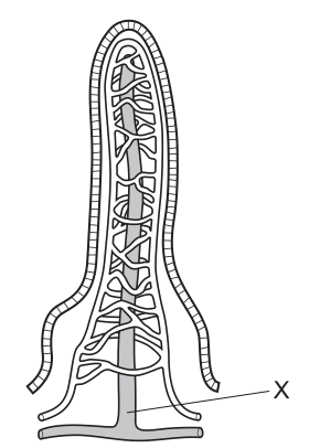
What is the main role of vessel X?
Options
A to carry amino acids to the liver for protein formation
B to deliver deoxygenated blood to the heart
C to supply oxygen to the cells of the villus
D to transfer fatty acids and glycerol to the lymph system
Working
The diagram shows a section through an intestinal villus. Vessel X is the central vessel that branches into a lymphatic vessel at the base, which identifies it as the lacteal.
The lacteal is a lymphatic capillary. Its role is to absorb and transport fatty acids and glycerol (which are reassembled into lipids within the epithelial cells) into the lymphatic system. The lymphatic fluid eventually drains into the bloodstream.
The surrounding network is the blood capillary network:
- Amino acids and glucose are absorbed into the blood capillaries and carried to the liver via the hepatic portal vein.
- Oxygen is supplied to the villus cells by the arterial blood in these capillaries.
- Deoxygenated blood is carried away by the venous blood in these capillaries.
Therefore, the main role of vessel X (the lacteal) is to transfer fatty acids and glycerol to the lymph system.
Answer
D
D
Walkthrough
- Identify structure X: The image description states that vessel X is the 'central lacteal vessel... branching into a lymphatic vessel at the base'. In an intestinal villus, the central vessel is the lacteal, surrounded by a network of blood capillaries.
- Recall nutrient absorption pathways: After digestion, nutrients are absorbed by the epithelial cells of the villus.
- Glucose and amino acids are small enough to enter the blood capillaries directly. They are carried via the hepatic portal vein to the liver.
- Fatty acids and glycerol enter the epithelial cells, where they are reassembled into triglycerides. These are too large to enter the blood capillaries directly, so they are packaged into chylomicrons and enter the lacteal (the lymphatic vessel).
- Evaluate the options:
- A: Carrying amino acids to the liver is the role of the blood capillaries and the hepatic portal vein, not the lacteal.
- B: Delivering deoxygenated blood to the heart is the role of the veins draining the blood capillary network.
- C: Supplying oxygen to the cells is the role of the arterial blood within the capillary network.
- D: Transferring fatty acids and glycerol to the lymph system is the specific function of the lacteal (vessel X).
Key Takeaways
- The intestinal villus contains two main transport systems: a blood capillary network and a central lacteal (lymphatic vessel).
- Water-soluble nutrients (glucose, amino acids, water-soluble vitamins, minerals) enter the blood capillaries.
- Lipid-soluble nutrients (fatty acids, glycerol, fat-soluble vitamins) enter the lacteal because the reassembled lipids are too large to pass through the blood capillary walls.
Common Mistakes
- Confusing the lacteal with blood capillaries: Students often assume all absorbed nutrients go into the blood. Remember that lipids enter the lymphatic system first.
- Misreading the diagram: Failing to identify X as the central lacteal rather than the surrounding capillary network.
- Incorrect nutrient routing: Thinking that amino acids or glucose enter the lymphatic system.
Things to Be Careful About
- Ensure you correctly identify the structure based on the diagram. Vessel X is explicitly described as branching into a lymphatic vessel, which is the defining feature of a lacteal.
- Do not confuse the role of the lacteal with the hepatic portal vein; the portal vein carries blood from the gut to the liver, but it carries blood-borne nutrients (glucose, amino acids), not lymph-borne lipids.
- Remember that fatty acids and glycerol are reassembled into lipids inside the epithelial cells before entering the lacteal; they do not enter the lacteal as free fatty acids and glycerol.
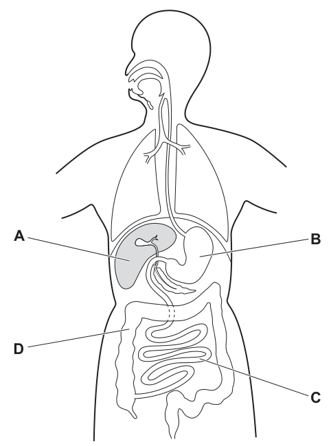
Which part of the human digestive system is a major region for assimilation of amino acids?
Options
A A
B B
C C
D D
Working
Amino acids absorbed from the small intestine are transported in the blood to the liver, where they are assimilated — used to build plasma proteins, or deaminated. On the diagram, A is the liver (the large organ beside the stomach), B is the stomach, C is the small intestine and D is the colon.
Answer
A
A
Walkthrough
The question asks where amino acids are assimilated. Assimilation is the uptake and use of absorbed nutrients by body cells. Amino acids are absorbed in the small intestine (labelled C), but they are not assimilated there — they travel in the hepatic portal vein to the liver (labelled A). In the liver, amino acids are used to build proteins such as plasma proteins, and excess amino acids are deaminated — the amino group is removed and converted to urea. So the major region of assimilation of amino acids is the liver.
Checking the labels on the figure: A points to the large organ on the left of the abdomen next to the stomach — the liver; B is the stomach; C is the coiled small intestine; D is the colon. The correct option is therefore A.
Key Takeaways
- Assimilation means the use of absorbed food molecules by cells, not their absorption.
- The liver is the major organ of assimilation: amino acids → plasma proteins; excess amino acids → deamination → urea.
- Glucose is assimilated by cells for respiration, but the liver also stores it as glycogen.
Common Mistakes
- Choosing C (small intestine): that is where amino acids are absorbed, not assimilated — a classic confusion between absorption and assimilation.
- Choosing B (stomach): the stomach digests protein with pepsin; it does not assimilate amino acids.
- Choosing D (colon): the colon absorbs water only.
Things to Be Careful About
- Read the question word precisely: "assimilation" points to the liver, while "absorption" would point to the small intestine.
- Learn the positions of the organs on an alimentary canal diagram so the labels can be identified quickly under exam conditions.
When the volume of the thorax increases, the pressure in the thorax is lowered. This results in air being taken into the body.
How is the volume of the thorax increased so that air is breathed in?
Options
A by the diaphragm and the external intercostal muscles contracting
B by the diaphragm relaxing and the external intercostal muscles contracting
C by the diaphragm and the internal intercostal muscles contracting
D by the diaphragm relaxing and the internal intercostal muscles contracting
Working
Breathing in (inhalation) requires the volume of the thorax to increase. This happens when the diaphragm contracts and flattens, and the external intercostal muscles contract, moving the ribs up and out.
- A is correct: both the diaphragm and the external intercostal muscles contract.
- B is wrong: the diaphragm contracts, it does not relax.
- C is wrong: the internal intercostal muscles are used in forced exhalation, not inhalation.
- D is wrong: both the diaphragm relaxes and the internal intercostal muscles contract, which is the opposite of what is needed.
Answer
A
A
Walkthrough
This question tests your knowledge of the mechanics of breathing. When you breathe in (inhalation), the volume of the thorax (the chest cavity) must increase. This is achieved by the contraction of two main muscle groups:
- The diaphragm: a dome-shaped muscle at the base of the thorax. When it contracts, it flattens and moves downwards.
- The external intercostal muscles: located between the ribs. When they contract, they pull the ribs upwards and outwards.
Both of these actions increase the volume of the thorax. This increase in volume causes the pressure inside the thorax to decrease (as the question states). Air then rushes in from outside to equalize the pressure.
Looking at the options:
- A correctly states that both the diaphragm and the external intercostal muscles contract.
- B is incorrect because the diaphragm must contract, not relax, to increase the thoracic volume.
- C is incorrect because it is the external intercostal muscles that are involved in quiet inspiration, not the internal intercostal muscles (which are used in forced expiration).
- D is incorrect because it gets both the diaphragm and the intercostal muscles wrong.
Key Takeaways
- Inhalation is an active process driven by muscle contraction.
- The diaphragm and external intercostal muscles contract to increase thoracic volume.
- Increasing volume decreases pressure, drawing air into the lungs.
- The internal intercostal muscles are used for forced exhalation, not quiet inhalation.
Common Mistakes
- Confusing internal and external intercostal muscles: The external intercostals are for inhalation; the internal intercostals are for forced exhalation.
- Thinking the diaphragm relaxes to breathe in: It contracts and flattens. Relaxation causes it to dome upwards for exhalation.
- Forgetting that inhalation is an active process: It requires energy for muscle contraction.
Things to Be Careful About
- The question specifically asks about increasing the volume of the thorax to breathe in. Focus on the muscles that cause this specific action.
- Note that the question is about the volume increase, which is the primary change; the pressure decrease is a consequence.
- Remember the specific roles: diaphragm contracts, external intercostals contract, ribs move up and out, volume increases, pressure decreases, air rushes in.
The diagram shows how some apparatus is set up to investigate respiration in germinating seeds.
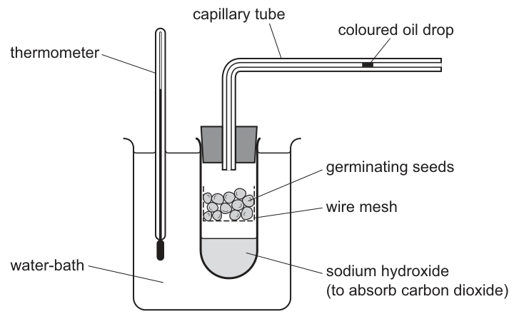
The coloured oil drop moves along the capillary tube.
What causes the movement of the coloured oil drop?
Options
A carbon dioxide released
B heat released
C oxygen used
D water used
Answer
C
C
Walkthrough
Germinating seeds carry out aerobic respiration, taking in oxygen and releasing carbon dioxide. The word equation for aerobic respiration is:
In this respirometer setup, the test-tube is sealed, so the total volume of gas can only change if the number of gas molecules changes. The sodium hydroxide solution at the bottom of the test-tube absorbs all the carbon dioxide produced by the seeds. Because the carbon dioxide is removed from the gas phase, the only gas that is consumed and not replaced is oxygen. As oxygen is used up, the total volume of gas inside the test-tube decreases, which causes the pressure inside to drop. The higher atmospheric pressure outside pushes the coloured oil drop along the capillary tube towards the test-tube. Therefore, the movement of the oil drop is caused by the oxygen being used.
Key Takeaways
- A respirometer with sodium hydroxide measures the rate of oxygen consumption.
- Sodium hydroxide absorbs carbon dioxide, so any change in gas volume is due solely to oxygen use.
- A decrease in gas volume leads to a decrease in pressure, drawing the oil drop towards the organism.
Common Mistakes
- Choosing A (carbon dioxide released): candidates forget that sodium hydroxide absorbs carbon dioxide, so it does not accumulate to push the oil drop.
- Choosing B (heat released): candidates ignore the water-bath, which keeps the temperature constant so that thermal expansion of the gas does not affect the oil drop.
- Choosing D (water used): water is a liquid product of respiration, not a gas, so its formation does not change the gas volume or pressure.
Things to Be Careful About
- Always check what the chemical at the bottom of the tube is. If it is sodium hydroxide (or potassium hydroxide), it absorbs carbon dioxide, and the setup measures oxygen uptake. If it is water, it measures the net change in gas volume (oxygen used minus carbon dioxide released).
- Remember that pressure changes drive the movement of the oil drop, not the physical presence of the gases themselves.
A ventricular septal defect is a hole in the septum between the left and right ventricles of the heart.
Which statement describes the effect on blood flow through the heart in a person with a ventricular septal defect?
Options
A Some deoxygenated blood flows into the pulmonary vein.
B Some oxygenated blood flows into the right atrium.
C Some oxygenated blood flows through the pulmonary artery.
D Some deoxygenated blood flows into the left atrium.
Working
A ventricular septal defect is a hole between the left and right ventricles. The left ventricle pumps at a higher pressure than the right, so oxygenated blood is forced from the left ventricle through the hole into the right ventricle. From there, this oxygenated blood is pumped out of the heart through the pulmonary artery.
Answer
C
C
Walkthrough
Let's trace the normal flow of blood through the heart. Deoxygenated blood returns to the right atrium, flows into the right ventricle, and is pumped to the lungs via the pulmonary artery. Oxygenated blood returns from the lungs via the pulmonary vein into the left atrium, flows into the left ventricle, and is pumped to the body via the aorta.
Now, a ventricular septal defect (VSD) is a hole in the septum — the wall between the left and right ventricles. The left ventricle is a thick, muscular chamber that generates high pressure to pump blood all the way around the body. The right ventricle generates much lower pressure. Because of this pressure difference, when there is a hole between them, oxygenated blood is forced from the high-pressure left ventricle through the hole into the low-pressure right ventricle. This is called a left-to-right shunt.
Once the oxygenated blood is in the right ventricle, it gets pumped out of the heart through the pulmonary artery, along with the normal deoxygenated blood. This means some oxygenated blood ends up flowing through the pulmonary artery, which normally only carries deoxygenated blood.
Let's check the options:
- A is wrong because the pulmonary vein normally carries oxygenated blood from the lungs, and a VSD doesn't push deoxygenated blood there.
- B is wrong because the defect is between the ventricles, not the atria, and blood doesn't flow backwards into the right atrium from this defect.
- C is correct because oxygenated blood from the left ventricle crosses the defect into the right ventricle and is then pumped through the pulmonary artery.
- D is wrong because the defect is not between the atria, and blood doesn't flow into the left atrium from this defect.
Key Takeaways
- The heart is a double pump: the right side deals with deoxygenated blood and the left side with oxygenated blood.
- Blood flows from high pressure to low pressure. The left ventricle is the highest-pressure chamber.
- A ventricular septal defect causes a left-to-right shunt, mixing oxygenated blood into the right side of the heart.
- The pulmonary artery normally carries deoxygenated blood; a VSD results in some oxygenated blood in it.
Common Mistakes
- Confusing a ventricular septal defect with an atrial septal defect (hole between the atria).
- Thinking the defect causes deoxygenated blood to mix into the left side of the heart. The pressure is higher on the left, so flow is left-to-right, not right-to-left.
- Assuming the defect directly involves the atria or the great vessels (aorta, pulmonary artery) rather than the ventricles.
Things to Be Careful About
- Remember the direction of blood flow is driven by pressure: high to low.
- The pulmonary artery is the only artery carrying deoxygenated blood; the pulmonary vein is the only vein carrying oxygenated blood. A VSD disrupts this clean separation.
- Read the options carefully — the question asks about the effect on blood flow, so you need to trace where the blood goes after crossing the defect.
Human blood is composed of plasma, red blood cells, white blood cells and platelets.
Which row states a function of each of these components?
Options
| plasma | red blood cells | white blood cells | platelets | |
|---|---|---|---|---|
| A | clotting | transporting carbon dioxide | transporting oxygen | producing antibodies |
| B | transporting carbon dioxide | transporting oxygen | producing antibodies | clotting |
| C | transporting carbon dioxide | transporting oxygen | clotting | producing antibodies |
| D | transporting oxygen | transporting carbon dioxide | producing antibodies | clotting |
Working
- Plasma transports dissolved substances, including carbon dioxide (as hydrogencarbonate ions).
- Red blood cells contain haemoglobin and transport oxygen.
- White blood cells defend the body by producing antibodies and engulfing pathogens.
- Platelets help blood to clot (clotting).
Row B correctly matches all four.
Answer
B
B
Walkthrough
This question asks you to match each of the four components of blood to its correct function. Let's go through each one:
-
Plasma is the liquid part of the blood. It is mostly water and it carries dissolved substances such as glucose, amino acids, urea, and importantly for this question, carbon dioxide (dissolved as hydrogencarbonate ions). So plasma is responsible for transporting carbon dioxide.
-
Red blood cells contain the red pigment haemoglobin, which binds to oxygen in the lungs and releases it to the tissues. Their main job is transporting oxygen.
-
White blood cells are part of the immune system. They defend the body by producing antibodies (which bind to antigens on pathogens) and by engulfing pathogens in a process called phagocytosis.
-
Platelets are small cell fragments that help blood to clot at the site of a wound, preventing blood loss and preventing entry of pathogens.
Now, look at each row:
- Row A is wrong because plasma does not clot (that's platelets) and red blood cells don't transport carbon dioxide (that's plasma).
- Row B matches all four correctly.
- Row C is wrong because white blood cells don't cause clotting (that's platelets) and platelets don't produce antibodies (that's white blood cells).
- Row D is wrong because plasma doesn't transport oxygen (that's red blood cells) and red blood cells don't transport carbon dioxide (that's plasma).
So the correct answer is B.
Key Takeaways
- Know the four main components of blood (plasma, red blood cells, white blood cells, platelets) and their functions.
- Plasma is the liquid carrier of dissolved substances, including carbon dioxide.
- Red blood cells transport oxygen via haemoglobin.
- White blood cells produce antibodies and engulf pathogens.
- Platelets are involved in blood clotting.
Common Mistakes
- Confusing plasma with red blood cells for carbon dioxide transport. Carbon dioxide is transported in the plasma, not in the red blood cells. Although a small amount of carbon dioxide does bind to haemoglobin, at 5090 level the answer is that plasma transports carbon dioxide.
- Confusing platelets with white blood cells for clotting. Platelets are the ones that clot blood, not white blood cells.
- Thinking red blood cells transport carbon dioxide. They transport oxygen; plasma transports carbon dioxide.
Things to Be Careful About
- Read each row carefully and check all four entries before deciding on the answer. A row might have one or two correct entries but be wrong overall.
- Pay attention to the exact wording of the functions. For example, "transporting carbon dioxide" is a function of plasma, not red blood cells.
- Remember that white blood cells produce antibodies; they don't just "fight infection" — the mark scheme would want the specific function.
How is malaria normally transmitted from person to person?
Options
A airborne droplets
B contaminated needles
C infected mosquitoes
D sexual intercourse
Working
Malaria is caused by a protozoan parasite, Plasmodium, which is carried from person to person by the bite of an infected female Anopheles mosquito. The mosquito is the vector. The other routes listed do not transmit malaria: it is not spread by airborne droplets, by contaminated needles, or by sexual intercourse.
Answer
C
C
Walkthrough
The question asks how malaria is normally transmitted from person to person. The key idea is that malaria is a vector-borne disease: the pathogen (Plasmodium) does not pass directly from one human to another. Instead, it is carried by a female Anopheles mosquito. When the mosquito bites an infected person, it takes up the parasite in the blood; when it later bites a healthy person, it injects the parasite in its saliva. So the mosquito is the vector, and the correct option is C, infected mosquitoes.
Now eliminate the distractors:
- A, airborne droplets — this is how influenza, the common cold and tuberculosis spread, when an infected person coughs or sneezes. Malaria is not spread this way.
- B, contaminated needles — this can transmit blood-borne pathogens such as HIV and hepatitis B, but not malaria as its normal route.
- D, sexual intercourse — this transmits sexually transmitted infections such as HIV and syphilis, not malaria.
Only C describes the normal route of transmission.
Key Takeaways
- Malaria is caused by a protozoan parasite, Plasmodium.
- The vector is the female Anopheles mosquito — the parasite is transmitted when the mosquito bites a person.
- A vector is an organism that carries a pathogen from one host to another without itself suffering the disease.
- Different diseases have different routes of transmission, and you should be able to match each disease to its route (airborne, water-borne, food-borne, vector-borne, blood-borne, sexual).
Common Mistakes
- Choosing A (airborne droplets) by confusing malaria with a respiratory infection. Malaria is not spread through the air.
- Choosing B (contaminated needles) because blood is involved. While malaria parasites are in the blood, needles are not the normal route of transmission.
- Writing the answer as "mosquito" without saying it is infected — the mark scheme wants the idea that the mosquito carries the parasite.
Things to Be Careful About
- The exact term "vector" is important in 5090 — the mosquito is the vector, not the pathogen.
- Note that only the female Anopheles mosquito transmits malaria, because it is the female that takes a blood meal.
- The question says "normally transmitted" — this points to the usual, everyday route (the mosquito bite), not unusual or rare routes such as blood transfusion or needle-stick injury.
Carbon monoxide is a poisonous gas that combines with haemoglobin to form carboxyhaemoglobin.
Which function of the blood will be affected if a person inhales carbon monoxide?
Options
A the ability of the blood to form clots
B the carriage of oxygen to the body's organs
C the formation of antibodies by lymphocytes
D the transport of adrenaline in the blood
Working
Haemoglobin in red blood cells carries oxygen from the lungs to the body's organs. Carbon monoxide combines with haemoglobin more strongly than oxygen does, forming carboxyhaemoglobin, so the blood can no longer carry oxygen properly.
- A is wrong — blood clotting depends on platelets and fibrinogen, not on haemoglobin.
- C is wrong — antibodies are made by lymphocytes, not by haemoglobin.
- D is wrong — adrenaline is transported dissolved in the plasma, not by haemoglobin.
Answer
B
B
Walkthrough
The question tells you that carbon monoxide combines with haemoglobin to form carboxyhaemoglobin. Your job is to work out which function of the blood depends on haemoglobin.
Haemoglobin is the red pigment packed inside red blood cells. Its job is to pick up oxygen in the lungs, where oxygen concentration is high, and release it to the tissues, where oxygen concentration is low. If carbon monoxide binds to haemoglobin instead, the haemoglobin is no longer free to carry oxygen, so the blood's ability to transport oxygen is reduced. That is exactly option B.
The other options are functions of the blood that do not involve haemoglobin:
- Clotting (A) is carried out by platelets and by fibrinogen in the plasma.
- Antibody formation (C) is done by lymphocytes, a type of white blood cell.
- Adrenaline transport (D) happens because adrenaline is a hormone carried dissolved in the plasma.
So the only affected function is the carriage of oxygen.
Key Takeaways
- Haemoglobin is the oxygen-carrying molecule in red blood cells.
- Carbon monoxide binds to haemoglobin more strongly than oxygen does, forming carboxyhaemoglobin and blocking oxygen transport.
- The blood has many functions, and each is carried out by a different component: red blood cells carry oxygen, platelets help clotting, lymphocytes make antibodies, and plasma transports dissolved substances such as hormones.
Common Mistakes
- Choosing A because carbon monoxide is a poisonous gas and the student assumes it affects clotting — clotting has nothing to do with haemoglobin.
- Choosing C because the student thinks of immunity as a blood function — but antibodies come from lymphocytes, not haemoglobin.
- Choosing D because hormones travel in blood — but adrenaline travels in plasma, not bound to haemoglobin.
Things to Be Careful About
- Read the question stem carefully: it specifically tells you that carbon monoxide forms carboxyhaemoglobin, which is the clue that oxygen carriage is the function affected.
- Remember that haemoglobin is inside red blood cells and its only role is gas transport, mainly oxygen (and a little carbon dioxide).
Which diseases can be cured with antibiotics?
Options
| lung cancer | HIV infection | cholera | |
|---|---|---|---|
| A | ✓ | ✓ | ✓ |
| B | ✓ | ✗ | ✓ |
| C | ✗ | ✓ | ✗ |
| D | ✗ | ✗ | ✓ |
key
✓ = can be cured with antibiotics
✗ = cannot be cured with antibiotics
Working
Antibiotics cure diseases caused by bacteria. Cholera is caused by a bacterium, so it can be cured with antibiotics. HIV is caused by a virus and lung cancer is uncontrolled cell division, so neither can be cured with antibiotics.
Answer
D
D
Walkthrough
The question asks which of the three listed diseases can be cured with antibiotics. Antibiotics are medicines that kill bacteria or stop them from reproducing, so they only work against bacterial diseases.
- Lung cancer is not an infection at all. It is uncontrolled cell division in the lungs, so antibiotics have no effect on it.
- HIV infection is caused by a virus. Viruses are not bacteria, so antibiotics do not cure HIV.
- Cholera is caused by the bacterium Vibrio cholerae. Because it is a bacterial infection, antibiotics can cure it.
The table shows a tick for cholera only in option D, so D is correct.
Key Takeaways
- Antibiotics target bacteria, not viruses, fungi or cancer cells.
- Cholera is a bacterial disease and can be treated with antibiotics.
- HIV is a viral disease and cannot be cured with antibiotics.
- Lung cancer is not an infectious disease and cannot be treated with antibiotics.
Common Mistakes
- Choosing an option that includes HIV: HIV is caused by a virus, and antibiotics do not kill viruses.
- Choosing an option that includes lung cancer: cancer is not caused by a pathogen, so antibiotics cannot cure it.
- Confusing cholera with a viral disease: cholera is caused by a bacterium, so it is the only one of the three that antibiotics can cure.
Things to Be Careful About
- Read the table carefully: the tick (✓) means "can be cured with antibiotics", not "is a disease".
- Only one option has a tick for cholera and crosses for both lung cancer and HIV.
- The mark scheme answer is D.
Vaccination helps to control the spread of transmissible diseases.
The MMR vaccine is given to young children and provides immunity against a disease called measles.
The graph shows the percentage of the population of a country vaccinated using the MMR vaccine and the number of cases of measles over time.
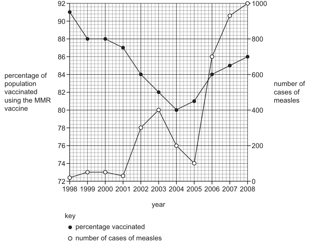
Which statement matches the data?
Options
A Each year, the percentage of the population vaccinated using the MMR vaccine decreases and the number of measles cases increases.
B The percentage of the population vaccinated against MMR decreased from in 1998 to in 2004. This may have led to the increase in the number of measles cases from 2001 to 2003 and from 2005 to 2008.
C The number of measles cases decreased from 80 in 2003 to 76 in 2005.
D The MMR vaccine does not give protection against measles because there is an increase in the vaccination rate from 2004 to 2008 and the number of measles cases increased from 2005 to 2008.
Working
The graph has two y-axes: the left axis shows the percentage of the population vaccinated (solid dots), and the right axis shows the number of measles cases (open circles). The x-axis shows the year from 1998 to 2008.
- A is incorrect. The percentage vaccinated decreases from 1998 to 2004, but then increases from 2004 to 2008. The number of measles cases also fluctuates (e.g., it decreases from 2003 to 2005), so neither variable changes in a single direction every year.
- B is correct. Reading the left axis, vaccination falls from in 1998 to in 2004. Reading the right axis, measles cases rise from roughly 50 in 2001 to 400 in 2003, and then rise again from roughly 100 in 2005 to 1000 in 2008. A drop in vaccination coverage reduces herd immunity, which can lead to increased disease incidence.
- C is incorrect. The number of measles cases in 2003 is approximately 400, not 80. The values 80 and 76 likely come from misreading the left y-axis (vaccination percentage) or confusing the axes.
- D is incorrect. While vaccination rates increased from 2004 to 2008 and cases increased from 2005 to 2008, this does not mean the vaccine is ineffective. The rise in cases is a lag effect from the significant drop in vaccination between 1998 and 2004, which allowed a large susceptible population to accumulate. Overall, when vaccination was high (1998–2001), cases were near zero, proving the vaccine provides protection.
Answer
B
B
Walkthrough
To answer this question, we must carefully read a dual-axis line graph and evaluate four statements against the data and biological principles.
- Read the graph axes and key: The left y-axis is the percentage of the population vaccinated (range 72–92%), plotted with solid dots. The right y-axis is the number of measles cases (range 0–1000), plotted with open circles. The x-axis is the year (1998–2008).
- Evaluate Option A: This statement claims a continuous inverse relationship every year. Looking at the graph, vaccination drops from 1998 to 2004 but then rises from 2004 to 2008. Measles cases also do not rise every year; they peak in 2003, drop in 2004 and 2005, and then rise again. Thus, A is false.
- Evaluate Option B: Check the specific values. In 1998, the solid dot is at 91% on the left axis. In 2004, it is at 80%. This matches the first part of the statement. Next, check measles cases: in 2001, cases are low (around 50), rising to a peak of 400 in 2003. From 2005 (around 100 cases) to 2008 (1000 cases), cases rise steeply. Biologically, a drop in vaccination coverage reduces herd immunity, allowing the virus to spread more easily. This statement is fully supported by the data and biological reasoning.
- Evaluate Option C: This option claims measles cases were 80 in 2003 and 76 in 2005. Looking at the right axis, cases in 2003 are ~400 and in 2005 are ~100. The numbers 80 and 76 are distractors, likely pulled from the left axis (vaccination percentages) to catch students misreading the graph.
- Evaluate Option D: This option observes that vaccination rose from 2004–2008 while cases rose from 2005–2008, and concludes the vaccine doesn't work. This is a classic trap. The increase in cases is a lag effect: the susceptible population built up during the vaccination dip (1998–2004). Even as vaccination rates recover, the large pool of unvaccinated individuals allows outbreaks to occur. The data from 1998–2001 (high vaccination, near-zero cases) proves the vaccine works. Thus, D is false.
Key Takeaways
- Dual-axis graphs require you to match each data series to its correct axis and key. Never assume both variables share the same scale or direction.
- Herd immunity and vaccination protect a population not just by protecting the vaccinated individual, but by breaking chains of transmission. A drop in vaccination coverage allows the disease to resurge.
- Lag effects are common in epidemiology. A recovery in vaccination rates will not immediately stop an outbreak if a large susceptible population already exists.
Common Mistakes
- Misreading the wrong axis: Option C is designed to catch students who read the left axis (vaccination %) instead of the right axis (measles cases) when answering about disease numbers.
- Ignoring fluctuations: Option A is a trap for students who only look at the broad trend (vaccination down, cases up) without noticing the years where the trends reverse or the cases dip.
- Misinterpreting correlation as causation against the vaccine: Option D uses a real observation (vaccination up, cases up) to draw a false biological conclusion. Students must understand that rising cases after a vaccination drop is expected due to the accumulation of susceptible individuals, not proof that the vaccine fails.
Things to Be Careful About
- Always check the key of the graph to ensure you are reading the correct line for the correct variable.
- When evaluating statements like D, apply biological knowledge (how herd immunity works, lag times in epidemiology) rather than just looking at a short-term correlation on the graph.
- Pay attention to the timeframes mentioned in the options (e.g., 2001 to 2003 vs. 2005 to 2008) to ensure the trend described actually matches that specific window on the x-axis.
The diagram shows the excretory system.
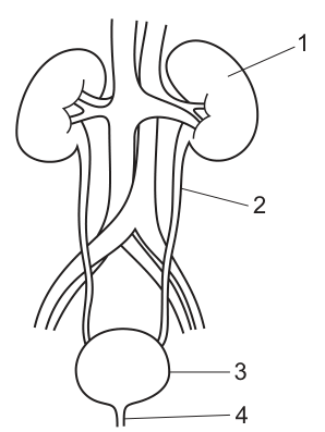
Which row identifies the names of the labelled structures?
Options
| 1 | 2 | 3 | 4 | |
|---|---|---|---|---|
| A | bladder | ureter | kidney | urethra |
| B | bladder | urethra | kidney | ureter |
| C | kidney | ureter | bladder | urethra |
| D | kidney | urethra | bladder | ureter |
Working
The diagram shows the human urinary system. We identify each labelled structure:
- Structure 1 is the kidney, where urine is formed by filtration.
- Structure 2 is the ureter, which carries urine from the kidney to the bladder.
- Structure 3 is the bladder (urinary bladder), which stores urine.
- Structure 4 is the urethra, which carries urine from the bladder to the outside of the body.
Matching these to the table:
- 1 = kidney
- 2 = ureter
- 3 = bladder
- 4 = urethra
This corresponds exactly to row C.
Answer
C
C
Walkthrough
The question asks to identify four structures in the human excretory system from a labelled diagram. We go through each label in order:
- Label 1 points to the bean-shaped organ on either side of the body that filters the blood to form urine. This is the kidney.
- Label 2 points to the tube that carries urine downwards from the kidney to the storage organ. This is the ureter.
- Label 3 points to the muscular sac that stores urine before it is passed out of the body. This is the bladder (or urinary bladder).
- Label 4 points to the final tube that carries urine from the bladder to the exterior. This is the urethra.
Reading down the columns in the options table, row C correctly lists kidney, ureter, bladder, and urethra for labels 1, 2, 3, and 4 respectively.
Key Takeaways
- The human excretory system consists of two kidneys, two ureters, one bladder, and one urethra.
- The pathway of urine is: kidney ureter bladder urethra.
- It is important not to confuse the ureter (kidney to bladder) with the urethra (bladder to outside).
Common Mistakes
- Confusing the ureter and the urethra. The ureter connects the kidney to the bladder, while the urethra connects the bladder to the outside of the body.
- Reversing the order of the bladder and urethra, or kidney and bladder.
- Misreading the labels on the diagram; a line pointing to the main sac is the bladder, while a line pointing to the small exit tube is the urethra.
Things to Be Careful About
- Always read the label lines carefully on a diagram; do not assume the bottom-most label is the last structure in the pathway without tracing the line.
- In MCQs, you can often eliminate options by getting just one label wrong. For example, knowing that 1 is the kidney immediately eliminates options A and B. Knowing that 4 is the urethra eliminates option D. This leaves C as the correct answer.
- Remember the spelling: ureter (with one 'h') and urethra (with an 'h').
As a result of an accident in a factory, a small piece of metal entered the spinal cord of an engineer and severed the tissue at X.
The diagram shows a section through the spinal cord in the neck at the point of the injury. A nervous pathway is also shown.
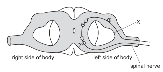
Which outcome from the accident is possible?
Options
A loss of all sensation in the head above the spinal cord injury
B loss of some sensation on the left side of the body
C loss of all sensation on the right side of the body
D some paralysis on the right side of the body
Working
The diagram shows a sensory neurone entering the spinal cord from the left side of the body. Its pathway is cut at X before it can cross over to the right side and ascend to the brain.
- Sensory neurones carry impulses towards the brain, so cutting this pathway causes loss of sensation (not paralysis), which eliminates D.
- The cut is below the neck injury only for the body — sensation in the head travels via cranial nerves directly to the brain, bypassing the spinal cord, so A is wrong.
- The severed fibres come from the left side of the body, not the right, so C is wrong.
- Only the affected sensory pathway is cut, so only some sensation on the left side is lost — B.
Answer
B
B
Walkthrough
Step 1: Identify what the diagram shows. A transverse section through the spinal cord has a sensory neurone arriving from the spinal nerve on the left side of the body. Its axon synapses with a relay neurone whose axon crosses over (decussates) to the right side and ascends towards the brain.
Step 2: Locate X. The cut at X severs the sensory pathway on the left side, before it crosses over. So impulses from receptors on the left side of the body can no longer reach the brain.
Step 3: Work out what kind of loss results.
- Sensory neurones carry information to the brain; motor neurones carry commands away. Cutting a sensory pathway causes loss of sensation, not movement — so option D (paralysis) is eliminated.
- The head above the injury is served by cranial nerves running straight to the brain, not through the spinal cord, so option A is impossible.
- The damaged fibres originate on the left side of the body, so loss must be on the left — option C is eliminated.
- Because only one specific tract is cut (other sensory tracts and other nerves are intact), the loss is of some sensation, not all — giving B.
Key Takeaways
- Sensory neurones carry impulses towards the CNS/brain; cutting them removes sensation but does not cause paralysis.
- Many sensory tracts cross from one side of the body to the other inside the spinal cord — where the cut sits relative to the crossing determines which side loses function.
- Sensation from the head travels by cranial nerves and is unaffected by a spinal cord injury in the neck.
- A single severed tract means partial ('some') loss, because other parallel pathways remain intact.
Common Mistakes
- Choosing C: assuming the side labelled on the diagram is reversed, or forgetting that the severed fibres enter from the left side of the body.
- Choosing D: confusing sensory with motor pathways — severing a sensory neurone cannot cause paralysis.
- Choosing A: forgetting that the face and head are innervated by cranial nerves that do not pass through the spinal cord.
- Saying 'loss of all sensation': one cut tract does not remove every sensory modality, since other tracts are unaffected.
Things to Be Careful About
- Read the labels 'right side of body' and 'left side of body' exactly as printed — they refer to the patient's sides, and the sensory input shown comes from the left.
- Note the position of X relative to the synapse and the crossing-over point: the cut is on the incoming (sensory) side, before decussation.
- Match the wording of the options precisely: 'some' versus 'all', 'sensation' versus 'paralysis'. These qualifiers are the discriminating features of the distractors.
The diagram shows a section through the eye.
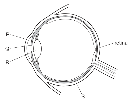
Which two structures focus light rays onto the retina?
Options
A P and Q
B P and R
C Q and R
D Q and S
Working
P is the cornea and R is the lens. Both are transparent and both refract (bend) light rays so that they are focused on the retina. Q is the iris, which controls the amount of light entering, and S is the sclera/choroid, which neither focuses light nor is transparent.
Answer
B
B
Walkthrough
The diagram labels four structures of the eye. P points to the transparent front surface of the eye — the cornea. R points to the biconvex, transparent body suspended behind the pupil — the lens. Light entering the eye is refracted mainly by the cornea, with fine focusing done by the lens changing shape during accommodation; together they bring light rays to a focus on the retina at the back of the eye. That matches option B.
Q is the iris — the pigmented ring that adjusts the size of the pupil to control how much light enters, not a focusing structure. S points to the outer/middle coat (sclera/choroid region), which protects the eye and supplies it with blood vessels but plays no part in bending light rays.
Key Takeaways
- The two focusing structures of the eye are the cornea (does most of the refraction) and the lens (fine adjustment).
- The iris controls light intensity entering the eye via the pupil; it does not focus light.
- The retina is where the focused image forms; it contains receptors.
Common Mistakes
- Choosing P and Q: the iris is often confused as a focusing structure because it sits right behind the cornea; its job is controlling light amount, not bending rays.
- Choosing Q and R: including the iris instead of the cornea — the cornea does most of the refraction, so omitting it loses the mark.
- Confusing the cornea with the conjunctiva or sclera when reading diagrams.
Things to Be Careful About
- Read the label lines carefully: P touches the front surface (cornea), R points inside the eye to the lens, Q points to the ring around the pupil (iris).
- 'Focus' means refract light to form an image on the retina — only transparent, curved structures can do this.
The diagram shows some of the endocrine glands of a human female.
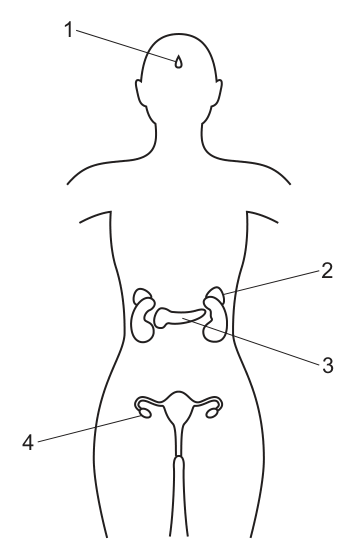
Which row matches the glands with the correct hormones?
Options
| 1 | 2 | 3 | 4 | |
|---|---|---|---|---|
| A | adrenaline | FSH | oestrogen | insulin |
| B | FSH | adrenaline | insulin | oestrogen |
| C | insulin | oestrogen | adrenaline | FSH |
| D | oestrogen | insulin | FSH | adrenaline |
Working
Identify each gland from the diagram:
- 1 = pituitary gland
- 2 = adrenal gland
- 3 = pancreas
- 4 = ovary
Recall the hormones produced by each:
- Pituitary gland produces FSH (and other hormones like LH, ADH).
- Adrenal gland produces adrenaline.
- Pancreas produces insulin (and glucagon).
- Ovary produces oestrogen (and progesterone).
Matching these to the columns:
- 1 → FSH
- 2 → adrenaline
- 3 → insulin
- 4 → oestrogen
This corresponds to row B.
Answer
B
B
Walkthrough
The question asks to match four labelled endocrine glands in a female human body diagram with the hormones they produce. We first identify each gland from the diagram:
- Label 1 points to the pituitary gland, located at the base of the brain.
- Label 2 points to the adrenal glands, located on top of the kidneys.
- Label 3 points to the pancreas, located in the abdominal cavity.
- Label 4 points to the ovary, located in the pelvis.
Next, we recall the primary hormones secreted by each gland:
- The pituitary gland (1) is the master gland and secretes several hormones, including follicle-stimulating hormone (FSH) and luteinising hormone (LH), which regulate the menstrual cycle.
- The adrenal glands (2) sit above the kidneys and secrete adrenaline (and cortisol) in response to stress or physical activity.
- The pancreas (3) has an endocrine function where the islets of Langerhans secrete insulin (to lower blood glucose) and glucagon (to raise it).
- The ovaries (4) are the primary female sex organs and secrete oestrogen (and progesterone), which regulate the menstrual cycle and develop secondary sexual characteristics.
Matching these to the columns in the table:
- Column 1 needs FSH.
- Column 2 needs adrenaline.
- Column 3 needs insulin.
- Column 4 needs oestrogen.
Row B is the only row that matches all four glands with their correct hormones.
Key Takeaways
- Endocrine glands are ductless glands that secrete hormones directly into the bloodstream.
- Each gland has a specific set of hormones: pituitary (FSH, LH, ADH, GH), adrenal (adrenaline), pancreas (insulin, glucagon), and ovaries (oestrogen, progesterone).
- Diagrams of the human endocrine system are frequently used to test the ability to locate these glands and link them to their functions.
Common Mistakes
- Confusing the adrenal gland with the kidney itself; the adrenal gland is a separate structure sitting on top of the kidney and produces adrenaline, not urine.
- Confusing the pancreas with the liver; the liver produces bile and handles detoxification, while the pancreas produces insulin and digestive enzymes.
- Attributing oestrogen to the pituitary gland; while the pituitary produces FSH and LH which stimulate the ovaries to produce oestrogen, the oestrogen itself is produced by the ovary.
- Confusing insulin with glucagon; both are produced by the pancreas, but insulin is the hormone that lowers blood glucose.
Things to Be Careful About
- Always read the label lines carefully on the diagram. Label 2 points to the adrenal gland, not the kidney below it. Label 3 points to the pancreas, not the liver above it.
- Remember that some glands produce multiple hormones. For example, the pancreas produces both insulin and glucagon, and the pituitary produces many hormones including FSH, LH, ADH, and growth hormone. The question provides one correct hormone per gland in the correct row, so match the specific hormone given to the correct gland.
- The word 'adrenaline' is specific to the adrenal medulla; do not confuse it with 'aldosterone' which is from the adrenal cortex.
When the body temperature is too high, which process causes the body temperature to return to normal?
Options
A contraction of hair erector muscles
B contraction of skeletal muscles, causing shivering
C secretion of sweat onto the surface of the skin
D vasoconstriction of arterioles supplying skin surface capillaries
Working
When the body temperature is too high, the body must lose heat. Sweat secreted onto the skin surface evaporates, taking heat away from the body and cooling it.
- A — contraction of hair erector muscles traps air as insulation, reducing heat loss: warms, not cools.
- B — shivering generates heat by muscle contraction: warms, not cools.
- C — sweating cools the body by evaporation: correct.
- D — vasoconstriction reduces blood flow to the skin, conserving heat: warms, not cools.
Answer
C
C
Walkthrough
The question asks which process returns the body temperature to normal when it is too high — in other words, which response cools the body down. This is an example of homeostasis: the body detects the temperature rise and brings about responses that lower it back to the normal range.
The only cooling response among the options is the secretion of sweat. Sweat is produced by sweat glands in the skin and reaches the surface through pores. As the sweat evaporates, it uses heat energy from the body, so the skin and the blood beneath it cool down. This is why sweating is the key cooling mechanism when the body is too hot.
Look at each of the other options to see why they are wrong:
- A — contraction of hair erector muscles makes the hairs stand up. In humans this has little effect, but the biological purpose is to trap a layer of insulating air next to the skin, which reduces heat loss. That is a warming response, used when the body is too cold, not too hot.
- B — shivering is the rapid contraction of skeletal muscles. Muscle contraction releases heat from respiration, so shivering raises body temperature. Again, this is a response to being too cold.
- D — vasoconstriction of arterioles supplying the skin capillaries reduces the blood flow to the skin surface. Less warm blood reaches the skin, so less heat is lost to the surroundings. This conserves heat and is a warming response, used when the body is too cold.
So the correct answer is C.
Key Takeaways
- When the body is too hot, the cooling responses are sweating (evaporation of sweat removes heat) and vasodilation of skin arterioles (more blood flows to the skin, so more heat is lost).
- When the body is too cold, the warming responses are shivering, vasoconstriction of skin arterioles, and contraction of hair erector muscles.
- These responses are coordinated by the thermoregulatory centre in the brain and are an example of negative feedback: a change in body temperature triggers responses that reverse the change.
Common Mistakes
- Choosing D (vasoconstriction) — this is a common confusion. Vasoconstriction reduces heat loss, so it is used when the body is too cold, not too hot. The cooling response is vasodilation, the opposite.
- Choosing B (shivering) — shivering generates heat, so it warms the body. It is a response to being too cold.
- Choosing A (hair erector muscles) — these raise hairs to trap insulating air, which reduces heat loss and warms the body.
Things to Be Careful About
- Read the question carefully: it asks for the process that cools the body when it is too high, not a general temperature-control response.
- Note the difference between vasoconstriction (reduces heat loss, warms) and vasodilation (increases heat loss, cools). The mark scheme for this type of question rewards the correct cooling mechanism.
- Sweating cools by evaporation — the heat needed to evaporate the water is taken from the body. This is the key idea behind option C.
Some seedlings were grown in a dark, warm and humid environment. The tips of the growing shoots were cut off and then replaced on one side of the shoot as shown. The shoots were then left to grow.
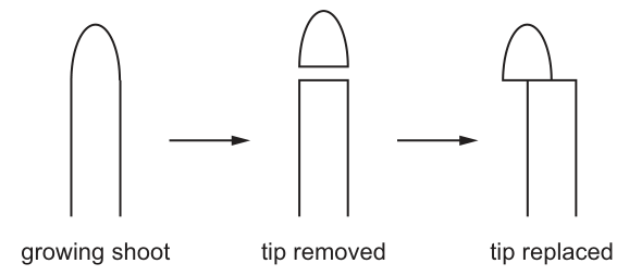
Which pattern of growth was then seen in the shoots?
Options
A The shoots stopped growing.
B The shoots grew straight up.
C The shoots grew with a curve to the left.
D The shoots grew with a curve to the right.
Working
The shoot tip produces auxin, which diffuses downwards and stimulates cell elongation. With the tip replaced on only the left side, auxin passes down the left side of the shoot only. The left side elongates faster than the right, so the shoot bends towards the side with less auxin — to the right.
Answer
D
D
Walkthrough
The tip of a growing shoot produces the plant hormone auxin. Auxin moves downwards from the tip and makes the cells behind the tip elongate — the more auxin a region of shoot receives, the faster it grows. In a normal, upright shoot the tip sits centrally, so auxin is distributed evenly and the shoot grows straight.
In this experiment the tip is cut off and placed back on only the LEFT half of the cut surface. Auxin can therefore only diffuse down the left side of the shoot. The left side receives auxin and its cells elongate; the right side receives none and stops elongating. Because one side grows faster than the other, the shoot bends away from the faster-growing side — it curves to the RIGHT.
This is the same mechanism behind phototropism: light on one side causes auxin to accumulate on the shaded side, which grows faster and bends the shoot towards the light. Here the 'unequal auxin' is created surgically instead of by light.
Key Takeaways
- Auxin is made in the shoot tip and promotes cell elongation in the region just behind the tip.
- Unequal distribution of auxin causes unequal growth, so the shoot bends towards the side with LESS auxin.
- Removing the tip stops elongation (no auxin source); replacing it on one side restores growth on that side only.
Common Mistakes
- Choosing A: the shoot does not stop growing — the replaced tip still supplies auxin to one side.
- Choosing B: assuming the tip 'repaired' the shoot so it grows straight — auxin is only delivered down one side.
- Choosing C: getting the direction inverted. Auxin makes the side it reaches grow FASTER, so the shoot bends away from the auxin side (left) and curves right.
- Confusing auxin's effect on shoots (stimulates elongation) with its effect on roots (inhibits elongation) — in a root the curve would be reversed.
Things to Be Careful About
- Read the figure carefully: the tip is placed on the LEFT half of the cut shoot, so the auxin-rich side is the left and the bend is to the right.
- The mark depends on knowing that auxin stimulates elongation in shoots — a vague idea that 'a hormone makes it grow' will not reliably get the direction correct.
- Remember the direction rule: shoot bends towards the side with less auxin (which is the slower-growing side).
Which statements about meiosis are correct?
Options
| meiosis produces genetically identical nuclei | meiosis produces haploid nuclei | |
|---|---|---|
| A | ✗ | ✗ |
| B | ✗ | ✓ |
| C | ✓ | ✗ |
| D | ✓ | ✓ |
key
✓ = yes
✗ = no
Working
Meiosis produces four genetically different haploid nuclei, not genetically identical ones. Genetically identical nuclei are produced by mitosis. Therefore the correct row is the one that says meiosis does not produce genetically identical nuclei (✗) and does produce haploid nuclei (✓).
Answer
B
B
Walkthrough
This question tests the two key outcomes of meiosis, so we need to recall exactly what meiosis does.
Meiosis is the type of cell division that produces gametes (sex cells). It involves two divisions and results in four daughter cells. Two things are true about these daughter cells:
- They are haploid — they contain half the number of chromosomes of the parent cell (one set instead of two).
- They are genetically different from each other and from the parent cell — because of the exchange of genetic material and the random separation of chromosomes.
Genetically identical nuclei are the product of mitosis, not meiosis. Mitosis produces two daughter cells with the same number of chromosomes as the parent and identical genetic information; it is used for growth, repair and asexual reproduction.
Now look at the two statements in the table:
- "meiosis produces genetically identical nuclei" — this is false (✗).
- "meiosis produces haploid nuclei" — this is true (✓).
The only row with ✗ in the first column and ✓ in the second column is B.
Key Takeaways
- Meiosis produces four genetically different haploid cells.
- Mitosis produces two genetically identical diploid cells.
- "Haploid" means having half the number of chromosomes; "diploid" means having the full set.
- When a table of ✓/✗ statements is given, judge each statement independently and then match the row.
Common Mistakes
- Choosing D (✓ ✓) — this happens when a student forgets that meiosis produces genetically different cells, confusing it with mitosis.
- Choosing A (✗ ✗) — forgetting that meiosis does produce haploid nuclei.
- Confusing meiosis with mitosis entirely; mitosis is the division that produces genetically identical cells.
Things to Be Careful About
- The mark scheme requires the letter B only; no explanation is needed on the answer sheet.
- Read the key carefully: ✓ means yes, ✗ means no. It is easy to misread the columns.
- Remember that "genetically identical" is the defining feature of mitosis, not meiosis. Meiosis is defined by producing haploid, genetically different cells.
What is a feature of asexual reproduction in plants?
Options
A It only needs one type of gamete.
B It requires two parents.
C It uses cell division by meiosis.
D It uses cell division by mitosis.
Working
Asexual reproduction involves only one parent, produces no gametes, and uses cell division by mitosis to produce genetically identical offspring.
- A is wrong: asexual reproduction does not use gametes at all.
- B is wrong: asexual reproduction needs only one parent.
- C is wrong: meiosis produces gametes for sexual reproduction, not asexual reproduction.
- D is correct: mitosis is the cell division used in asexual reproduction.
Answer
D
D
Walkthrough
The question asks for a feature of asexual reproduction in plants. Think about what asexual reproduction means: it is reproduction that involves only one parent and produces offspring that are genetically identical to that parent (clones). No gametes are formed and no fertilisation takes place.
The key biological point is the type of cell division involved. Mitosis produces two genetically identical daughter cells and is the division used for growth, repair and asexual reproduction. Meiosis, by contrast, halves the chromosome number and produces genetically different gametes, so it is the division used in sexual reproduction.
Now look at each option:
- A — "It only needs one type of gamete." This is wrong because asexual reproduction does not involve gametes at all. Gametes are only produced for sexual reproduction.
- B — "It requires two parents." This is the opposite of the truth; asexual reproduction requires only one parent.
- C — "It uses cell division by meiosis." Meiosis produces gametes for sexual reproduction, so this cannot be a feature of asexual reproduction.
- D — "It uses cell division by mitosis." This is correct. Mitosis produces genetically identical cells, which is exactly what asexual reproduction needs.
So the answer is D.
Key Takeaways
- Asexual reproduction involves one parent, produces no gametes, and uses mitosis, giving genetically identical offspring.
- Sexual reproduction involves two parents (or two types of gamete), uses meiosis to form gametes, and gives genetically varied offspring.
- Knowing which type of cell division belongs to which kind of reproduction is a core 5090 distinction.
Common Mistakes
- Choosing C because of confusing meiosis with mitosis — meiosis is for gamete production in sexual reproduction, not for asexual reproduction.
- Choosing A because of thinking a single gamete is involved — asexual reproduction uses no gametes at all.
- Choosing B because of confusing asexual with sexual reproduction — asexual needs only one parent.
Things to Be Careful About
- The question asks for a feature of asexual reproduction, so any option mentioning gametes, two parents or meiosis is a signal that it belongs to sexual reproduction.
- Remember the precise wording: mitosis produces genetically identical cells, which is the defining outcome of asexual reproduction.
The seeds of some plants will not normally germinate until they have been in the soil for several months.
Some seeds were collected from a plant of this type.
Some students wanted to find out whether a chemical in the testa prevented germination.
The seeds were divided into three groups.
Each group of seeds was put into a shallow dish and covered with water.
The lid that was used was loose fitting so that oxygen could reach the seeds.
Group 1 were whole seeds.
Group 2 were seeds from which the testas had been removed.
Group 3 were seeds from which the testas had been removed, but the testas were placed separately in the same dish.

Only the seeds in group 2 germinated.
What would be the most logical extension of this experiment?
Options
A change the water of the seeds in group 3 every day to see if they germinate
B change the apparatus so that oxygen cannot reach the seeds
C repeat the experiment at several different temperatures
D repeat the experiment using different species of seed
Working
Group 2 (testas removed) germinated, so the embryo can germinate when the testa is absent. Group 3 (testas removed but present in the same water) did not germinate, so the chemical in the testa diffuses into the water and still prevents germination. The logical next step is to test whether this chemical can be removed from the water: change the water in group 3 daily and see if the seeds then germinate.
B would remove a variable (oxygen) needed for germination, C and D change other variables rather than testing the chemical already shown to inhibit germination.
Answer
A
A
Walkthrough
The students' hypothesis is that a chemical in the testa prevents germination. The three groups are designed to test this:
- Group 1 (whole seeds) did not germinate — consistent with the testa blocking germination.
- Group 2 (testas removed) germinated — the embryos are viable and will germinate without the testa, so the delay is not due to the embryo itself.
- Group 3 (testas removed but left in the same dish of water) did not germinate — this is the crucial result. The testa is no longer touching the seed, yet germination is still prevented. The only way the testa can still act is if its chemical dissolves into the surrounding water and then reaches the seed.
So the evidence points to a water-soluble inhibitor that diffuses out of the testa into the water. The most logical extension is to test this directly: change the water in group 3 every day, which would wash the inhibitor away, and see whether the seeds then germinate. If they do, that confirms the chemical is water-soluble and was the cause of dormancy.
Option B removes oxygen, which seeds need for aerobic respiration during germination — that would stop all groups germinating and tells us nothing about the testa chemical. Options C and D change temperature or species; neither follows from the specific result obtained, because the question being investigated is the role of the testa chemical, not the general conditions for germination.
Key Takeaways
- A good experimental extension follows directly from the result obtained, testing the hypothesis one step further.
- Group 3 is the key control: testa absent from the seed but present in the water still blocks germination, implying a diffusible, water-soluble inhibitor.
- Seeds need water and oxygen (and a suitable temperature) to germinate; removing oxygen would invalidate the experiment.
- Changing the water is a way of removing a dissolved chemical — a standard technique in germination experiments.
Common Mistakes
- Choosing C (different temperatures) or D (different species): these are general 'make it more reliable' answers, but they do not test the hypothesis about the testa chemical and so are not the most logical extension.
- Choosing B: forgetting that oxygen is required for germination, so excluding it would prevent germination in all groups and destroy the experiment.
- Misreading group 3 as showing the chemical is not the cause — in fact it strengthens the case, because the testa still inhibits even when separated from the seed.
- Thinking that changing the water is 'just repeating' — it is a deliberate test of whether the inhibitor is water-soluble and removable.
Things to Be Careful About
- 'Most logical extension' means the next step that follows from the specific result — always link your choice back to what groups 1, 2 and 3 showed.
- Keep the controlled variables in mind: the loose-fitting lid supplying oxygen is mentioned deliberately, which rules out option B.
- The reasoning chain is: group 2 germinates → embryo is viable; group 3 does not → something from the testa in the water inhibits germination; therefore test whether removing that water (and its dissolved chemical) allows germination.
The diagram shows the changes in thickness of the uterus lining during one menstrual cycle.
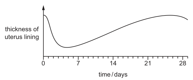
When would the levels of progesterone and LH be highest?
Options
| progesterone | LH | |
|---|---|---|
| A | between days 12 and 16 | between days 25 and 28 |
| B | between days 19 and 23 | on day 14 |
| C | on day 5 | between days 1 and 5 |
| D | on day 13 | on day 10 |
Answer
B
B
Walkthrough
The graph shows the thickness of the uterus lining over a 28-day menstrual cycle. To answer the question, we must recall the hormonal changes that drive these physical changes.
- Days 1 to 5 (Menstruation): The uterus lining breaks down. Both progesterone and oestrogen levels are low.
- Days 6 to 13 (Follicular phase): The lining rebuilds under the influence of rising oestrogen.
- Day 14 (Ovulation): A sudden surge in Luteinising Hormone (LH) from the pituitary gland triggers the release of an egg from the ovary. This is the point where LH is at its highest.
- Days 15 to 28 (Luteal phase): The empty follicle transforms into the corpus luteum, which secretes large amounts of progesterone. Progesterone maintains and thickens the uterus lining to prepare for a fertilised egg. Progesterone levels rise after ovulation and peak between days 19 and 23, corresponding to the thickest part of the uterine lining on the graph.
Matching these timings to the options, progesterone is highest between days 19 and 23, and LH is highest on day 14. This matches option B.
Key Takeaways
- The menstrual cycle is regulated by hormones from the pituitary gland and the ovaries.
- LH triggers ovulation and has a sharp peak on day 14.
- Progesterone is produced by the corpus luteum after ovulation and peaks in the second half of the cycle (around days 19–23) to maintain the uterine lining.
Common Mistakes
- Confusing the timing of LH and progesterone peaks: LH peaks just before ovulation (day 14), while progesterone peaks after ovulation (days 19–23).
- Assuming progesterone is high during menstruation (days 1–5) or the follicular phase; it is low during these times.
- Misreading the graph and assuming the highest uterine lining thickness at the end of the cycle (day 28) corresponds to the highest progesterone level. Progesterone actually starts to fall before day 28 if fertilisation does not occur, which triggers menstruation.
Things to Be Careful About
- Remember the exact timing of the LH surge (day 14) and the progesterone peak (days 19–23).
- The uterine lining is thickest around day 21–23, which corresponds to the peak of progesterone.
- Option A has progesterone between 12–16 (too early, ovulation just happened) and LH between 25–28 (wrong, LH is low then).
- Option C has day 5 (menstruation, low hormones).
- Option D has day 13 (just before ovulation, oestrogen is high but the LH surge is day 14).
In the umbilical cord, the blood flowing from the fetus to the placenta in the umbilical artery has
Options
A a higher concentration of carbon dioxide than the blood in the umbilical vein.
B a higher concentration of oxygen than the blood in the umbilical vein.
C a lower concentration of urea than the blood in the umbilical vein.
D the same concentration of glucose as the blood in the umbilical vein.
Working
The umbilical artery carries blood from the fetus to the placenta. At the placenta, carbon dioxide and urea diffuse out of the fetal blood into the mother's blood, while oxygen and glucose diffuse from the mother's blood into the fetal blood.
So the blood in the umbilical artery (going to the placenta) has a higher concentration of carbon dioxide than the blood in the umbilical vein (returning from the placenta).
- B is wrong: the umbilical artery has a lower oxygen concentration than the vein.
- C is wrong: the umbilical artery has a higher urea concentration than the vein.
- D is wrong: the umbilical artery has a lower glucose concentration than the vein.
Answer
A
A
Walkthrough
The placenta is the exchange surface between the mother's blood and the fetus's blood. The fetus's blood travels to the placenta along the umbilical artery and returns along the umbilical vein — note that this is the reverse of the usual rule, where arteries carry blood away from the heart and veins carry blood towards it. Here, the artery carries blood away from the fetus's heart (towards the placenta) and the vein carries blood back towards the fetus's heart.
At the placenta, the fetal blood gives up its waste products — carbon dioxide and urea — which diffuse into the mother's blood to be removed. The fetal blood also picks up oxygen and glucose from the mother's blood. So the blood in the umbilical artery, on its way to the placenta, is carrying the waste it has not yet unloaded: it has a higher concentration of carbon dioxide than the blood in the umbilical vein, which has already offloaded the carbon dioxide and picked up oxygen and glucose.
That makes A correct. Option B is wrong because the artery is poorer in oxygen than the vein. Option C is wrong because the artery carries more urea, not less, than the vein. Option D is wrong because the artery carries less glucose, not the same amount, than the vein.
Key Takeaways
- The placenta is the site of exchange between fetal and maternal blood: oxygen and glucose move into the fetal blood; carbon dioxide and urea move out of it.
- The umbilical artery carries blood from the fetus to the placenta; the umbilical vein carries blood from the placenta to the fetus.
- In the umbilical cord the naming is the opposite of the usual circulation: the artery carries deoxygenated, waste-laden blood and the vein carries oxygenated, nutrient-rich blood.
Common Mistakes
- Assuming that an artery always carries oxygenated blood. In the umbilical cord the artery carries deoxygenated blood — this is the classic trap in this question.
- Confusing the direction of flow: swapping the artery and vein leads to choosing B or C.
- Thinking urea is removed from the mother's blood into the fetus — the fetus produces urea as a waste product and must get rid of it through the placenta.
Things to Be Careful About
- Learn the direction of blood flow in the umbilical vessels and the substances exchanged at the placenta as a single linked fact.
- The question asks about concentration in the artery compared with the vein, so compare each substance in the correct direction: artery (to placenta) has more carbon dioxide and urea, less oxygen and glucose, than the vein.
DNA is divided into sections called
Options
A chromosomes.
B genes.
C nuclei.
D proteins.
Working
DNA is a long molecule. Each section of DNA that codes for a characteristic (usually a protein) is called a gene. Chromosomes are made of DNA, nuclei contain the chromosomes, and proteins are the products made using the information in genes.
Answer
B
B
Walkthrough
The question asks what the sections of DNA are called. In the 5090 syllabus, a gene is defined as a section of DNA that codes for a particular protein (which in turn controls a characteristic). So the answer is genes (option B).
Look at each option to see why the others are wrong:
- A — chromosomes: A chromosome is a long, thread-like structure made of DNA wound around proteins. A chromosome contains many genes, but it is not a section of DNA — it is the structure that carries the DNA. So this is not the name for a section of DNA.
- B — genes: Correct. Each gene is one section of the DNA molecule that carries the code for one protein.
- C — nuclei: The nucleus is the organelle inside the cell that contains the chromosomes (and therefore the DNA). It is not a section of DNA at all.
- D — proteins: Proteins are the molecules that genes code for. They are made from amino acids and are not sections of DNA.
So the only option that correctly names a section of DNA is B.
Key Takeaways
- A gene is a section of DNA that codes for a protein.
- A chromosome is a long molecule of DNA (with proteins) that carries many genes.
- The nucleus contains the chromosomes.
- Proteins are the end products made using the information in genes.
Common Mistakes
- Confusing a gene with a chromosome. A chromosome is the whole DNA molecule; a gene is only one section of it. The mark scheme rejects "chromosomes" here because chromosomes are made of DNA, not sections of it.
- Confusing a gene with a protein. A gene is the code; a protein is what the code makes.
Things to Be Careful About
- The definition must be precise: a gene is a section of DNA that codes for a protein. This exact wording is what the syllabus expects.
- In this MCQ, only one option names a section of DNA, so once you recall the definition the answer is immediate.
A man and his wife have three children. The first two are both girls; one has blood group A and one has blood group O.
The children's father has blood group B.
What is the probability that the third child is a boy with blood group B?
Options
A
B
C
D
Working
The girl with blood group O has genotype , so both parents must carry the allele. The father has blood group B and carries , so his genotype is . The mother must provide an allele for the child with blood group A, so the mother is .
Cross: .
Gametes: father , ; mother , .
| (AB) | (B) | |
| (A) | (O) |
Probability of blood group B = .
Probability of a boy = .
Probability of a boy with blood group B = .
Answer
A
A
Walkthrough
This is a genetics probability problem. We need to work out the parents' genotypes from the information given, then find the chance that a third child is a boy with blood group B.
Step 1 — Work out the father's genotype. The father has blood group B. Blood group B can be or . One child has blood group O, which only happens with genotype . That child must have received an allele from each parent, so the father must carry an allele. His genotype is therefore .
Step 2 — Work out the mother's genotype. The mother has a child with blood group A, which requires the allele. So the mother must carry . She also must carry an allele (because of the O child). Her genotype is therefore , and her blood group is A.
Step 3 — Set up the cross. Father produces gametes and , each with probability . Mother produces gametes and , each with probability .
Step 4 — Punnett square. The four equally likely combinations are (blood group AB), (blood group B), (blood group A) and (blood group O). Each has probability . So the probability that a child has blood group B is .
Step 5 — Sex of the child. The chance of any child being a boy is , and this is independent of blood group.
Step 6 — Combine the probabilities. Because sex and blood group are independent, multiply: . The correct option is A.
Key Takeaways
- Blood group O means genotype , which forces both parents to carry the allele.
- Blood groups A and B each have two possible genotypes; the children's blood groups let you pin down which one each parent has.
- The A and B alleles are codominant, so gives blood group AB.
- Sex is determined independently of blood group, so you multiply the two probabilities.
- The probability of two independent events both happening is the product of their individual probabilities.
Common Mistakes
- Assuming the father is just because he has blood group B — the O child rules this out, because an O child needs an allele from both parents.
- Forgetting that the O child requires an allele from each parent, and so failing to deduce the father's genotype.
- Not realising the mother must be ; some candidates think the mother could be anything, but the A child requires her to carry .
- Confusing the probability of blood group B () with the final answer — forgetting to multiply by the chance of being a boy.
- Adding probabilities instead of multiplying them.
Things to Be Careful About
- The answer required is the option letter A.
- In this cross, blood group B offspring have genotype , one of four equally likely outcomes.
- The probability of a boy is always taken as at this level.
- ; check your arithmetic.
- The first two children being girls is a red herring — each birth is independent, so the third child's sex is still .
Which outcomes might farmers want to achieve by using artificial selection?
Options
| increased | decreased | |
|---|---|---|
| A | fertiliser use | pesticide use |
| B | growth rate | yield |
| C | pesticide use | growth rate |
| D | yield | fertiliser use |
Working
Artificial selection is used to breed organisms with desirable characteristics. A farmer wants outcomes that improve profit and reduce costs.
- Increased yield — desirable (more produce to sell).
- Decreased fertiliser use — desirable (lower cost, less pollution).
Option D pairs these two desirable outcomes.
The other rows each contain at least one undesirable outcome:
- A: increased fertiliser use (costly).
- B: decreased yield (less produce).
- C: increased pesticide use (costly) and decreased growth rate (slower production).
Answer
D
D
Walkthrough
The question asks which outcomes a farmer might want to achieve by using artificial selection (selective breeding). Artificial selection is when humans choose which plants or animals to breed, based on desirable characteristics, so that the offspring inherit those characteristics.
A farmer wants outcomes that increase profit and reduce costs. So we judge each option row by row:
- A: increased fertiliser use is not desirable — fertiliser costs money. Decreased pesticide use is desirable, but the row only scores if BOTH outcomes are wanted.
- B: increased growth rate is desirable, but decreased yield is not — yield is the amount of crop produced, and a farmer wants more of it.
- C: increased pesticide use is costly and decreased growth rate slows production — both are undesirable.
- D: increased yield means more crop to sell, and decreased fertiliser use means lower costs. Both are desirable, so D is correct.
Key Takeaways
- Artificial selection is the process of breeding organisms with desirable traits so those traits appear in the next generation.
- A farmer's goals usually involve increasing output (yield, growth rate) and decreasing input costs (fertiliser, pesticide).
- In a table question, every column must match the condition — here, both the 'increased' and 'decreased' columns must be outcomes a farmer would want.
Common Mistakes
- Choosing A because 'decreased pesticide use' is good, while ignoring that 'increased fertiliser use' is bad.
- Choosing B because 'increased growth rate' is good, while missing that 'decreased yield' is bad.
- Confusing yield with growth rate — yield is the amount of crop harvested, not how fast it grows.
Things to Be Careful About
- Read both columns of the table together — the correct row must have a desirable outcome in BOTH columns.
- Remember that artificial selection aims for characteristics that benefit the farmer, not just any change.
- The mark scheme accepts only D; there is no partial credit for a row that is half right.
Single-cell protein can be produced by growing a fungus in a fermenter.
Why is it necessary for the fermenter to have a cooling unit?
Options
A Fungi produce protein faster at low temperatures.
B The air bubbles generate heat as they float upwards.
C The glucose syrup is too warm when it is added to the fermenter.
D The fungus releases heat as it respires.
Working
The fungus growing in the fermenter carries out aerobic respiration, and respiration releases heat. This heat would raise the temperature inside the fermenter above the optimum for the fungus and its enzymes, so a cooling unit is needed to remove it.
- A is wrong: low temperatures slow enzyme activity, they do not speed up protein production.
- B is wrong: air bubbles do not generate heat as they rise.
- C is wrong: the glucose syrup is not the reason a permanent cooling unit is fitted.
Answer
D
D
Walkthrough
This question asks why a fermenter used to grow a fungus for single-cell protein must have a cooling unit. The key biology is that living organisms respire, and respiration is an exothermic process — it releases heat energy. In a large fermenter, millions of fungus cells are respiring at once, so a lot of heat builds up. If the temperature rises too high, the fungus's enzymes would be denatured and the fungus would die or stop growing. The cooling unit removes this excess heat and keeps the temperature at the optimum for the fungus.
Now look at the options:
- A says fungi produce protein faster at low temperatures. This is backwards — enzyme-controlled reactions slow down at low temperatures because the molecules have less kinetic energy, so fewer successful collisions happen. Fungi do not work faster when cold.
- B says the air bubbles generate heat as they float upwards. Bubbles of air are pumped in to supply oxygen for aerobic respiration; they do not themselves generate heat.
- C says the glucose syrup is too warm when added. Even if the syrup were warm, that would only be a brief initial effect, not the reason for having a permanent cooling unit throughout the run.
- D says the fungus releases heat as it respires. This is the correct reason — respiration is a heat-releasing (exothermic) process, and the cooling unit removes that heat.
So the answer is D.
Key Takeaways
- Respiration releases heat energy — it is an exothermic process.
- In a fermenter, the growing microorganism respires and produces heat, so a cooling unit is needed to keep the temperature at the optimum for the organism and its enzymes.
- Fermenters control several conditions: temperature (cooling unit), oxygen supply (air pumped in), pH, and nutrients.
Common Mistakes
- Choosing A because it sounds plausible that "cooling helps". Remember that low temperatures slow down enzyme activity; the cooling unit is not there to speed up the fungus, it is there to counteract the heat the fungus itself produces.
- Choosing C because the syrup is warm. The question asks why the fermenter has a cooling unit as a permanent feature, not about the temperature of one input.
- Confusing the purpose of the air supply (to provide oxygen for respiration) with heat generation.
Things to Be Careful About
- The mark scheme requires the idea that the fungus respires and that respiration releases heat. The word respires is the key term — do not just say "the fungus gets hot" without linking it to respiration.
- Note that this is a one-mark multiple-choice question, so only the letter is needed in the final answer.
The diagram shows the pyramid of energy for the following food chain:
A pyramid of energy shows the total quantity of energy stored in the biomass of organisms at each trophic level in the food chain per year.
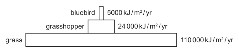
To one decimal place, what percentage of energy is transferred from the producer to the herbivore?
Options
A
B
C
D
Working
Percentage energy transfer =
From producer (grass) to herbivore (grasshopper):
Rounded to one decimal place: .
Answer
D
D
Walkthrough
The question asks for the percentage of energy transferred from the producer (grass) to the herbivore (grasshopper). The pyramid of energy gives the energy stored in the biomass at each trophic level per year: grass = , grasshopper = , and bluebird = .
To find the percentage transfer between two levels, divide the energy at the receiving level by the energy at the supplying level, then multiply by 100.
Transfer from grass to grasshopper = .
Rounded to one decimal place, this is , which matches option D.
Key Takeaways
- A pyramid of energy shows the total energy stored in biomass at each trophic level.
- Energy transfer efficiency between trophic levels is calculated as .
- Typical energy transfer efficiency is around 10%, but it varies and must be calculated from the data given rather than assumed.
Common Mistakes
- Calculating the transfer from the wrong pair of levels: option C () is the transfer from herbivore to carnivore (), and option B () is the transfer from producer to carnivore ().
- Forgetting to multiply by 100 to convert the decimal to a percentage.
- Rounding incorrectly before the final step or to the wrong number of decimal places.
Things to Be Careful About
- Read the question carefully to identify which two trophic levels are being compared ("from the producer to the herbivore").
- Ensure the answer is rounded to the correct number of decimal places as requested ("to one decimal place").
- Use the exact numerical values from the diagram; do not estimate from the relative sizes of the bars.
Which statements about the carbon cycle are correct?
- Burning wood and fossil fuels adds carbon dioxide to the atmosphere.
- Decay and photosynthesis remove carbon dioxide from the atmosphere.
- Photosynthesis and respiration add carbon dioxide to the atmosphere.
Options
A 1 and 2
B 1 only
C 2 only
D 3 only
Working
Statement 1: burning (combustion) of wood and fossil fuels releases into the atmosphere — correct.
Statement 2: decay releases (decomposers respire); only photosynthesis removes . The statement claims both remove it — incorrect.
Statement 3: photosynthesis removes ; only respiration adds it. The statement claims both add it — incorrect.
Only statement 1 is correct.
Answer
B
B
Walkthrough
This question asks you to judge three statements about the carbon cycle and then pick the option that lists the correct ones. The carbon cycle is all about carbon dioxide moving into and out of the atmosphere, so the key skill is knowing which processes add and which remove it.
Statement 1 — Burning wood and fossil fuels adds carbon dioxide to the atmosphere. When anything burns, it is a combustion reaction: the carbon in the fuel combines with oxygen to form carbon dioxide. This is definitely correct.
Statement 2 — Decay and photosynthesis remove carbon dioxide from the atmosphere. This is a trap because it joins two processes with "and", and they do opposite things. Photosynthesis does remove from the air — that is one of its raw materials. But decay (decomposition) does the reverse: decomposers such as bacteria and fungi break down dead organisms and release through their own respiration. So decay adds , it does not remove it. Because one half of the statement is wrong, the whole statement is wrong.
Statement 3 — Photosynthesis and respiration add carbon dioxide to the atmosphere. Again the two processes are joined, and again they work in opposite directions. Respiration does add , but photosynthesis removes it. So this statement is also wrong.
Only statement 1 is correct, so the answer is B (1 only).
Key Takeaways
- The carbon cycle is a balance between processes that add carbon dioxide to the atmosphere and processes that remove it.
- Add : respiration (including decomposers decaying dead matter), burning/combustion of wood and fossil fuels.
- Remove : photosynthesis.
- When a statement joins two processes with "and", both halves must be correct for the statement to be true.
Common Mistakes
- Thinking decay removes carbon dioxide. Decay is carried out by decomposers, which respire and therefore release .
- Thinking photosynthesis adds carbon dioxide. Photosynthesis uses up as a raw material.
- Choosing option A (1 and 2) because statement 2 contains the word "photosynthesis", which does remove — forgetting that decay does the opposite.
Things to Be Careful About
- Read statements 2 and 3 as whole sentences. A statement with "and" is only correct if both processes behave as described.
- Remember the direction of each process: respiration and combustion add ; photosynthesis removes it.
- The mark scheme gives only the letter B — there is no partial credit, so be certain before choosing.
Aphids are small insects that feed on plants by sucking fluids from plant cells.
The population of aphids feeding on a bean plant increases rapidly.
Which row shows a combination of factors that explains this rapid growth?
Options
| competition | food supply | |
|---|---|---|
| A | high | high |
| B | high | low |
| C | low | high |
| D | low | low |
Working
A population grows rapidly when more individuals survive and reproduce than die. This is most likely when there is plenty of food available and little competition for that food.
- High food supply → more energy for growth and reproduction.
- Low competition → less sharing of resources, so more aphids survive.
Row C shows low competition and high food supply, which explains the rapid growth.
Answer
C
C
Walkthrough
The question asks which combination of competition and food supply explains a rapid increase in the aphid population.
For a population to grow rapidly, birth rate must be greater than death rate. Two important factors affect this:
- Food supply – aphids feed by sucking fluids from plant cells. If food is plentiful, each aphid is more likely to survive and reproduce, so the population can grow quickly.
- Competition – if competition is low, aphids do not have to fight each other for food or space. More individuals survive, and the population increases faster.
So the best combination is low competition and high food supply, which is row C.
Looking at the other rows:
- A: high competition, high food – even if food is plentiful, high competition would slow population growth because resources are shared among many individuals.
- B: high competition, low food – both factors limit growth, so the population would not increase rapidly.
- D: low competition, low food – low competition helps, but a shortage of food would limit survival and reproduction.
Therefore, C is the correct answer.
Key Takeaways
- Population growth is affected by the availability of resources such as food.
- Competition for resources limits population growth.
- Rapid population growth is most likely when resources are abundant and competition is low.
Common Mistakes
- Choosing A because it includes high food supply, while ignoring that high competition would limit growth.
- Choosing D because it includes low competition, while ignoring that low food supply would prevent rapid growth.
- Reading the table columns incorrectly and confusing which row has low competition and high food supply.
Things to Be Careful About
- Check both columns before selecting an answer.
- The correct answer must satisfy both conditions: low competition and high food supply.
- In the final answer, give the option letter, not just the words.
A lake has been polluted by sewage.
How will the water in this lake compare with unpolluted water?
Options
| bacteria | nitrates | oxygen | |
|---|---|---|---|
| A | more | more | more |
| B | more | more | less |
| C | more | less | more |
| D | less | more | more |
Working
Sewage is broken down by decomposing bacteria, so the bacterial population increases. Decomposition releases nitrates into the water. The large bacterial population respires aerobically, using up the dissolved oxygen, so the oxygen concentration falls.
Answer
B
B
Walkthrough
Sewage is rich in organic waste. Bacteria that decompose this waste find abundant food, so their numbers rise sharply — that fixes the first column: more bacteria, which eliminates option D.
As the bacteria decompose the sewage, proteins and other nitrogen-containing compounds in it are broken down and nitrates are released into the water. So the nitrate concentration also rises — this eliminates option C.
The extra bacteria respire aerobically. Aerobic respiration consumes oxygen, and with so many bacteria respiring, the dissolved oxygen in the lake drops well below that of unpolluted water. So oxygen is less — this eliminates option A and leaves B.
This is the classic sequence of eutrophication: nutrient enrichment → bacterial growth → oxygen depletion, which can ultimately kill fish and other aerobic organisms in the lake.
Key Takeaways
- Sewage pollution feeds decomposer bacteria, so bacterial numbers increase.
- Decomposition of nitrogenous waste releases nitrates into the water.
- Aerobic respiration by the enlarged bacterial population uses up dissolved oxygen.
- This chain of events is the mechanism behind eutrophication and the death of aquatic life in polluted water.
Common Mistakes
- Choosing A (oxygen "more"): forgetting that bacteria respire aerobically and consume the dissolved oxygen.
- Choosing C (nitrates "less"): forgetting that decomposition of sewage releases nitrates into the water.
- Choosing D (bacteria "less"): forgetting that sewage provides food for decomposers, so their population grows.
- Thinking plants add oxygen: any photosynthesis by algae is outweighed by bacterial respiration in heavily polluted water, and algal blooms eventually die and add to the bacterial load.
Things to Be Careful About
- Work through the table column by column: fixing bacteria as "more" first eliminates D, then nitrates as "more" eliminates C, then oxygen as "less" eliminates A.
- Remember the direction of each change is relative to unpolluted water, as the question states.
- The mark scheme gives only the letter B, so in the exam you must supply the full reasoning chain yourself: sewage → bacterial growth → decomposition releases nitrates → bacterial respiration depletes oxygen.
Your score so far
Answer a question to start scoring
Your marks add up here as you work through the paper.