Biology 5090/11 — October/November 2024
Cambridge O-Level · Multiple Choice · answer key with instant marking and worked solutions
Topics Disease and Immunity · Coordination and Control · Organisms and Their Environment · Movement Into and Out of Cells · Transport in Humans · Inheritance · +14 more
Tap an option under each question to check it — your score builds as you go.
Which structures are present in plant cells but not in animal cells?
Options
A cell membrane, cytoplasm, chloroplasts
B cellulose cell wall, chloroplasts, sap vacuole
C cellulose cell wall, cell membrane, cytoplasm
D cytoplasm, nucleus, chloroplasts
Working
The cell membrane, cytoplasm and nucleus are present in both plant and animal cells, so any option containing them cannot be correct.
- A contains cell membrane and cytoplasm (both cell types) — wrong.
- C contains cell membrane and cytoplasm (both cell types) — wrong.
- D contains cytoplasm and nucleus (both cell types) — wrong.
Only B lists three structures found exclusively in plant cells: cellulose cell wall, chloroplasts and sap vacuole.
Answer
B
B
Walkthrough
The question asks which structures are present in plant cells but not in animal cells. Start by recalling the differences between the two cell types.
Animal cells have a cell membrane, cytoplasm, nucleus, mitochondria and ribosomes. Plant cells have all of those too, but they also have three extra structures: a cellulose cell wall, chloroplasts (in the parts that photosynthesise) and a large sap vacuole. These three are the structures found only in plant cells.
Now check each option:
- A — cell membrane, cytoplasm, chloroplasts. The cell membrane and cytoplasm are present in both cell types, so A is wrong.
- B — cellulose cell wall, chloroplasts, sap vacuole. All three are plant-only structures, so B is correct.
- C — cellulose cell wall, cell membrane, cytoplasm. The cell membrane and cytoplasm are in both cell types, so C is wrong.
- D — cytoplasm, nucleus, chloroplasts. Cytoplasm and nucleus are in both cell types, so D is wrong.
Only B lists exactly the three structures that are unique to plant cells.
Key Takeaways
- The three structures present in plant cells but not in animal cells are the cellulose cell wall, chloroplasts and sap vacuole.
- The cell membrane, cytoplasm and nucleus are present in both plant and animal cells.
- When a question asks for structures "not" found in one cell type, every structure in the chosen option must be absent from that cell type.
Common Mistakes
- Choosing A or C because they contain chloroplasts or the cell wall, while forgetting that the cell membrane and cytoplasm are found in both cell types.
- Confusing the cell membrane (present in both) with the cellulose cell wall (plant only).
- Choosing D because it contains chloroplasts, while overlooking that cytoplasm and nucleus are in animal cells too.
Things to Be Careful About
- Read the wording carefully: the question wants structures present in plant cells but not in animal cells. Each item in the correct option must be plant-only.
- The cell membrane is not the same as the cellulose cell wall — the wall is the rigid outer layer found only in plant cells.
- A large sap vacuole is a feature of mature plant cells; animal cells do not have a large central vacuole of this type.
The wobbegong, or carpet shark, is a type of shark that spends its time resting on the sea floor. There are twelve different species of wobbegong. The largest is Orectolobus maculatus, the spotted wobbegong, growing to about three metres in length.
What is the genus name of the largest wobbegong?
Options
A carpet shark
B maculatus
C Orectolobus
D wobbegong
Working
The binomial name of the spotted wobbegong is Orectolobus maculatus.
In the binomial system, the first part of the name is the genus name and is written with a capital letter. The second part is the species name and is written with a lower-case letter.
Therefore, the genus name is Orectolobus.
Answer
C
C
Walkthrough
The question gives the scientific name of the largest wobbegong: Orectolobus maculatus. A scientific name in the binomial system always has two parts. The first part, written with a capital letter, is the genus name. The second part, written with a lower-case letter, is the species name. Here, Orectolobus is the genus and maculatus is the species.
The question asks for the genus name, so the correct answer is Orectolobus.
Look at the options:
- A carpet shark — this is a common name, not a scientific name, so it cannot be the genus.
- B maculatus — this is the species name, not the genus.
- C Orectolobus — this is the genus name. Correct.
- D wobbegong — this is another common name for the group of sharks, not a scientific genus name.
Key Takeaways
- In the binomial system, every species has a two-part scientific name.
- The first part is the genus name and starts with a capital letter.
- The second part is the species name and starts with a lower-case letter.
- Common names such as "wobbegong" or "carpet shark" are not used as scientific genus names.
Common Mistakes
- Choosing B because it is part of the scientific name, without remembering that the genus is the first part, not the second.
- Choosing A or D because they are names used in everyday language, but they are not scientific names.
- Thinking that the species name must be the genus name because it is the more specific part. In binomial nomenclature, the genus name comes first.
Things to Be Careful About
- The genus name is always written with a capital letter: Orectolobus.
- The species name is always written with a lower-case letter: maculatus.
- When a binomial name is printed, both parts are usually written in italics.
- Read the question carefully: it asks for the genus name, not the species name or a common name.
The bacterium Vibrio cholerae that causes the disease cholera acts on the cells of the intestinal wall. It causes chloride ions to move out of the cells into the small intestine.
Which process will then lead to the production of dangerously high amounts of watery diarrhoea?
Options
A active transport
B active uptake
C diffusion
D osmosis
Working
Vibrio cholerae produces a toxin that causes chloride ions to move into the small intestine. These ions lower the water potential of the fluid in the intestinal lumen. The fluid in the blood and tissue cells has a higher water potential, so water moves across the membranes into the intestine by osmosis.
Active transport and active uptake refer to the movement of ions or molecules against a concentration gradient, not the movement of water. Water moving through a partially permeable membrane down a water potential gradient is called osmosis, which is more precise than diffusion.
Answer
D
D
Walkthrough
This question tests a precise definition and asks you to follow a cause-and-effect sequence.
First, identify what the cholera bacterium does. The pathogen Vibrio cholerae releases a toxin that affects the cells lining the small intestine. These cells then release chloride ions into the intestinal lumen. The chloride ions are solutes, so the fluid in the small intestine is less watery as more chloride ions accumulate — in O Level terms, the intestinal fluid has a low water potential.
Now apply the principle of osmosis: water moves from a region of higher water potential to a region of lower water potential, through a partially permeable membrane. The blood in the capillaries and the cells of the intestinal wall have a higher water potential than the intestinal contents, so water moves by osmosis into the small intestine. Large volumes of water accumulate in the gut and are lost as watery diarrhoea, which is what happens in severe cholera.
Now look at each option.
- A, active transport – this moves substances against a concentration gradient and uses energy from respiration. It is not the process by which water leaves the cells.
- B, active uptake – this means the same general idea as active transport, and it is still about moving solutes, not water.
- C, diffusion – water can be said to move down a gradient, but the precise O Level term for the movement of water through a partially permeable membrane down a water potential gradient is osmosis. Choosing osmosis shows the examiner that you know the precise biological term.
- D, osmosis — correct, because this is exactly the movement of water into the intestine after the chloride ions have lowered its water potential.
The answer is D.
Key Takeaways
- Cholera causes chloride ions to move into the small intestine.
- Chloride ions lower the water potential of the intestinal contents.
- Water moves into the intestinal contents by osmosis, not by active transport or diffusion, and this produces watery diarrhoea.
- The precise term for water movement across a membrane down a water potential gradient is osmosis.
Common Mistakes
- Choosing A or B because the chloride movement involves the intestine: the question asks for the process that happens after the chloride ions are in the small intestine and that produces watery diarrhoea, which is the movement of water, not ions.
- Choosing C, diffusion, because water moves down a gradient. This is too broad: on partial permeable membranes, the movement of water in an water potential gradient is called osmosis.
- Confusing the movement of the solute (chloride) with the movement of the solvent (water). The solute movement may be active, but the final damaging fluid movement is osmosis.
Things to Be Careful About
- Always answer in the exact terms the mark scheme wants. Here the correct word is osmosis, grounded in the idea of water potential, not “water moving” or simply “diffusion.”
- Consider the order of events: the bacterium causes chloride to move first; then the water follows by osmosis.
- On a report or in an explanation, do not say that water moves because of the concentration of water. This is accepted, but O Level answers are marked using water potential: water moves down the water potential gradient through a partially permeable membrane. That is the precise phrase that earns the mark.
Which movement of a substance in a plant requires the plant to provide energy?
Options
A absorption of carbon dioxide by a palisade cell
B absorption of oxygen by a mesophyll cell
C nitrate uptake by root hair cells
D transport of water up through the xylem
Working
Active transport is the only process listed that moves substances against a concentration gradient, so it is the only one requiring energy from the plant.
- A: carbon dioxide diffuses into a palisade cell during photosynthesis, down its concentration gradient — passive.
- B: oxygen diffuses into a mesophyll cell, down its concentration gradient — passive.
- C: nitrate ions are usually at a lower concentration in the soil than in the root hair cell, so they are taken up by active transport, which requires energy — correct.
- D: water moves up the xylem by transpiration pull, a passive process driven by evaporation from the leaves.
Answer
C
C
Walkthrough
The question asks which movement needs energy supplied by the plant. Energy is only needed for active transport — movement of a substance against its concentration gradient. Every other option is a passive process:
- Carbon dioxide enters a palisade cell by diffusion: during photosynthesis the cell uses CO₂, so its internal CO₂ concentration stays low and CO₂ diffuses in down the gradient.
- Oxygen enters a mesophyll cell by diffusion, again down a concentration gradient (produced in the leaf, used in respiration elsewhere).
- Water moves up the xylem passively, pulled by transpiration — evaporation of water from the leaves creates the pull; no metabolic energy is spent by the xylem itself.
- Nitrate ions, however, are commonly more concentrated inside the root hair cell than in the soil water, so they cannot diffuse in. Root hair cells use active transport, using energy from respiration, to pump nitrate ions in against the gradient. This is why root hair cells contain many mitochondria.
Key Takeaways
- Active transport = movement against the concentration gradient, requiring energy from respiration.
- Diffusion (of gases like CO₂ and O₂) and transpiration-driven water movement are passive — no energy needed.
- Root hair cells are the classic 5090 example of active ion uptake.
Common Mistakes
- Choosing D, thinking the plant 'pumps' water up the xylem — the movement is passive, driven by transpiration.
- Choosing A or B, forgetting that gas exchange in leaves is diffusion down a gradient.
- Writing 'the plant pumps ions in' without naming active transport or mentioning the concentration gradient — the precise terms carry the marks.
Things to Be Careful About
- The mark scheme requires the process that needs energy from the plant — respiration provides that energy, so active transport is the only fit.
- Remember the direction: active transport moves substances against the concentration gradient; diffusion moves them down it.
Which statements are correct?
- A molecule of DNA consists of many nucleotides.
- A molecule of glycogen consists of many glucose molecules.
- A molecule of lipid consists of glycerol and amino acid molecules.
Options
A 1, 2 and 3
B 1 and 2 only
C 1 and 3 only
D 2 and 3 only
Working
Statement 1 is correct: DNA is a polymer made of many nucleotide monomers.
Statement 2 is correct: glycogen is a polymer made of many glucose molecules.
Statement 3 is incorrect: a lipid is made of glycerol and fatty acids, not amino acids — amino acids are the monomers of proteins.
Only statements 1 and 2 are correct.
Answer
B
B
Walkthrough
This question checks that you know the small building blocks (monomers) that make up three large biological molecules.
Statement 1 — DNA. DNA is a nucleic acid. It is a polymer built from many smaller units called nucleotides. Each nucleotide contains a sugar (deoxyribose), a phosphate group and a base. So statement 1 is correct.
Statement 2 — Glycogen. Glycogen is a carbohydrate and a storage polysaccharide found in the liver and muscles of animals. It is made of many glucose molecules joined together. So statement 2 is correct.
Statement 3 — Lipid. Lipids (fats and oils) are made of glycerol and fatty acids. Amino acids are the building blocks of proteins, not of lipids. So statement 3 is wrong.
Since statements 1 and 2 are correct and statement 3 is not, the answer is B (1 and 2 only).
Key Takeaways
- Large biological molecules are built from smaller repeating units: DNA from nucleotides, carbohydrates (starch, glycogen, cellulose) from glucose, proteins from amino acids, and lipids from glycerol and fatty acids.
- Each type of large molecule has its own characteristic monomers — keep them straight.
Common Mistakes
- Assuming statement 3 is correct because amino acids are a familiar building block — but amino acids build proteins, not lipids. Lipids are built from glycerol and fatty acids.
- Confusing glycogen with DNA: both are large polymers, but glycogen is made of glucose units while DNA is made of nucleotides.
Things to Be Careful About
- Judge each statement on its own before looking at the options.
- Recognise the option format: each option lists a combination of the numbered statements, so you must work out which statements are correct and then match that set to a letter.
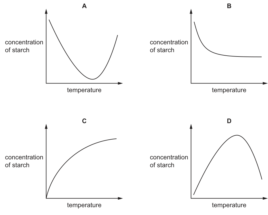
Some students investigated the effect of temperature on the digestion of starch to form maltose. They added equal volumes of amylase and starch solution to a number of test-tubes. The test-tubes were left for 30 minutes at a range of temperatures between and . They then measured the concentration of starch in each test-tube.
Which graph was produced from these results?
Options
Answer
A
A
Walkthrough
- Understand the reaction: Amylase is an enzyme that digests (breaks down) starch into maltose. The experiment measures the concentration of starch remaining after 30 minutes at different temperatures.
- Low temperatures (e.g., 10 °C): At low temperatures, molecules have low kinetic energy. Collisions between amylase and starch are infrequent, so the rate of reaction is very slow. Very little starch is digested, meaning the concentration of starch remaining is high.
- Increasing temperature towards the optimum: As temperature rises, kinetic energy increases, and the rate of reaction increases. More starch is digested, so the concentration of starch remaining decreases.
- Optimum temperature: Around 37 °C (for human amylase), the enzyme works at its maximum rate. The most starch is digested, so the concentration of starch remaining is at its minimum.
- High temperatures (e.g., 60 °C): Above the optimum temperature, the enzyme amylase denatures. Its active site changes shape, and it can no longer bind to starch. The rate of reaction drops sharply, so less starch is digested. The concentration of starch remaining increases again.
- Match to the graph: The curve for starch concentration against temperature must start high, fall to a minimum at the optimum temperature, and then rise again. This is a U-shaped curve, which is Graph A.
- Why not Graph D? Graph D is an inverted U-shape. This would be the correct graph if the y-axis was "concentration of maltose" (product formed) or "rate of reaction". Students often memorise the bell-curve shape for enzyme activity and forget to check what is actually plotted on the axes.
Key Takeaways
- Enzyme activity vs. temperature: Activity is low at low temperatures (low kinetic energy), highest at the optimum temperature, and low at high temperatures (enzyme denaturation).
- Substrate remaining vs. product formed: When plotting the substrate remaining (starch) against temperature, the graph is U-shaped (high → low → high). When plotting the product formed (maltose) or the rate of reaction, the graph is an inverted U-shape (low → high → low).
- Always read the y-axis label carefully. 5090 frequently tests this inversion to ensure students understand what is actually being measured, rather than just memorising the shape of an enzyme curve.
Common Mistakes
- Choosing Graph D: Selecting the classic bell-curve graph without noticing that the y-axis is "concentration of starch" (substrate remaining) rather than "rate of reaction" or "concentration of maltose".
- Misunderstanding low temperatures: Thinking that the enzyme is denatured at 10 °C. Enzymes are not denatured by cold; they are simply inactive due to low kinetic energy. The substrate concentration is high because little reaction has occurred, not because the enzyme is destroyed.
- Confusing substrate and product: Failing to recognise that as the reaction proceeds, substrate decreases and product increases. The two graphs are mirror images of each other across a horizontal line.
Things to Be Careful About
- Axis labels: The question specifically asks for the graph of "concentration of starch". If it asked for "concentration of maltose" or "amount of starch digested", the answer would be D.
- Denaturation vs. low kinetic energy: At 60 °C, the high starch concentration is due to denaturation (the enzyme is permanently damaged). At 10 °C, the high starch concentration is due to low kinetic energy (the enzyme is intact but slow). Both result in a high y-value, but for completely different biological reasons.
- The minimum point: The lowest point on Graph A corresponds to the optimum temperature of the enzyme, where digestion is most complete.
The graph shows the rate of photosynthesis in a plant in full sunlight at two different temperatures and at different concentrations of carbon dioxide.
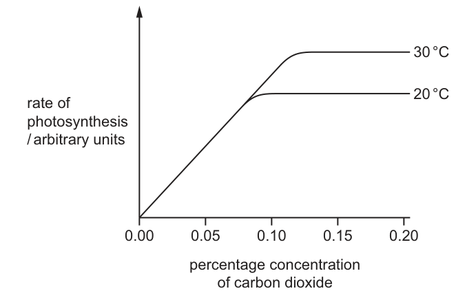
At normal atmospheric carbon dioxide concentrations, what limits the rate of photosynthesis?
Options
A carbon dioxide concentration
B light intensity
C temperature
D water availability
Working
Normal atmospheric carbon dioxide concentration is approximately 0.04%. On the graph, this value falls on the steep, linear portion of the curve (between 0.00% and 0.08%). At this point, an increase in carbon dioxide concentration leads to a proportional increase in the rate of photosynthesis. Therefore, carbon dioxide concentration is the factor limiting the rate of photosynthesis.
Note that both the 20 °C and 30 °C curves overlap at this low concentration, meaning temperature is not limiting here. The question also states the plant is in "full sunlight", so light intensity is not limiting.
Answer
A
A
Walkthrough
The question asks what limits the rate of photosynthesis at normal atmospheric carbon dioxide concentrations. Normal atmospheric CO₂ is about 0.04%.
Look at the graph: the x-axis is the percentage concentration of carbon dioxide, ranging from 0.00% to 0.20%. The value 0.04% is near the left side of the graph, in the region where the curve is rising steeply and linearly. In this region, as carbon dioxide concentration increases, the rate of photosynthesis increases. By definition, the factor that is increasing and causing the rate to increase is the limiting factor. Therefore, carbon dioxide concentration is limiting the rate of photosynthesis at 0.04%.
Why not temperature? The graph shows two curves for 20 °C and 30 °C. At low carbon dioxide concentrations (up to about 0.08%), the two curves overlap perfectly. This means that changing the temperature from 20 °C to 30 °C does not change the rate of photosynthesis. If temperature were the limiting factor, the higher temperature curve would be above the lower one. Since they overlap, temperature is not limiting at this concentration.
Why not light intensity? The question stem explicitly states the plant is in "full sunlight", which means light is not in short supply and therefore cannot be the limiting factor.
Why not water? Water is not plotted on the graph, and there is no indication that water is limiting. The graph only provides information about carbon dioxide and temperature.
Key Takeaways
- A limiting factor is any variable that, when increased, causes an increase in the rate of a biological process.
- On a graph of rate against a variable, if the curve is still rising, that variable is the limiting factor.
- When multiple curves are plotted for different values of a second variable (like temperature), and the curves overlap at a certain range of the first variable, the second variable is NOT limiting in that range.
- Normal atmospheric carbon dioxide concentration is approximately 0.04%, which is very low compared to the concentrations used in experiments (often up to 0.10% or more).
Common Mistakes
- Choosing temperature (C): Students often see two temperature curves and assume temperature must be the answer. However, at low CO₂ concentrations, the curves overlap, proving temperature is not limiting. Temperature only becomes limiting at higher CO₂ concentrations where the curves separate and plateau at different levels.
- Choosing light intensity (B): Students might recall that light is a limiting factor for photosynthesis and choose it without reading the stem. The stem explicitly says "full sunlight", removing light as a limiting factor.
- Misreading the graph: Assuming that because the graph has a plateau, CO₂ is never limiting. Students must read the specific concentration asked about (normal atmospheric, ~0.04%) and check the graph at that exact point.
Things to Be Careful About
- Always recall or estimate the normal atmospheric concentration of CO₂ (~0.04%). Even without knowing the exact number, 0.04% is clearly in the first quarter of the x-axis (0.00 to 0.20), where the curve is still rising.
- Do not be distracted by the two temperature curves when answering about low carbon dioxide concentrations; overlapping curves mean the differing variable is not limiting.
- Read the question stem carefully for conditions that remove other factors from consideration, such as "full sunlight" removing light intensity as a limiting factor.
The diagram shows a section through a dicotyledonous leaf.
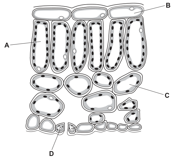
Which cell cannot photosynthesise?
Options
A A
B B
C C
D D
Answer
B
B
Walkthrough
The question asks to identify the cell in a dicotyledonous leaf cross-section that cannot photosynthesise. Photosynthesis requires chloroplasts, which contain the pigment chlorophyll.
- A (palisade mesophyll cell): These cells are located just below the upper epidermis. They are elongated and packed with many chloroplasts, making them the main site of photosynthesis in the leaf.
- B (upper epidermal cell): The epidermis is the outer protective layer of the leaf. Epidermal cells are transparent and contain no chloroplasts. This transparency is an adaptation that allows maximum light to pass through to the photosynthesising mesophyll cells below.
- C (spongy mesophyll cell): These cells are located below the palisade layer. They are irregularly shaped with large air spaces between them, but they still contain chloroplasts and can photosynthesise.
- D (guard cell): Guard cells surround the stomata in the lower epidermis. Unlike other epidermal cells, guard cells contain chloroplasts and can photosynthesise to help regulate stomatal opening.
Since cell B (the upper epidermal cell) lacks chloroplasts, it cannot photosynthesise. The correct answer is B.
Key Takeaways
- Photosynthesis occurs only in cells that contain chloroplasts.
- In a leaf cross-section, palisade mesophyll, spongy mesophyll, and guard cells contain chloroplasts.
- Upper and lower epidermal cells (except guard cells) are transparent and lack chloroplasts to allow light transmission.
Common Mistakes
- Choosing D (guard cell): Students sometimes assume all epidermal cells lack chloroplasts. Guard cells are the exception; they do contain chloroplasts.
- Choosing A or C: Students may misread the diagram or forget that both palisade and spongy mesophyll cells contain chloroplasts.
- Ignoring the diagram details: The diagram shows dots inside cells A, C, and D representing chloroplasts, but cell B is empty. Reading the diagram carefully prevents this error.
Things to Be Careful About
- Always check whether the question asks which cell can or cannot photosynthesise. This is a negative question.
- In leaf cross-section diagrams, chloroplasts are often drawn as small dark dots or ovals inside the cell. Epidermal cells will be blank inside.
- Remember that only guard cells among the epidermal cells contain chloroplasts.
A leafy stem was cut from a plant and placed in a red dye solution for 18 hours.
The diagram shows a transverse section of the stem after 18 hours.
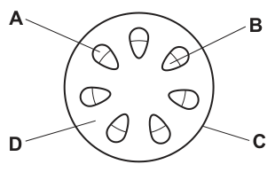
Which tissue will be stained red?
Options
A A
B B
C C
D D
Working
Red dye dissolved in water is carried upwards in the xylem vessels, which transport water and dissolved ions. In a transverse section of a dicotyledonous stem, the vascular bundles are arranged in a ring; within each bundle the xylem lies on the inner side and the phloem on the outer side. Label B points to the inner part of a vascular bundle — the xylem — so this is the tissue stained red. Label C is the phloem, A is the cortex, and D is the pith; none of these conduct water.
Answer
B
B
Walkthrough
When a leafy stem stands in red dye, the dye is simply water with a coloured solute. Water is absorbed and pulled up the stem only through the xylem vessels — dead, hollow tubes whose open lumen and lack of end walls make them a continuous pipe for water. So after 18 hours the only tissue stained red is the xylem.
Now read the diagram. It shows a transverse section of a dicotyledonous stem, in which the vascular bundles are arranged in a ring near the edge. Within each bundle the arrangement is fixed: xylem towards the centre of the stem, phloem towards the outside. In the figure, B points to the inner part of a vascular bundle (the xylem), C points to the outer part (the phloem), A points to the cortex between the bundles, and D points to the central pith. The stained tissue is therefore B.
Key Takeaways
- Water and dissolved mineral ions travel up the stem exclusively in the xylem; the phloem transports sugars (translocation), so dye experiments stain xylem only.
- In a dicotyledonous stem, vascular bundles form a ring, and within each bundle xylem is on the inner side, phloem on the outer side.
- This xylem-inside/phloem-outside arrangement is reversed in the root, where xylem forms a central star.
Common Mistakes
- Choosing C (the phloem): students often confuse the two transport tissues. Remember phloem carries sucrose, not water from the soil.
- Confusing the positions within the bundle: in the stem xylem is inner, phloem outer — the reverse of the root.
- Choosing A or D: the cortex and pith are packing/ground tissues and conduct nothing.
Things to Be Careful About
- The mark scheme gives only the letter, so the discrimination is entirely in knowing which label is xylem — read the pointer carefully; B points to the inner half of the bundle.
- 'Stained red' means the dye travelled in the water stream; it does not indicate living tissue. Xylem vessels are dead cells, which is exactly why they can carry water freely.
The diagram shows the movement of water through part of a leaf.
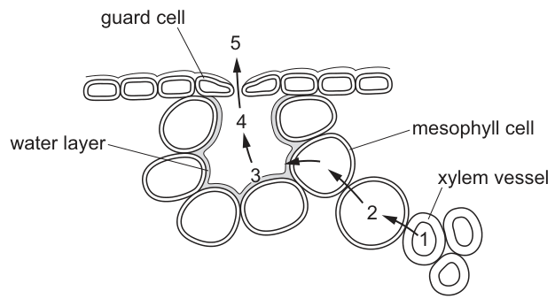
Which processes are involved in the movement of water at these stages?
Options
| 1–2 | 3–4 | 4–5 | |
|---|---|---|---|
| A | diffusion | evaporation | osmosis |
| B | evaporation | diffusion | osmosis |
| C | osmosis | diffusion | evaporation |
| D | osmosis | evaporation | diffusion |
Working
- Stage 1–2: water moves from the xylem vessel into the mesophyll cells across their partially permeable membranes — this is osmosis.
- Stage 3–4: liquid water from the thin water layer on the mesophyll cell surfaces changes into water vapour in the air space — this is evaporation.
- Stage 4–5: water vapour moves down its concentration gradient from the air space, through the stoma, into the outside air — this is diffusion.
Answer
D
D
Walkthrough
This question tests whether you can name the three different processes that carry water along the transpiration stream through a leaf. The key is to look at what the water is at each stage and where it is moving:
-
Stage 1–2 — xylem vessel to mesophyll cell. The water is still liquid, and it crosses living cell membranes. Movement of water across a partially permeable membrane from a higher water potential to a lower water potential is osmosis. This eliminates options A and B immediately.
-
Stage 3–4 — water layer to air space. Here liquid water on the surface of the spongy mesophyll cells turns into water vapour inside the air space. A liquid becoming a gas is evaporation, not diffusion or osmosis. This points to option D.
-
Stage 4–5 — air space out of the stoma. Water vapour now moves from a region of high concentration (inside the leaf) to low concentration (the outside air), down a concentration gradient, through the open stoma between the guard cells. That movement of molecules down a gradient is diffusion.
So the sequence is osmosis → evaporation → diffusion, which is option D.
Key Takeaways
- The transpiration stream has three distinct steps in the leaf: osmosis out of the xylem into mesophyll cells, evaporation from wet cell surfaces into the air spaces, and diffusion of water vapour out through the stomata.
- Osmosis always involves water crossing a partially permeable membrane; diffusion involves any molecules moving down a concentration gradient without a membrane being crossed here; evaporation is a change of state from liquid to gas.
- Identifying the physical state of the water at each stage (liquid inside cells → vapour in air spaces) tells you which process applies.
Common Mistakes
- Choosing option C by putting 'osmosis' first but then swapping the last two stages — remember stage 3–4 is a change of state (evaporation), while stage 4–5 is movement of already-formed vapour (diffusion).
- Calling stage 1–2 'active transport' or 'transpiration' — water leaves the xylem passively by osmosis; no energy is spent.
- Confusing evaporation with diffusion: evaporation happens at the wet cell surface; diffusion carries the vapour through the stoma.
- Thinking water vapour exits by osmosis — osmosis requires a membrane and liquid water; the stoma is an open pore.
Things to Be Careful About
- Read the diagram carefully: the numbers mark positions along the pathway, and the question asks about the movement between consecutive numbered points (1–2, 3–4, 4–5).
- Use the precise terms: 'osmosis' (not 'water moving in'), 'evaporation' (not 'turning to gas'), 'diffusion' (not 'spreading out').
- In MCQs like this, eliminate one column at a time: knowing stage 1–2 must be osmosis removes A and B at once, leaving only C and D to decide.
The diagram shows part of the human digestive system.
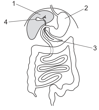
Which parts produce bile and store bile?
Options
A 1 and 2
B 1 and 4
C 2 and 4
D 3 and 4
Working
The diagram labels the following structures:
- 1: gall bladder
- 2: stomach
- 3: pancreas
- 4: liver
Bile is produced by the liver (4) and stored in the gall bladder (1). Therefore, the parts that produce and store bile are 1 and 4.
Answer
B
B
Walkthrough
- Identify the labelled structures: From the diagram and its description, label 1 points to the gall bladder, 2 to the stomach, 3 to the pancreas, and 4 to the liver.
- Recall the role of bile: Bile is a digestive juice that emulsifies fats, breaking large lipid droplets into smaller ones to increase the surface area for the enzyme lipase. It is not an enzyme itself.
- Match production and storage to organs:
- The liver (4) is the large organ that continuously produces bile.
- The gall bladder (1) is a small sac that stores and concentrates the bile between meals, releasing it into the duodenum when fat is present.
- Evaluate the options: The question asks for the parts that produce and store bile. These are the liver (4) and the gall bladder (1). The pair is 1 and 4, which matches option B.
Key Takeaways
- The liver produces bile, and the gall bladder stores it. Do not confuse their roles.
- Bile is an emulsifier, not an enzyme; it physically breaks down fats rather than chemically digesting them.
- The pancreas (3) produces digestive enzymes (like lipase) and bicarbonate to neutralise stomach acid, but it does not produce bile.
Common Mistakes
- Reversing production and storage: Students often think the gall bladder produces bile and the liver stores it. Remember: the liver is the factory (producer), and the gall bladder is the warehouse (storage).
- Confusing the pancreas with the liver/gall bladder: The pancreas (3) is involved in digestion via enzymes, but it has no role in bile production or storage.
- Misreading the diagram labels: Carefully trace the pointer lines. Label 4 points to the large, shaded organ (liver), while label 1 points to the small sac attached to it (gall bladder).
Things to Be Careful About
- Order of the pair: The question asks for "produce" and "store" in that order, but the options list the numbers in no particular order (e.g., "1 and 4"). Ensure you select the option that contains both correct numbers, regardless of their order in the option text.
- Bile vs. enzymes: Bile does not contain enzymes. It is often tested alongside enzymes, so be clear that bile emulsifies fats while lipase digests them chemically.
- Diagram literacy: In 5090 diagrams, pointer lines can be tricky. Always follow the line from the number to the exact structure it indicates. Here, 4 clearly indicates the large liver mass, and 1 indicates the small gall bladder nestled beneath it.
Five test-tubes containing cooked egg white are set up as shown. Cooked egg white contains a protein. Protease solutions of different pH are added to each tube.
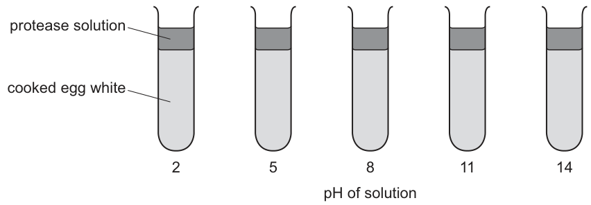
Which diagram shows the results of this experiment for a protease from the stomach?
Options
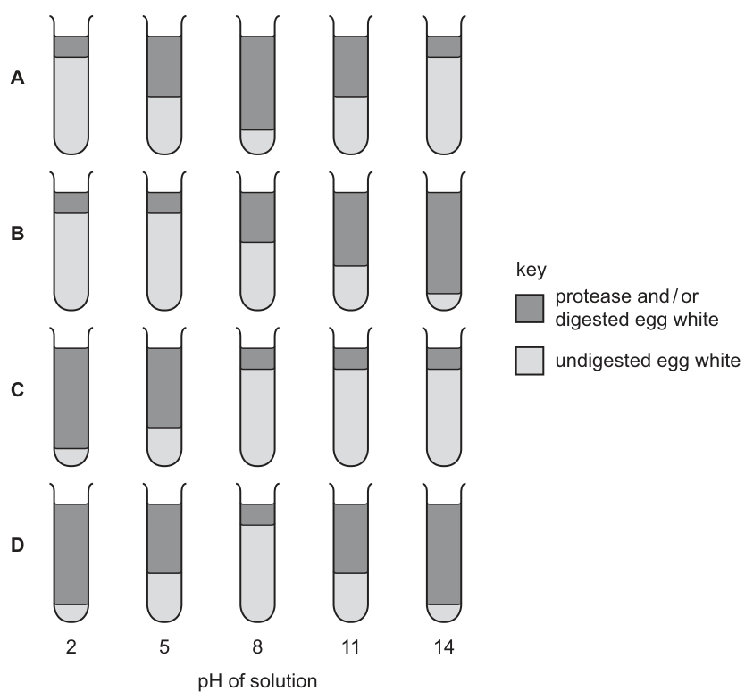
Working
A protease from the stomach is pepsin, which works best at the acidic pH of the stomach, about pH 2. At higher pH values the enzyme is denatured or inactive, so little or no digestion occurs in tubes at pH 5, 8, 11 and 14.
- A shows digestion increasing towards neutral pH — wrong for a stomach enzyme.
- B shows digestion at pH 5–8 — wrong; the enzyme is inactive there.
- D shows digestion at every pH — impossible.
- C shows most digestion at pH 2, less at pH 5, and almost none above — this matches a stomach protease.
Answer
C
C
Walkthrough
The experiment tests how well a protease digests cooked egg white (a protein) at different pH values. The key point is knowing WHERE the enzyme comes from: the question specifies a protease from the stomach. The stomach enzyme is pepsin, and it is adapted to work in strongly acidic conditions — its optimum pH is about 2.
At pH values far from the optimum, the shape of the enzyme's active site changes (the enzyme is denatured), so the substrate no longer fits and digestion stops. So:
- pH 2: near optimum → rapid digestion of egg white → tube mostly 'protease and/or digested egg white'.
- pH 5: some activity but much reduced → partial digestion.
- pH 8, 11, 14: far too alkaline → enzyme denatured/inactive → egg white remains undigested.
Only option C shows this pattern: complete digestion at pH 2, mostly digested at pH 5, and little or no digestion from pH 8 upwards. Option A would suit an enzyme with a neutral optimum (like one from the small intestine); B suggests activity around pH 5–8; D suggests a pH-independent enzyme, which does not exist.
Key Takeaways
- Each enzyme has an optimum pH at which its active site fits the substrate best.
- Pepsin, the stomach protease, has an optimum around pH 2 because the stomach contains hydrochloric acid.
- Away from the optimum pH, enzymes are denatured — the active site loses its shape — so the reaction rate falls sharply.
- Proteases in different parts of the gut have different optima: pepsin (acidic) versus trypsin in the small intestine (slightly alkaline).
Common Mistakes
- Choosing A, which describes an enzyme with a neutral/alkaline optimum — that would fit a protease from the small intestine, not the stomach.
- Assuming enzymes work at all pH values (option D) — every enzyme has a limited pH range.
- Confusing denaturation by pH with simply 'slowing down': at extreme pH the active site's shape is permanently changed.
- Forgetting that the question names the source (stomach) — the answer depends entirely on matching the enzyme to its natural environment.
Things to Be Careful About
- Read the stem carefully: 'a protease from the stomach' fixes the optimum at about pH 2. If it had said 'from the pancreas/small intestine', the answer pattern would be reversed.
- Use the key correctly: shaded = digested (or just protease solution present), unshaded = undigested egg white. In C, the shaded region grows as the unshaded egg white shrinks.
- Digestion at pH 5 in option C is only partial — that is correct, because pH 5 is closer to the optimum than pH 8 but still not optimal.
Which diagram shows the absorption of different food molecules into a villus?
Options
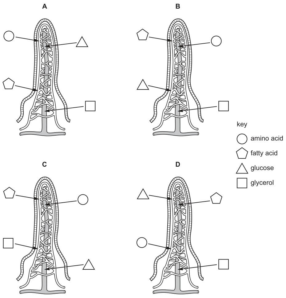
Answer
D
In the intestinal villus, digested food molecules are absorbed into either the blood capillaries or the central lacteal:
- Glucose and amino acids are absorbed into the blood capillaries.
- Fatty acids and glycerol are absorbed into the lacteal.
Diagram D correctly shows glucose (triangle) and amino acid (circle) entering the blood capillaries, and fatty acid (pentagon) and glycerol (square) entering the lacteal.
D
Walkthrough
The question asks to identify the correct diagram showing the absorption of digested food molecules into an intestinal villus. To solve this, we must recall the specific absorption pathways for the main products of digestion:
- Identify the structures in the villus diagram: The diagram shows an intestinal villus with an outer network of blood capillaries and a central lymphatic vessel called the lacteal.
- Recall the absorption pathways:
- Carbohydrates are digested into glucose, which is absorbed into the blood capillaries.
- Proteins are digested into amino acids, which are also absorbed into the blood capillaries.
- Lipids (fats) are digested into fatty acids and glycerol. These are absorbed into the lacteal (lymphatic vessel) because they are reassembled into triglycerides and packaged into chylomicrons, which are too large to enter the blood capillaries.
- Examine the key and diagrams:
- Circle = amino acid
- Pentagon = fatty acid
- Triangle = glucose
- Square = glycerol
- Evaluate the options:
- Diagram A: amino acid, glucose, and fatty acid go to blood; glycerol goes to lacteal. (Incorrect)
- Diagram B: fatty acid, amino acid, and glucose go to blood; glycerol goes to lacteal. (Incorrect)
- Diagram C: fatty acid, amino acid, and glycerol go to blood; glucose goes to lacteal. (Incorrect)
- Diagram D: glucose (triangle) and amino acid (circle) enter the blood capillaries; fatty acid (pentagon) and glycerol (square) enter the lacteal. (Correct)
Key Takeaways
- Glucose and amino acids are absorbed into the blood capillaries in the intestinal villus.
- Fatty acids and glycerol are absorbed into the lacteal (lymphatic vessel) in the intestinal villus.
Common Mistakes
- Confusing which molecules go into the blood capillaries versus the lacteal. A common error is thinking that all digested molecules enter the blood capillaries, or that lipids enter the blood directly.
- Misreading the key symbols (circle, pentagon, triangle, square) and their corresponding molecules, leading to incorrect matching.
Things to Be Careful About
- Ensure you are matching the correct symbol from the key to the correct molecule and tracing the arrow to the correct vessel (blood capillary vs. lacteal).
- Remember that fatty acids and glycerol enter the lacteal, not the blood capillaries, because they are reassembled into triglycerides and packaged into chylomicrons, which are too large to enter the blood capillaries but can enter the larger lacteal.
The table shows the changes that occur in the thorax during breathing.
Which changes occur during inspiration?
Options
| diaphragm | external intercostal muscles | internal intercostal muscles | pressure in thorax | |
|---|---|---|---|---|
| A | contracts | contract | relax | falls and then rises |
| B | relaxes | relax | contract | rises and then falls |
| C | relaxes | contract | relax | rises and then falls |
| D | contracts | relax | contract | falls and then rises |
Working
During inspiration:
- the diaphragm contracts and flattens;
- the external intercostal muscles contract to raise the ribs;
- the internal intercostal muscles relax;
- the volume of the thorax increases, so the pressure in the thorax falls below atmospheric pressure, drawing air in, then rises again as air enters.
This matches row A exactly.
Answer
A
A
Walkthrough
This question tests the mechanics of breathing. Inspiration (breathing in) is an active process. The diaphragm is a sheet of muscle below the lungs; when it contracts it flattens and moves downwards, increasing the volume of the thorax. At the same time the external intercostal muscles contract, pulling the ribs upwards and outwards, which also increases the volume of the thorax. The internal intercostal muscles do the opposite job — they pull the ribs down and inwards — so they must relax during inspiration.
Increasing the volume of a closed space decreases the pressure inside it (Boyle's law, though you don't need the name). So the pressure in the thorax falls below atmospheric pressure. Air then flows in from outside down the pressure gradient until the pressures equalise, at which point the pressure has risen back to atmospheric. That is why the pressure "falls and then rises".
Now check the rows:
- A — diaphragm contracts, external intercostals contract, internal intercostals relax, pressure falls then rises. This is exactly right.
- B — diaphragm relaxes and internal intercostals contract describe expiration, not inspiration.
- C — diaphragm relaxes is wrong, and the pressure change is wrong too.
- D — external intercostals relax and internal intercostals contract are both wrong for inspiration.
So A is the only row where every entry matches inspiration.
Key Takeaways
- Inspiration is active: the diaphragm contracts and flattens, the external intercostal muscles contract to raise the ribs, and the internal intercostal muscles relax.
- Increasing the volume of the thorax lowers the pressure inside it, so air moves in from higher atmospheric pressure.
- Expiration is mostly passive: the diaphragm relaxes (moves up), the external intercostals relax, the internal intercostals may contract, the volume decreases, and the pressure rises, pushing air out.
- The pressure in the thorax falls during inspiration and then rises again as air enters and equalises the pressure.
Common Mistakes
- Confusing the two sets of intercostal muscles: external intercostals contract to raise the ribs (inspiration), internal intercostals contract to lower the ribs (forced expiration).
- Thinking the diaphragm relaxes during inspiration — it actually contracts and flattens.
- Mixing up the pressure change: during inspiration the pressure falls (not rises) because the volume increases.
Things to Be Careful About
- Every entry in the row must be correct — one wrong entry eliminates the option.
- The pressure change is described as "falls and then rises" because the pressure drops below atmospheric and then returns to atmospheric as air flows in. Do not choose a row that says only "rises" or only "falls".
- Remember that the diaphragm is a muscle and "contracts" is the precise term the mark scheme expects.
Scientists have concluded that absorption of mineral ions in plants requires energy from respiration.
Which observation best supports this conclusion?
Options
A Carbohydrate is stored in the roots.
B Living roots give off carbon dioxide.
C The root hairs have a large surface area.
D Uptake of mineral ions is reduced in lower oxygen concentrations.
Working
Mineral ions are taken up by root hair cells by active transport, which requires energy from respiration. Respiration needs oxygen and releases carbon dioxide.
- A: storage of carbohydrate in roots does not show respiration is linked to uptake.
- B: living roots giving off carbon dioxide shows respiration occurs, but not that its energy is used for ion uptake.
- C: large surface area of root hairs aids absorption generally, not the need for energy.
- D: if oxygen concentration falls, respiration slows, less energy is available, and ion uptake falls — this directly links ion uptake to respiration.
Answer
D
D
Walkthrough
The question asks which observation best supports the idea that absorbing mineral ions needs energy from respiration. Mineral ions are usually at a higher concentration inside the root hair cell than in the soil water, so they cannot simply diffuse in — they must be moved against their concentration gradient by active transport. Active transport requires energy, and that energy comes from respiration.
Now test each observation against the conclusion:
- A — Carbohydrate stored in roots tells you the root has a food reserve; it says nothing about how ions are absorbed.
- B — Living roots releasing carbon dioxide proves respiration is happening (respiration produces carbon dioxide), but it does not connect that respiration to ion uptake specifically.
- C — A large surface area on root hairs speeds absorption by diffusion as well as active transport; it supports absorption in general, not the energy requirement.
- D — Lower oxygen means slower aerobic respiration, so less energy is released. If ion uptake drops when oxygen drops, the two processes are directly linked: the energy from respiration powers the uptake. This is exactly the evidence needed.
Key Takeaways
- Mineral ions are absorbed by root hair cells by active transport, moving them against their concentration gradient.
- Active transport requires energy released by respiration.
- Respiration uses oxygen and releases carbon dioxide, so reducing oxygen reduces the energy supply.
- A good piece of supporting evidence must link the two variables in the conclusion — here, respiration and ion uptake — not just show one of them exists.
Common Mistakes
- Choosing B because it mentions carbon dioxide and therefore 'respiration' — but it never links respiration to ion uptake.
- Choosing C because it sounds like an adaptation for absorption — but surface area helps diffusion too, so it is not specific to energy-requiring uptake.
- Confusing active transport with diffusion: diffusion is passive and needs no energy, so evidence about surface area or gradients would not support the energy conclusion.
Things to Be Careful About
- In 'which observation best supports...' questions, eliminate options that are true statements but irrelevant to the specific conclusion.
- The key logical chain is: less oxygen → less respiration → less energy → less ion uptake. Option D captures this chain directly.
When people travel in an aircraft and sit still in their seats for long periods of time, their lower legs may swell up.
Which statement explains why this may happen?
Options
A Fewer leg muscle contractions occur to push blood along the veins.
B The rate of the heartbeat is reduced as no exercise is happening.
C There is little or no arterial blood pressure to force the blood upwards.
D Valves in the veins do not fill up with blood to prevent its backflow.
Working
Veins carry blood back to the heart at low pressure. Blood in the veins of the lower legs must flow upwards against gravity, and the veins have no strong pump of their own. Two features solve this: valves that prevent backflow, and contractions of the surrounding skeletal muscles, which squeeze the veins and push blood along. When a person sits still for a long time, leg muscles barely contract, so this 'muscle pump' stops working and blood pools in the lower legs, making them swell.
- A is correct: fewer leg muscle contractions occur to push blood along the veins.
- B is incorrect: the heart rate does not necessarily drop just because a person is sitting still, and even a slower heart rate would not directly cause blood to pool in the legs.
- C is incorrect: arterial blood pressure is not what pushes blood up the veins; veins are a low-pressure system and rely on the muscle pump and valves.
- D is incorrect: valves in the veins do fill with blood and close to prevent backflow; the statement is false.
Answer
A
A
Walkthrough
This question is about venous return — how blood gets back to the heart from the lower parts of the body.
Blood leaving the heart travels in arteries under high pressure from the pumping of the heart. By the time blood has passed through the capillaries and reached the veins, that pressure has mostly been lost. Veins carry blood back to the heart at low pressure, so something else must help push the blood along, especially in the legs where blood has to flow upwards against gravity.
The body uses two main mechanisms:
- Valves inside the veins — these are one-way flaps that stop blood from flowing backwards. They close if blood tries to fall back down the leg.
- Skeletal muscle contractions — when the leg muscles contract (for example when walking), they squeeze the veins running between them, pushing blood upwards. This is often called the skeletal muscle pump.
When someone sits still in an aircraft seat for hours, their leg muscles barely contract. The muscle pump stops working, so blood is not pushed efficiently up the veins. Blood pools in the veins of the lower legs, fluid leaks out into the tissues, and the legs swell up.
Now look at each option:
- A — "Fewer leg muscle contractions occur to push blood along the veins." This exactly describes the loss of the muscle pump. Correct.
- B — "The rate of the heartbeat is reduced as no exercise is happening." Sitting still does not necessarily reduce heart rate, and even if it did, a slower heart would not specifically cause the lower legs to swell. The heart is not the structure that pushes blood up the leg veins. Incorrect.
- C — "There is little or no arterial blood pressure to force the blood upwards." Arterial blood pressure is the pressure in the arteries, not the veins. Blood in the veins is not pushed by arterial pressure; it is pushed by the muscle pump and helped by valves. Incorrect.
- D — "Valves in the veins do not fill up with blood to prevent its backflow." This is factually wrong. Valves do fill with blood and close to prevent backflow. That is their whole job. Incorrect.
Key Takeaways
- Veins carry blood back to the heart at low pressure.
- Blood returns from the legs against gravity with the help of valves (prevent backflow) and skeletal muscle contractions (squeeze blood along).
- When muscles are inactive for a long time, blood pools in the lower legs, causing swelling.
- This is a classic structure-and-function idea: the vein's structure (valves) and its surroundings (skeletal muscles) work together to solve the problem of low-pressure return.
Common Mistakes
- Choosing C because of confusing arterial pressure with venous return. Arterial pressure pushes blood away from the heart; it does not push blood up the leg veins.
- Choosing B because of thinking 'no exercise means slower heart'. The heart rate does not necessarily drop, and the swelling is not caused by heart rate.
- Choosing D because of misreading the negative: the statement says valves do not fill with blood, which is false — valves fill and close to prevent backflow.
Things to Be Careful About
- Read the question stem carefully: the key clue is "sit still... for long periods" — this points directly to lack of muscle contraction, not to heart rate or arterial pressure.
- Remember that the mark scheme requires the precise idea: the swelling happens because fewer leg muscle contractions occur, so blood is not pushed along the veins. The word "muscle contractions" is the essential idea.
- Do not confuse the functions of arteries and veins: arteries are high-pressure, veins are low-pressure and rely on the muscle pump.
The diagram shows a blood cell as seen under a light microscope.

What is the type of blood cell shown?
Options
A phagocyte
B lymphocyte
C red blood cell
D platelet
Working
The diagram shows a white blood cell with a multi-lobed nucleus. Comparing this to the options:
- Red blood cells lack a nucleus.
- Lymphocytes have a large, single, round nucleus.
- Platelets are small, irregular cell fragments with no nucleus.
- Phagocytes (such as neutrophils) are characterised by a multi-lobed nucleus.
The cell shown matches the description of a phagocyte.
Answer
A
A
Walkthrough
The question asks to identify a blood cell from a microscopic drawing. The key distinguishing feature visible in the diagram is the nucleus. We evaluate each option based on its nuclear characteristics:
- Red blood cell (C): Mature mammalian red blood cells are anucleate (they have no nucleus) and are biconcave discs. The diagram clearly shows a nucleus, so this is incorrect.
- Platelet (D): Platelets are small, irregular fragments of cytoplasm derived from megakaryocytes. They are much smaller than red blood cells and have no nucleus. This is incorrect.
- Lymphocyte (B): Lymphocytes are a type of white blood cell, but they are characterised by a large, single, round nucleus that occupies most of the cell volume, with only a thin rim of cytoplasm. The nucleus in the diagram is not round; it is lobed. This is incorrect.
- Phagocyte (A): Phagocytes (specifically polymorphonuclear leukocytes like neutrophils) are white blood cells that engulf pathogens. Their defining morphological feature under a light microscope is a multi-lobed (segmented) nucleus connected by thin chromatin strands. This matches the diagram perfectly.
Key Takeaways
- Blood cells can be identified under a light microscope primarily by the presence, size, and shape of their nucleus.
- Red blood cells and platelets have no nucleus.
- Lymphocytes have a single, large, round nucleus.
- Phagocytes (neutrophils) have a multi-lobed nucleus.
Common Mistakes
- Confusing lymphocytes and phagocytes: Students often remember that white blood cells have nuclei but forget the specific shape. A lymphocyte has a round nucleus; a phagocyte has a lobed nucleus. Selecting B instead of A is a common error here.
- Misidentifying red blood cells: Remember that mature mammalian red blood cells lose their nucleus during maturation to make more room for haemoglobin. If a cell has a nucleus, it is not a red blood cell.
- Forgetting platelets are fragments: Platelets are not whole cells; they are small cytoplasmic fragments and are significantly smaller than the other blood cells shown in typical diagrams.
Things to Be Careful About
- Always look at the nucleus first when identifying blood cells in diagrams. The shape of the nucleus is the most reliable distinguishing feature at this level.
- Ensure you read the diagram carefully: a multi-lobed nucleus is the hallmark of a phagocyte (neutrophil), not a lymphocyte.
- In Paper 1, you do not need to explain your reasoning in the answer space; simply select the correct option letter.
Which statements about mosquitoes are correct?
Options
| where mosquitoes lay their eggs | sex of the adult mosquito that sucks blood | |
|---|---|---|
| A | in decaying organic material | female |
| B | in decaying organic material | male |
| C | in stagnant water | female |
| D | in stagnant water | male |
Working
Mosquitoes lay their eggs in stagnant water. The adult mosquito that sucks blood is the female, which needs the blood for egg development.
Answer
C
C
Walkthrough
This question is about the mosquito as a vector of malaria. Two facts are needed:
-
Where mosquitoes lay their eggs: mosquitoes lay eggs in stagnant water, not in decaying organic material. This eliminates options A and B.
-
Which sex of adult mosquito sucks blood: only the female mosquito sucks blood. The male mosquito feeds on nectar and plant juices. The female needs the blood meal to obtain proteins for developing her eggs. This eliminates option D.
The only option that combines both correct facts is C: stagnant water and female.
Key Takeaways
- The female Anopheles mosquito is the vector of malaria.
- Female mosquitoes suck blood because they need the nutrients to produce eggs.
- Mosquitoes breed in stagnant water, which is why removing stagnant water helps control malaria.
Common Mistakes
- Choosing B or D because a student thinks male mosquitoes bite. In fact, only females take blood meals.
- Choosing A or B because a student thinks mosquitoes lay eggs in decaying organic material, which is true for some insects but not mosquitoes.
- Confusing the mosquito with other insects; the mark scheme wants the specific facts about mosquitoes.
Things to Be Careful About
- Read both columns of the table together: the correct option must be right in BOTH columns.
- The mark scheme gives the correct answer as C, so ensure both parts of the statement match.
- No need to mention malaria in the answer; the question only asks about mosquitoes.
The spread of HIV was reduced in many countries between 1990 and 2010.
Which action assisted this reduction?
Options
A development of an effective vaccine
B health education
C treating patients with new antibiotics
D using drugs that slow the appearance of symptoms of HIV
Working
- A is incorrect: there is no effective vaccine against HIV.
- C is incorrect: antibiotics act on bacteria, not on viruses, so they cannot treat or prevent HIV.
- D is incorrect: drugs that slow the appearance of symptoms help the infected person but do not stop the virus being passed on.
- B is correct: health education teaches people how HIV is transmitted and how to avoid infection, so it directly reduces the spread of the virus.
Answer
B
B
Walkthrough
This question asks which action helped reduce the spread of HIV between 1990 and 2010. The key idea is that to reduce the spread of a disease, you must either stop the pathogen passing from person to person, or prevent people from becoming infected in the first place.
Look at each option in turn.
A — development of an effective vaccine. A vaccine gives active immunity by making the body produce antibodies before a person meets the pathogen. However, despite many years of research, there is still no effective vaccine against HIV. So this could not have been the cause of the reduction.
B — health education. This is the correct answer. Health education campaigns teach people how HIV is transmitted — through sexual contact, contaminated blood and needles, and from mother to baby — and how to reduce the risk, for example by using condoms or clean needles. When people know the routes of transmission, they can change their behaviour and avoid infection, so the spread of the virus falls.
C — treating patients with new antibiotics. Antibiotics kill bacteria or stop them reproducing. HIV is a virus, not a bacterium, so antibiotics have no effect on it. This option is wrong.
D — using drugs that slow the appearance of symptoms of HIV. These drugs (antiretroviral drugs) keep an infected person healthier for longer and delay the onset of AIDS. However, they do not remove the virus from the body, and an infected person can still pass HIV on. Slowing symptoms is not the same as stopping spread, so this option is wrong.
Key Takeaways
- HIV is a virus spread through body fluids; it is controlled by preventing transmission, not by treating the infected person alone.
- Health education is a key control measure for HIV because it changes behaviour and prevents new infections.
- Antibiotics act only on bacteria, never on viruses.
- No effective vaccine against HIV exists, so vaccination cannot be credited as the cause of reduced spread.
Common Mistakes
- Choosing D — confusing treatment that slows symptoms with prevention of spread. Treating an infected person does not stop them transmitting the virus.
- Choosing C — forgetting that antibiotics are useless against viruses. HIV is a virus, so antibiotics cannot help.
- Choosing A — assuming a vaccine exists for every disease. There is no effective HIV vaccine, so this option cannot be the cause.
Things to Be Careful About
- Read the question as asking about reducing the spread of HIV, not about helping people who already have it. That distinction immediately rules out D.
- Remember that preventing a viral disease requires stopping transmission (education, barrier methods, clean needles) — antibiotics are never the answer for a virus.
Which statement about the immune system is correct?
Options
A A pathogen has its own antigens which will have the same shape as antibodies that bind to them.
B Antigens stimulate an immune response by phagocytes which produce antibodies.
C Active immunity involves acquiring antibodies from another individual and provides short-term protection against a pathogen.
D Antibodies are protein molecules that bind to antigens either leading to the destruction of pathogens or marking a pathogen for destruction by lymphocytes.
Working
A is incorrect: a pathogen has its own antigens, but antibodies are made by lymphocytes and have a shape complementary to the antigen, not the same shape.
B is incorrect: phagocytes engulf and destroy pathogens; antibodies are produced by lymphocytes.
C is incorrect: active immunity is when the body produces its own antibodies after exposure to a pathogen. Acquiring antibodies from another individual is passive immunity, which gives short-term protection.
D is correct: antibodies are protein molecules that bind to antigens, either destroying pathogens directly or marking them for destruction.
Answer
D
D
Walkthrough
This question asks you to identify the one correct statement about the immune system. Let us look at each option in turn.
Option A says a pathogen has its own antigens which have the same shape as the antibodies that bind to them. This is wrong. A pathogen does have antigens on its surface, but antibodies are not the same shape as antigens. An antibody is made by a lymphocyte and has a binding site whose shape is complementary to the antigen, rather like a lock and key. The antibody fits onto the antigen, but it is not identical in shape.
Option B says antigens stimulate an immune response by phagocytes which produce antibodies. This mixes up the roles of two types of white blood cell. Phagocytes engulf and digest pathogens. Antibodies are produced by lymphocytes, not by phagocytes. So B is incorrect.
Option C says active immunity involves acquiring antibodies from another individual and provides short-term protection. This is actually a description of passive immunity. In active immunity, the body makes its own antibodies after being exposed to an antigen, for example through an infection or a vaccine. Passive immunity, such as antibodies passed from mother to baby, does provide short-term protection. So C is incorrect.
Option D says antibodies are protein molecules that bind to antigens either leading to the destruction of pathogens or marking a pathogen for destruction by lymphocytes. This is correct. Antibodies are proteins. They bind to specific antigens. This binding can neutralise or destroy the pathogen directly, or it can mark the pathogen so that other immune cells destroy it.
The correct answer is therefore D.
Key Takeaways
- Antigens are molecules, often on the surface of pathogens, that trigger an immune response.
- Antibodies are proteins produced by lymphocytes. Each antibody has a shape complementary to a specific antigen.
- Phagocytes engulf and destroy pathogens; they do not produce antibodies.
- Active immunity: the body makes its own antibodies after exposure to an antigen. It provides long-term protection.
- Passive immunity: antibodies are received from another individual. It provides short-term protection.
Common Mistakes
- Confusing phagocytes with lymphocytes: phagocytes engulf pathogens, lymphocytes produce antibodies.
- Confusing active and passive immunity: active immunity involves the body producing its own antibodies; passive immunity involves receiving antibodies from another source.
- Thinking antibodies and antigens have the same shape: they have complementary shapes, like a lock and key.
Things to Be Careful About
- Read each statement carefully and check every part of it. A statement is only correct if all of it is correct.
- Remember the precise terms: antibodies are made by lymphocytes, not phagocytes.
- Know the difference between active and passive immunity, including the source of the antibodies and the duration of protection.
For which disease can antibiotics be used as an effective treatment?
Options
A a bacterial infection
B coronary heart disease
C malaria
D a viral infection
Working
Antibiotics are drugs that kill bacteria or stop them from reproducing. They have no effect on viruses, on the protozoan that causes malaria, or on non-infectious conditions such as coronary heart disease. Therefore antibiotics can be used effectively only against a bacterial infection.
Answer
A
A
Walkthrough
This question tests what antibiotics actually do. Antibiotics are substances that kill bacteria or prevent them from multiplying. They are only useful against diseases caused by bacteria.
Look at each option:
- A bacterial infection — this is exactly what antibiotics target, so this is correct.
- B coronary heart disease — this is not an infection at all; it is a condition of the blood vessels supplying the heart, so antibiotics cannot treat it.
- C malaria — malaria is caused by a protozoan parasite, not a bacterium. Antibiotics do not kill protozoa, so they are not an effective treatment for malaria.
- D a viral infection — viruses are not bacteria. Antibiotics do not work against viruses, so this is incorrect.
Therefore the only disease for which antibiotics can be used effectively is a bacterial infection.
Key Takeaways
- Antibiotics are specific to bacteria: they kill bacteria or stop them reproducing.
- Antibiotics do not work against viruses, protozoa, fungi, or non-infectious diseases.
- Malaria is caused by a protozoan, not a bacterium.
- Using antibiotics unnecessarily, such as for viral infections, contributes to antibiotic resistance.
Common Mistakes
- Choosing C because malaria is an infectious disease — but it is caused by a protozoan parasite, not a bacterium.
- Choosing D because a viral infection is an infection — but antibiotics have no effect on viruses.
- Thinking antibiotics are general "infection killers"; they are only effective against bacterial infections.
Things to Be Careful About
- The mark scheme requires the single correct option, A.
- Do not confuse "disease" with "bacterial disease". Antibiotics treat only bacterial infections.
- Remember that antibiotic resistance is a major concern, so antibiotics should not be used for viral infections.
The diagram shows part of the human urinary system.
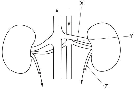
What are structures X, Y and Z?
Options
| X | Y | Z | |
|---|---|---|---|
| A | artery | vein | ureter |
| B | artery | vein | urethra |
| C | vein | artery | ureter |
| D | vein | artery | urethra |
Working
The diagram shows the human urinary system with the major blood vessels and tubes leading to and from the kidneys.
- Structure X: Points to a blood vessel branching from the central vessel with a downward-pointing arrow. The downward flow indicates blood moving away from the heart (this central vessel is the aorta). Blood vessels carrying blood away from the heart to organs are arteries. Specifically, this is the renal artery.
- Structure Y: Points to a blood vessel leaving the kidney and joining the central vessel with an upward-pointing arrow. The upward flow indicates blood moving towards the heart (this central vessel is the vena cava). Blood vessels carrying blood back to the heart are veins. Specifically, this is the renal vein.
- Structure Z: Points to a tube leaving the bottom of the kidney and heading downwards. This tube carries urine from the kidney to the bladder, so it is the ureter. The urethra would carry urine from the bladder to the outside of the body, which is not shown here.
Summary:
- X = artery
- Y = vein
- Z = ureter
This matches option A.
Answer
A
A
Walkthrough
To answer this question, we must identify three structures (X, Y, and Z) on a diagram of the human urinary system. We do this by using the direction of blood flow and the anatomical position of the tubes.
Step 1: Identify X and Y (blood vessels)
Look at the two central vertical vessels. One has an arrow pointing up, the other has an arrow pointing down.
- Blood is pumped out of the heart via the aorta, which runs downwards through the abdomen. The central vessel with the downward arrow is the aorta. Branches coming off the aorta to supply organs with oxygenated blood are arteries. Structure X is a branch going into the right kidney, so it is the renal artery.
- Deoxygenated blood returns to the heart via the vena cava, which runs upwards. The central vessel with the upward arrow is the vena cava. Branches from organs returning blood to the vena cava are veins. Structure Y is a branch leaving the right kidney and entering the vena cava, so it is the renal vein.
Step 2: Identify Z (tube)
Structure Z is a tube leaving the lower end of the kidney. In the urinary system, urine is produced in the kidneys and travels down tubes called ureters to be stored in the bladder. The urethra is the tube that carries urine from the bladder to the outside of the body. Since Z connects the kidney to the bladder (not the bladder to the outside), it is the ureter.
Step 3: Match to options
- X: artery
- Y: vein
- Z: ureter
This corresponds exactly to option A.
Key Takeaways
- Arteries vs. veins: Arteries carry blood away from the heart (down the aorta to the kidneys), while veins carry blood towards the heart (up the vena cava from the kidneys). Always check the arrows on the main vessels to confirm flow direction.
- Ureter vs. urethra: The ureter connects the kidney to the bladder. The urethra connects the bladder to the exterior of the body. Confusing these two is a common mistake.
Common Mistakes
- R urethra: Candidates often misidentify the ureter as the urethra. Remember the urethra is lower down, leaving the bladder, not the kidney.
- Confusing artery and vein: Some candidates guess based on whether the vessel is on the left or right side of the diagram, or based on thickness. The correct method is to follow the arrows: flow away from the heart = artery, flow towards the heart = vein.
- Ignoring the arrows: Without noting the upward and downward arrows on the central vessels, it is impossible to definitively say which is the aorta and which is the vena cava.
Things to Be Careful About
- Always look at the direction of flow (arrows) on the major blood vessels before naming branches. The aorta always carries blood away from the heart; the vena cava always carries it back.
- Read the options carefully. Options B, C, and D all contain at least one incorrect structure (urethra instead of ureter, or swapped artery/vein labels).
- In 5090, precise terminology is required: "ureter" not "urethra", "artery" not just "blood vessel".
Nerve impulses in neurones can travel:
- away from the central nervous system
- towards the central nervous system
- within the central nervous system.
In which direction do impulses in sensory and in relay neurones travel?
Options
| sensory neurones | relay neurones | |
|---|---|---|
| A | 1 | 2 |
| B | 1 | 3 |
| C | 2 | 1 |
| D | 2 | 3 |
Working
Sensory neurones carry impulses towards the central nervous system, so they match direction 2.
Relay neurones lie entirely within the central nervous system, so they match direction 3.
Therefore the correct row is sensory = 2, relay = 3.
Answer
D
D
Walkthrough
In a reflex arc, a stimulus is detected by a receptor. The impulse then travels along a sensory neurone towards the central nervous system (CNS), which is the brain and spinal cord. Inside the CNS, the impulse passes to a relay neurone, which connects the sensory neurone to a motor neurone. The relay neurone therefore carries the impulse only within the CNS. Finally, the motor neurone carries the impulse away from the CNS to an effector, such as a muscle.
The question gives three possible directions:
- away from the central nervous system
- towards the central nervous system
- within the central nervous system
Sensory neurones carry impulses towards the CNS, so they match direction 2. Relay neurones carry impulses within the CNS, so they match direction 3. This gives the row sensory = 2, relay = 3, which is option D.
Key Takeaways
- Sensory neurones carry impulses from receptors towards the CNS.
- Relay neurones carry impulses within the CNS, connecting sensory and motor neurones.
- Motor neurones carry impulses away from the CNS to effectors.
- Knowing the direction of impulse travel is essential for understanding reflex arcs.
Common Mistakes
- Choosing A or B (sensory = 1) confuses sensory neurones with motor neurones. Motor neurones carry impulses away from the CNS, not sensory neurones.
- Choosing C (relay = 1) confuses relay neurones with motor neurones. Relay neurones do not leave the CNS.
- Thinking that relay neurones carry impulses towards the CNS; they are entirely inside the CNS.
Things to Be Careful About
- Read the table columns carefully: the first column is sensory neurones and the second is relay neurones.
- Direction 1 is only correct for motor neurones, which are not asked about here.
- The mark scheme requires the correct pairing: sensory = 2, relay = 3, so the answer is D.
The diameter of the pupil of the eye was measured at a range of different light intensities.
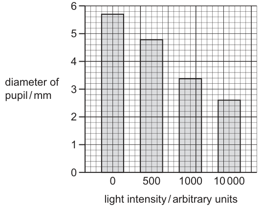
What is the percentage decrease in pupil diameter (rounded to 1 decimal place) when a person moves from an area of complete darkness to an area of light intensity arbitrary units?
Options
A 45.6%
B 53.4%
C 54.3%
D 54.4%
Working
From the bar chart:
- Pupil diameter at 0 arbitrary units (complete darkness) =
- Pupil diameter at arbitrary units =
Calculate the decrease in diameter:
Calculate the percentage decrease:
Rounding to 1 decimal place gives .
Answer
D
D
Walkthrough
The question asks for the percentage decrease in pupil diameter when a person moves from complete darkness to a light intensity of arbitrary units. First, read the pupil diameter values from the bar chart at the two specified light intensities. At 0 arbitrary units (complete darkness), the bar reaches . At arbitrary units, the bar reaches .
Next, find the absolute decrease in diameter by subtracting the final value from the initial value: . Then, apply the percentage decrease formula, dividing the decrease by the original (initial) value and multiplying by 100: . Finally, round this result to 1 decimal place as instructed, giving . This matches option D.
Key Takeaways
- Graph reading: always extract values directly from the axes or grid lines of the provided chart.
- Percentage change: remember to divide the change by the original value, not the new value. The formula is .
- Rounding: follow the rounding instruction precisely. rounds up to , not down to .
Common Mistakes
- Calculating percentage remaining instead of percentage decrease: , which is option A. The question asks for the decrease, so you must subtract from 100% or use the decrease value in the numerator.
- Incorrect rounding: truncating to (option C) instead of rounding to the nearest tenth. Since the hundredths digit is 8, the tenths digit rounds up.
- Using the wrong base value: dividing by the new value () or the difference () instead of the original value ().
Things to Be Careful About
- Read the graph carefully: the y-axis is pupil diameter in mm, and the x-axis is light intensity in arbitrary units. Ensure you are reading the correct bar for and not misreading the grid lines (each small square on the y-axis represents ).
- Pay attention to the command word and rounding instruction: "rounded to 1 decimal place" means you must look at the second decimal digit (8) to decide whether to round up or down.
When a person is frightened, which substance causes an increase in the blood sugar level?
Options
A adrenaline
B carbon dioxide
C insulin
D lactic acid
Working
When a person is frightened, the body prepares for action. The adrenal glands release adrenaline, which causes the liver to convert glycogen to glucose, so the blood sugar level rises. Insulin lowers blood sugar, carbon dioxide is a waste gas and lactic acid is a product of anaerobic respiration, so they do not raise blood sugar.
Answer
A
A
Walkthrough
The question asks which substance raises blood sugar when a person is frightened. Fear is a stress that triggers the body's 'fight or flight' response. The adrenal glands release the hormone adrenaline into the blood. Adrenaline prepares the body for action by increasing heart rate and blood pressure, and it also makes the liver break down stored glycogen into glucose. This glucose is released into the blood, so the blood sugar level rises, giving the muscles more fuel.
Now look at the other options. Insulin (C) does the opposite: it lowers blood sugar by helping glucose enter cells and be stored as glycogen. Carbon dioxide (B) is a waste product of respiration and has no direct role in raising blood sugar. Lactic acid (D) is produced during anaerobic respiration in muscles, especially during vigorous exercise, and is not involved in increasing blood sugar. Therefore the correct answer is A, adrenaline.
Key Takeaways
- Adrenaline is a hormone released by the adrenal glands during stress or fright.
- Adrenaline raises blood glucose by stimulating the conversion of glycogen to glucose in the liver.
- Insulin lowers blood glucose, so it is the opposite of adrenaline in blood glucose control.
- The 'fight or flight' response supplies extra glucose to muscles for rapid action.
Common Mistakes
- Choosing insulin because it is associated with blood sugar. Insulin actually lowers blood sugar, not raises it.
- Thinking carbon dioxide or lactic acid could raise blood sugar. These are not hormones and do not control blood glucose.
- Confusing adrenaline with other hormones. Adrenaline is specifically linked to stress and the 'fight or flight' response.
Things to Be Careful About
- Use the exact term 'adrenaline' as in the mark scheme.
- Remember that adrenaline increases blood sugar, while insulin decreases it.
- The question asks for the substance that causes an increase, so focus on the effect on blood sugar, not on other effects of adrenaline such as heart rate.
Raynaud’s syndrome is a condition that can be triggered by stress. It causes the hands and feet to react as if they were affected by extreme cold.
Which reaction will take place in the hands and feet of a person with Raynaud’s syndrome?
Options
A vasoconstriction – blood vessels at the skin surface narrow, reducing blood flow
B vasoconstriction – blood vessels at the skin surface widen, increasing blood flow
C vasodilation – blood vessels at the skin surface narrow, reducing blood flow
D vasodilation – blood vessels at the skin surface widen, increasing blood flow
Working
Raynaud's syndrome makes the hands and feet behave as if they were in extreme cold. In cold conditions the body conserves heat by vasoconstriction: the blood vessels at the skin surface narrow, reducing blood flow to the skin.
- A is correct: vasoconstriction, vessels narrow, blood flow reduced.
- B is wrong: vasoconstriction narrows vessels, it does not widen them.
- C and D are wrong: the reaction is vasoconstriction, not vasodilation.
Answer
A
A
Walkthrough
The question gives you a condition — Raynaud's syndrome — and tells you it makes the hands and feet react as if they were affected by extreme cold. So the task is really: what does the body do to the skin's blood vessels when it is very cold?
When the body is cold, it needs to conserve heat. It does this by vasoconstriction: the muscles in the walls of the blood vessels near the skin surface contract, making the vessels narrower. Narrower vessels carry less blood, so less warm blood reaches the skin surface and less heat is lost to the surroundings. This is the opposite of vasodilation, where the vessels widen, more blood flows near the skin, and more heat is lost.
Because Raynaud's syndrome triggers the same response as extreme cold, the hands and feet undergo vasoconstriction — the vessels narrow and blood flow is reduced. That matches option A exactly.
Option B mixes the two ideas wrongly: it says vasoconstriction but then claims the vessels widen and blood flow increases, which is the opposite of what vasoconstriction means. Options C and D both name vasodilation, which is the warm-body response, not the cold-body response.
Key Takeaways
- Vasoconstriction = blood vessels at the skin surface narrow, reducing blood flow — this conserves heat and happens when the body is cold.
- Vasodilation = blood vessels at the skin surface widen, increasing blood flow — this releases heat and happens when the body is hot.
- The body controls its core temperature by adjusting the diameter of the blood vessels near the skin surface, a part of homeostasis.
Common Mistakes
- Confusing vasoconstriction with vasodilation — remember that "constriction" means narrowing, "dilation" means widening.
- Matching vasoconstriction with "widen, increasing blood flow". Vasoconstriction always means narrower vessels and reduced blood flow; vasodilation always means wider vessels and increased blood flow.
- Forgetting that the question is really asking about the body's response to cold, and instead choosing a "stress" response such as increased blood flow.
Things to Be Careful About
- Read both halves of each option: the term (vasoconstriction/vasodilation) AND the description (narrow/widen, reduce/increase blood flow). Both halves must be correct for the option to score.
- The mark scheme requires the exact terms "vasoconstriction" and "narrow"/"reducing blood flow" — a vague answer such as "blood vessels get smaller" would not be credited on a written paper, so use the precise wording.
Which flow diagram describes how auxin affects growth in negative gravitropism?
Options
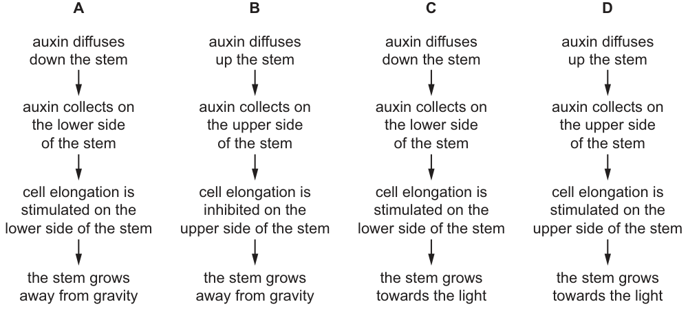
Working
In negative gravitropism a shoot grows upwards, away from gravity. Auxin is made in the shoot tip and diffuses down the stem; gravity causes it to accumulate on the lower side. Auxin stimulates cell elongation in shoots, so the lower side elongates more than the upper side, and the shoot bends and grows away from gravity.
- B is wrong: auxin diffuses down the stem, not up, and collects on the lower side.
- C is wrong: the final outcome is growth away from gravity, not towards the light (that is phototropism).
- D is wrong: auxin moves down and collects on the lower side, and the outcome is not towards the light.
Answer
A
A
Walkthrough
The question asks for the flow diagram of negative gravitropism — the growth of a shoot upwards, against the pull of gravity. Work through the mechanism step by step:
- Auxin is produced in the shoot tip and diffuses down the stem. This immediately rules out B and D, which say auxin diffuses up the stem.
- Gravity causes auxin to settle on the lower side of the shoot. So auxin collects on the lower side — this confirms A and C and eliminates B and D again (they place auxin on the upper side).
- Auxin stimulates cell elongation in shoots. Unlike in roots, where high auxin inhibits growth, in a shoot more auxin means more elongation. So the lower side elongates more.
- Because the lower side grows faster, the shoot bends upwards — it grows away from gravity. This is the definition of negative gravitropism. Option C ends with 'grows towards the light', which describes phototropism, a different response to a different stimulus.
Only option A has all four steps correct: auxin diffuses down → collects on the lower side → stimulates elongation on the lower side → stem grows away from gravity.
Key Takeaways
- Auxin is made in the shoot tip and moves down the stem; gravity makes it accumulate on the shaded/lower side in a horizontal shoot.
- Auxin stimulates cell elongation in shoots (but inhibits it in roots) — this asymmetry explains both positive gravitropism in roots and negative gravitropism in shoots.
- Negative gravitropism = growth away from gravity (shoots); positive gravitropism = growth towards gravity (roots). Phototropism is growth towards light and must not be confused with it.
Common Mistakes
- Choosing C: the first three steps are correct, but the final outcome 'towards the light' describes phototropism, not negative gravitropism. Always check the stimulus named in the question.
- Choosing B or D: auxin does not diffuse up the stem, and it collects on the lower (not upper) side of a horizontal shoot.
- Reversing the effect of auxin: in shoots auxin stimulates elongation; saying it inhibits elongation on the lower side gives the wrong bending direction.
Things to Be Careful About
- Read the last box of each flow diagram carefully — the question tests whether you know the stimulus (gravity) and the direction of growth (away from it).
- Remember the shoot-specific rule: auxin stimulates elongation in shoots. The root behaves oppositely, so do not transfer root logic here.
- 'Negative gravitropism' means the response is negative with respect to the gravity vector — i.e. growth upwards, away from gravity.
The diagram shows some stages during the asexual reproduction of a single-celled organism.
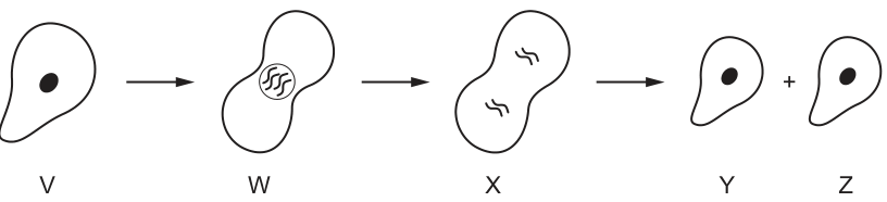
Which row shows the relative amounts of DNA in each of the cells V, W, X, Y and Z?
Options
| V | W | X | Y | Z | |
|---|---|---|---|---|---|
| A | 1 | 1 | 1 | 2 | 2 |
| B | 1 | 1 | 2 | 1 | 1 |
| C | 1 | 2 | 2 | 1 | 1 |
| D | 2 | 1 | 1 | 2 | 2 |
Working
- Stage V: parent cell with unreplicated DNA. Relative DNA = 1.
- Stage W: DNA has replicated (chromosomes are condensing). Relative DNA = 2.
- Stage X: cell is dividing but all replicated DNA is still within the cell. Relative DNA = 2.
- Stages Y and Z: daughter cells, each with unreplicated DNA. Relative DNA = 1.
The sequence of relative DNA amounts is 1, 2, 2, 1, 1. This matches row C.
Answer
C
C
Walkthrough
The diagram illustrates asexual reproduction (binary fission or mitosis) in a single-celled organism. To find the relative amounts of DNA at each stage, we track what happens to the genetic material:
- Stage V is the parent cell before division begins. Its DNA is in its normal, unreplicated state. We assign this a relative amount of 1.
- Stage W shows the cell after DNA replication has occurred (visible as condensing chromosomes). Because the DNA has doubled in preparation for division, the relative amount is now 2.
- Stage X shows the cell actively dividing. Although the nucleus is splitting, the cell as a whole still contains all the replicated DNA. The relative amount remains 2.
- Stages Y and Z are the two identical daughter cells formed after division is complete. Each daughter cell receives one complete set of unreplicated DNA. The relative amount in each is 1.
Putting it together, the sequence of relative DNA amounts for V, W, X, Y, and Z is 1, 2, 2, 1, 1. Checking the table, this corresponds exactly to row C.
Key Takeaways
- DNA replication occurs before cell division, temporarily doubling the DNA content of the cell.
- The cell's total DNA content remains doubled (relative amount 2) until the cytoplasm and nucleus actually divide into two separate daughter cells.
- Daughter cells return to the normal, unreplicated DNA state (relative amount 1) immediately after division.
Common Mistakes
- Assuming W has DNA = 1: Forgetting that DNA must replicate before a cell can divide, meaning the DNA content doubles during the preparation stage.
- Assuming X has DNA = 1: Thinking that because the cell is splitting, the DNA has already halved. In reality, the cell does not lose DNA until the division is complete and two separate cells exist.
- Confusing the stages: Misreading the diagram and thinking Y and Z have doubled DNA, or that V has doubled DNA.
Things to Be Careful About
- The question asks for relative amounts, not absolute quantities. Use a simple baseline (1) for the normal unreplicated state.
- Ensure you track the DNA through the entire sequence: replication (V to W), division (W to X), and separation into daughter cells (X to Y and Z). Missing one step will give the wrong sequence.
- In multiple-choice questions like this, eliminating options by checking just the first or last stage can quickly narrow down the answer (e.g., V must be 1 and Y/Z must be 1, which eliminates D immediately).
Asexual reproduction involves the cell cycle in which the cell divides and then growth and DNA synthesis take place before the cell is ready to divide again.
The diagram shows the stages in one complete cell cycle.
This cell cycle takes 1.75 hours to complete.
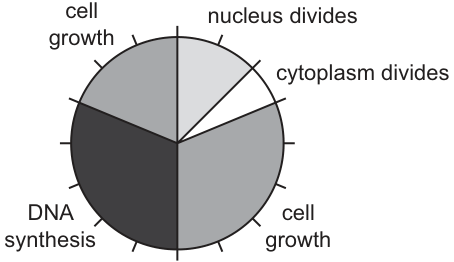
Using the diagram, which activity in the cycle takes 52.5 minutes in total?
Options
A cell growth
B cytoplasm divides
C nucleus divides
D DNA synthesis
Working
Total time for one cell cycle = 1.75 hours.
Convert to minutes: minutes.
The pie chart is divided into 10 equal sectors.
Time taken per sector = minutes.
We need to find the activity that takes 52.5 minutes.
Number of sectors required = sectors.
From the diagram:
- nucleus divides = 1 sector
- cytoplasm divides = 1 sector
- DNA synthesis = 3 sectors
- cell growth = 3 sectors (G1) + 2 sectors (G2) = 5 sectors.
Total cell growth takes 5 sectors, which is minutes.
Answer
A
A
Walkthrough
The question asks which activity in the cell cycle takes 52.5 minutes, given that the entire cycle takes 1.75 hours and is represented by a pie chart divided into 10 equal sectors.
First, convert the total time from hours to minutes to match the target unit:
Next, determine the time represented by each sector on the pie chart. Since there are 10 equal sectors:
Then, calculate how many sectors correspond to the target time of 52.5 minutes:
Finally, count the sectors for each activity in the diagram:
- nucleus divides: 1 sector
- cytoplasm divides: 1 sector
- DNA synthesis: 3 sectors
- cell growth: 3 sectors (G1 phase) + 2 sectors (G2 phase) = 5 sectors
Since cell growth occupies 5 sectors, it takes minutes. The correct option is A.
Key Takeaways
- Always convert units to be consistent before calculating (hours to minutes).
- When interpreting a pie chart divided into equal sectors, calculate the value of one sector first.
- Sum related sectors if an activity is split across multiple phases (e.g., cell growth in G1 and G2 phases of the cell cycle).
Common Mistakes
- Forgetting to convert hours to minutes, leading to an incorrect time per sector and a wrong final answer.
- Only counting one of the cell growth phases (G1 or G2) instead of both, resulting in 3 sectors instead of the required 5.
- Miscounting the total number of sectors on the pie chart.
Things to Be Careful About
- Ensure all time units are in the same format (minutes) before dividing.
- Read the diagram carefully: cell growth is split into two distinct phases (G1 and G2) on the pie chart, and both must be added together to get the total time for cell growth.
- Verify the arithmetic: must equal exactly 5, confirming the 5 sectors of cell growth.
Which diagram matches each type of flower with its features?
Options
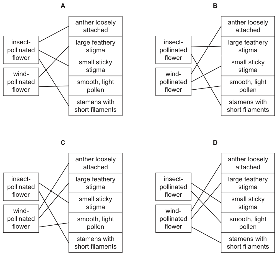
Working
Insect-pollinated flowers have adaptations for insects to carry their pollen: a small sticky stigma to trap pollen from insects, and stamens with short filaments so the anthers are inside the flower for insects to brush against.
Wind-pollinated flowers have adaptations for wind dispersal of pollen: anthers loosely attached so pollen is easily released into the air, a large feathery stigma to catch airborne pollen, and smooth, light pollen to be carried by the wind.
Diagram C correctly matches insect-pollinated flowers to 'small sticky stigma' and 'stamens with short filaments', and wind-pollinated flowers to 'anther loosely attached', 'large feathery stigma', and 'smooth, light pollen'.
Answer
C
C
Walkthrough
The question asks to match structural features to the correct type of flower based on their pollination mechanism. We evaluate each feature against the two flower types:
- Insect-pollinated flowers rely on insects to transfer pollen. To ensure the insect contacts the pollen, the anthers are positioned inside the flower, which requires stamens with short filaments. To ensure the insect picks up or deposits pollen, the stigma must be small and sticky.
- Wind-pollinated flowers rely on air currents. To release pollen into the wind, the anthers are loosely attached so they break open easily. To catch pollen from the air, the stigma is large and feathery. To travel long distances in the wind, the pollen is smooth and light.
Looking at the diagrams, only Diagram C correctly connects insect-pollinated flowers to 'small sticky stigma' and 'stamens with short filaments', and wind-pollinated flowers to 'anther loosely attached', 'large feathery stigma', and 'smooth, light pollen'.
Key Takeaways
- Insect-pollinated flowers adapt to insects with a small, sticky stigma and short filaments keeping the anthers inside the flower.
- Wind-pollinated flowers adapt to the wind with a large, feathery stigma, loosely attached anthers, and smooth, light pollen.
Common Mistakes
- Confusing the stigma adaptations: stating that wind-pollinated flowers have a sticky stigma or that insect-pollinated flowers have a feathery stigma.
- Confusing the pollen adaptations: stating that insect-pollinated flowers have smooth, light pollen.
- Confusing the filament length: stating that wind-pollinated flowers have short filaments (they actually have long filaments to expose the anthers to the air).
Things to Be Careful About
- Ensure every feature is matched correctly. Diagram C is the only option with all five connections correct. Diagrams A, B, and D contain at least one incorrect line (e.g., connecting 'large feathery stigma' to an insect-pollinated flower).
What is the sequence of organs that a sperm cell must pass through so it can fertilise an egg cell?
Options
A sperm duct → urethra → vagina → cervix → uterus → oviduct
B sperm duct → vagina → urethra → oviduct → cervix → uterus
C uterus → sperm duct → vagina → urethra → cervix → oviduct
D urethra → sperm duct → uterus → vagina → oviduct → cervix
Working
A sperm cell is produced in the testes and travels through the sperm duct, then the urethra, and is released into the vagina during ejaculation. Inside the female, the sperm must pass through the cervix, then the uterus, and finally reach the oviduct where fertilisation occurs.
Option A gives the correct sequence: sperm duct → urethra → vagina → cervix → uterus → oviduct.
- B is wrong because it places the urethra after the vagina and puts the oviduct before the cervix and uterus.
- C is wrong because it begins with the uterus, a female organ, before the male organs.
- D is wrong because it puts the urethra before the sperm duct and scrambles the female order.
Answer
A
A
Walkthrough
This question tests your knowledge of the pathway a sperm cell takes from production in the male to fertilisation in the female. Let's trace it step by step.
In the male:
- Sperm are produced in the testes.
- They pass through the sperm duct (also called the vas deferens).
- From the sperm duct, they enter the urethra, the tube that runs through the penis.
- During ejaculation, sperm are released from the penis into the vagina of the female.
In the female:
- The sperm enter the vagina.
- They must pass through the cervix, the narrow opening at the lower end of the uterus.
- Then they enter the uterus (womb).
- Finally, they reach the oviduct (Fallopian tube), where fertilisation with the egg cell occurs.
So the full sequence is: sperm duct → urethra → vagina → cervix → uterus → oviduct. This matches option A.
Now let's look at why the other options are wrong:
- B (sperm duct → vagina → urethra → oviduct → cervix → uterus): This puts the urethra after the vagina, but the urethra is in the male and comes before the vagina in the female. It also puts the oviduct before the cervix and uterus, which is wrong — the sperm must pass through the cervix and uterus before reaching the oviduct.
- C (uterus → sperm duct → vagina → urethra → cervix → oviduct): This starts with the uterus, a female organ, before any male organs. The sperm cannot start in the uterus.
- D (urethra → sperm duct → uterus → vagina → oviduct → cervix): This puts the urethra before the sperm duct (wrong order in the male) and scrambles the female order (uterus before vagina, oviduct before cervix).
Key Takeaways
- The pathway of sperm: testes → sperm duct → urethra → (out of penis) → vagina → cervix → uterus → oviduct.
- Fertilisation occurs in the oviduct (Fallopian tube), so the oviduct is always the last organ in the sequence.
- The male reproductive tract (sperm duct, urethra) comes first, then the female reproductive tract (vagina, cervix, uterus, oviduct).
Common Mistakes
- Confusing the order of the cervix and uterus: the cervix is the lower part of the uterus, so sperm pass through the cervix first, then the uterus.
- Placing the oviduct before the cervix/uterus — the oviduct is the final destination where fertilisation happens.
- Confusing the sperm duct with the urethra — the sperm duct carries sperm from the testes, while the urethra carries sperm (and urine) out through the penis.
- Forgetting that the urethra is part of the male tract and therefore comes before the vagina in the sequence.
Things to Be Careful About
- The question asks for the sequence of organs the sperm passes through — both male and female organs are included, so you must know both tracts in order.
- The urethra is in the male, so it must come before the vagina.
- The oviduct is where fertilisation occurs, so it is the last organ in the sequence — any option ending elsewhere is wrong.
The diagram shows the changes in the thickness of the uterus lining of a woman during her menstrual cycle.
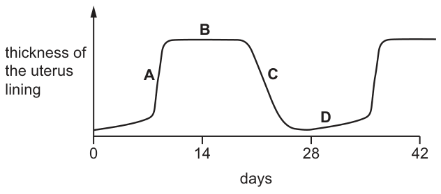
At which time is the woman most likely to be fertile?
Options
A A
B B
C C
D D
Working
Fertility is greatest around ovulation, which occurs about day 14 of the menstrual cycle. At this time the uterus lining is thick and vascular (the plateau on the graph), ready to receive an embryo if fertilisation occurs.
- A — lining still thickening, before the plateau
- B — plateau of maximal thickness around day 14–20, spanning ovulation
- C — lining breaking down (menstruation)
- D — lining at its thinnest after menstruation
Answer
B
B
Walkthrough
The graph shows how the thickness of the uterus (endometrium) lining changes across a 28-day menstrual cycle. The cycle has three phases:
- Days 1–5: menstruation — the lining breaks down and is lost; it is at its thinnest just afterwards (point D).
- Days 6–13: repair/proliferative phase — oestrogen from the developing follicle causes the lining to build up again (point A is during this rise).
- Around day 14: ovulation — an egg is released from the ovary. From day 14 to about day 28, progesterone maintains the lining in its thick, spongy, well-supplied-with-blood capillaries state (point B, the plateau), ready for implantation of an embryo.
- If no fertilisation occurs, progesterone falls, the lining breaks down again (point C) and menstruation begins.
The woman is most likely to be fertile around ovulation (day 14), because that is when an egg is available to be fertilised and the uterus lining is fully prepared to accept an embryo. On the graph, point B sits on the plateau of maximal thickness that spans day 14, so B is the correct answer.
Point A is tempting because the lining is growing rapidly there, but growth alone does not indicate fertility — the key event is ovulation at day 14, which lies within the plateau marked B.
Key Takeaways
- Ovulation occurs at about day 14 of a 28-day menstrual cycle, and this is the most fertile time.
- The uterus lining builds up before ovulation and is maintained thick by progesterone after ovulation, ready for implantation.
- If fertilisation does not occur, progesterone levels fall and the lining breaks down (menstruation).
- Reading a graph means matching each labelled point to a phase of the cycle, not just picking the highest or steepest part.
Common Mistakes
- Choosing A because the lining is increasing fastest — but rapid thickening happens before ovulation; the fertile window is at/after day 14.
- Thinking the woman is most fertile when the lining is thickest at its very maximum late in the plateau — the question asks about likelihood of conception, tied to ovulation at day 14, which point B covers.
- Confusing menstruation (lining breaking down, C) with a fertile time.
- Forgetting that the egg survives only about 1–2 days after ovulation, so fertility is tightly linked to day 14 rather than any other point.
Things to Be Careful About
- Anchor your answer to ovulation at day 14, not simply to 'thick lining' in general — the mark scheme credits the point that spans day 14.
- Read the x-axis carefully: the cycle repeats every 28 days, so check which labelled point actually coincides with day 14 of each cycle.
- In MCQs like this, eliminate options by naming the phase each point represents before choosing — it prevents guessing between A and B.
What is an example of discontinuous variation in humans?
Options
A body weight
B height
C blood group
D skin colour
Working
Discontinuous variation produces distinct categories with no intermediates. Body weight, height and skin colour are continuous traits that show a range of values, so they are continuous variation. Blood group has separate, discrete categories, so it is discontinuous variation.
Answer
C
C
Walkthrough
Variation means the differences between individuals of the same species. It can be divided into two types:
- Continuous variation – there is a complete range of values between two extremes, with many small differences. Examples include height, body weight and skin colour. These features are usually controlled by many genes and are also affected by the environment, such as diet.
- Discontinuous variation – individuals fall into distinct categories with no intermediates. Examples include blood group, sex, and the ability to roll the tongue. These features are usually controlled by one or a small number of genes and are not significantly affected by the environment.
Here, the question asks for an example of discontinuous variation. Blood group is the only option that gives separate categories: a person is blood group A, B, AB or O, with no in-between types. Height, body weight and skin colour all show a continuous range, so they are examples of continuous variation.
Therefore, the correct answer is C.
Key Takeaways
- Continuous variation shows a range of values with no clear categories.
- Discontinuous variation shows distinct, separate categories.
- Human examples of continuous variation: height, body weight, skin colour.
- Human examples of discontinuous variation: blood group, sex, tongue rolling.
- Continuous traits are usually influenced by many genes and the environment; discontinuous traits are usually controlled by one or a few genes.
Common Mistakes
- Choosing A or B: body weight and height are continuous, not discontinuous.
- Choosing D: skin colour shows a continuous range, not separate categories.
- Confusing the two terms: some students think "discontinuous" means "not shown continuously throughout life" rather than "having no intermediates". The key idea is the presence or absence of distinct categories.
Things to Be Careful About
- Read the question carefully: it asks for an example of discontinuous variation, not continuous variation.
- Remember that a single trait can be affected by many genes, but the defining feature of discontinuous variation is the absence of intermediate forms.
- In the exam, use the precise terms "continuous variation" and "discontinuous variation" as required by the mark scheme.
Night-blindness is an inherited condition in which people have unusually poor vision when light levels are low.
The diagram shows the inheritance of night-blindness in three generations of a family.
Couple 4 and 5 are expecting their second child, individual 7.
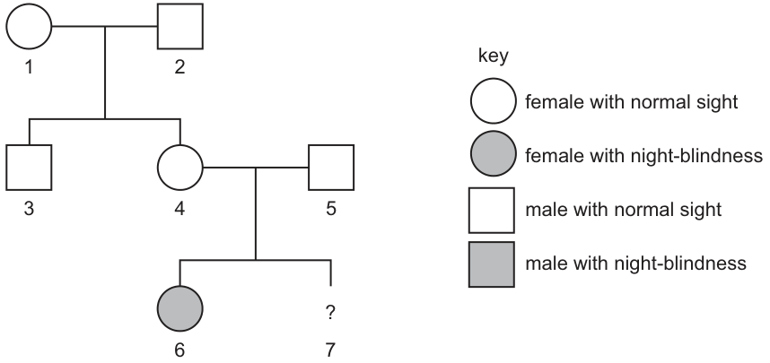
What is the probability that individual 7 will be a male and will also show night-blindness as his phenotype?
Options
A 0.500
B 0.300
C 0.250
D 0.125
Working
- Deduce the inheritance pattern: Individuals 4 and 5 have normal sight, but their daughter (individual 6) has night-blindness. This means the condition is recessive. Because the affected child is female and her father (individual 5) has normal sight, the condition cannot be X-linked recessive (an affected female would require an affected father ). Therefore, night-blindness is autosomal recessive.
- Determine parental genotypes: Let be the dominant normal allele and be the recessive night-blindness allele. Since individuals 4 and 5 are normal but have an affected child (), both must be heterozygous ().
- Probability of night-blindness: A cross between gives a 1/4 () probability of the offspring being (night-blind).
- Probability of being male: The probability of having a male child is 1/2 ().
- Combined probability: The probability of individual 7 being both male and night-blind is .
Answer
D
D
Walkthrough
Step 1: Determine the mode of inheritance from the pedigree.
Look at parents 4 and 5. Both have normal sight, but they have a daughter (individual 6) who is affected (shaded). When unaffected parents produce an affected child, the trait must be recessive. The parents are carriers, hiding the recessive allele.
Next, check if it is X-linked or autosomal. If it were X-linked recessive, an affected female (individual 6) would have the genotype . She must inherit one from her mother and one from her father. This means her father (individual 5) would have to be and therefore affected. But individual 5 is normal (unshaded square). This contradiction rules out X-linked recessive inheritance. Thus, the trait is autosomal recessive.
Step 2: Assign genotypes to the parents.
Let represent the dominant normal allele and represent the recessive night-blindness allele. Because individuals 4 and 5 are phenotypically normal but produced an affected child (), both must carry one copy of the recessive allele. Their genotypes are both heterozygous: .
Step 3: Calculate the probability of the child being night-blind.
Perform a monohybrid cross for :
- (normal sight) = 1/4
- (normal sight) = 2/4
- (night-blindness) = 1/4
The probability of individual 7 inheriting the genotype and showing the night-blind phenotype is or .
Step 4: Calculate the probability of the child being male.
Sex determination in humans is independent of autosomal traits. The probability of any child being male is or .
Step 5: Combine the probabilities.
The question asks for the probability that individual 7 is both male and night-blind. Since these are independent events, multiply their probabilities:
This matches option D.
Key Takeaways
- Pedigree deduction rules: Unaffected parents with an affected child always indicate a recessive trait. An affected female with a normal father always rules out X-linked recessive inheritance, pointing to autosomal recessive.
- Autosomal vs. sex-linked: Real-world night-blindness (congenital stationary night blindness) can be X-linked, but in genetics problems, you must strictly follow the pedigree data rather than prior knowledge. The pedigree here definitively proves autosomal recessive.
- Combined probabilities: When a question asks for two independent conditions (e.g., "male AND affected"), calculate each probability separately and multiply them.
Common Mistakes
- Assuming X-linked inheritance: Students often guess X-linked recessive for vision conditions or because they see a pattern in the pedigree. However, the affected female (6) with a normal father (5) is the classic textbook proof that it cannot be X-linked recessive. If a student assumes X-linked, they might incorrectly conclude the probability is 0 or calculate it wrongly.
- Ignoring the "male" condition: Many students correctly calculate the probability of the child being night-blind () and then select option C (), forgetting to multiply by the probability of the child being male.
- Misreading the pedigree: Confusing the generations or misidentifying which individuals are the parents of individual 7. Individual 7 is the child of 4 and 5, not 1 and 2.
Things to Be Careful About
- Deduce, don't assume: Always deduce the inheritance pattern from the pedigree provided. Do not let real-world biological knowledge override the logic of the diagram. The diagram is the ground truth for the exam.
- Read the question carefully: The phrase "male and will also show night-blindness" is a compound probability question. Break it into two parts: and .
- Genetic notation: When writing out the cross, use clear notation (e.g., and ) to avoid confusion with sex-linked notation (, ). Since it is autosomal, sex chromosomes are irrelevant to the trait's inheritance.
- Decimal precision: The options are given to three decimal places (, , , ). Ensure your final multiplication yields the exact decimal match ().
What is essential for natural selection to occur?
Options
| competition | variation | |
|---|---|---|
| A | ✓ | ✓ |
| B | ✓ | ✗ |
| C | ✗ | ✓ |
| D | ✗ | ✗ |
key
✓ = yes
✗ = no
Working
Natural selection requires individuals in a population to show variation, so some are better adapted than others. It also requires competition for limited resources, so that the better-adapted individuals survive and reproduce more successfully. Both are essential.
Answer
A
A
Walkthrough
Natural selection is the process by which organisms better adapted to their environment tend to survive and produce more offspring. For this to happen, there must first be variation: individuals in a population differ in their characteristics, often because of different alleles. Without variation, all individuals are identical and there is nothing for selection to act on. Second, there must be competition: resources such as food, water, space and mates are limited, so not all individuals can survive and reproduce. When they compete, those with the advantageous variation are more likely to survive and pass on their alleles. Therefore both variation and competition are essential. In the table, option A marks both with ✓, so A is correct.
Key Takeaways
- Natural selection needs variation and competition.
- Variation provides the raw material; competition provides the pressure.
- Advantageous alleles increase in frequency over generations.
Common Mistakes
- Choosing C (variation only): variation alone is not enough; without competition, all individuals could survive and selection would not occur.
- Choosing B (competition only): competition alone cannot select anything if all individuals are identical.
- Confusing natural selection with artificial selection, where a human chooses which organisms breed.
Things to Be Careful About
- Read the key: ✓ means yes and ✗ means no.
- The question asks what is essential, so both conditions must be present.
- Use the exact terms "variation" and "competition" when explaining natural selection.
The gene for human insulin production can be inserted into bacterial DNA to enable the industrial production of insulin.
What is an advantage of using this type of insulin to treat a patient with diabetes?
Options
A Any offspring of the patient will be protected against developing diabetes.
B The patient’s pancreas will start producing insulin when it is needed by the body.
C The insulin gene will be replaced in the patient’s DNA in cells in the pancreas.
D The patient will not suffer any side effects from using insulin produced in animals.
Working
The insulin made by the genetically modified bacteria is human insulin, identical to the insulin produced by a person's own pancreas. When injected, it is recognised as 'self' by the patient's immune system, so it does not trigger the allergic reactions that animal insulin (from pigs or cattle) can cause.
- A is wrong: treating a patient with insulin does not alter their gametes or protect their offspring from inheriting diabetes.
- B is wrong: injected insulin does not repair the pancreas or make it start producing insulin.
- C is wrong: the insulin gene is not inserted into the patient's own DNA; the gene was inserted into the bacteria used to manufacture the insulin.
Answer
D
D
Walkthrough
This question is about the medical use of insulin made by genetically modified bacteria. The gene for human insulin is cut out of human DNA and inserted into the DNA of bacteria. The bacteria then multiply in a fermenter and each one makes human insulin, which is collected and purified for injection.
The key point is that this insulin is human insulin — its amino acid sequence is exactly the same as the insulin a person's own pancreas makes. Before this technology existed, insulin for diabetics came from the pancreas of pigs and cattle. That animal insulin is slightly different in structure from human insulin, and some patients' immune systems reacted against it, causing side effects.
Now look at each option:
- A — Injecting insulin treats the patient; it cannot change the genes in their egg or sperm cells, so it can have no effect on any offspring. Also, Type 1 diabetes is not usually inherited in a simple way, and treatment certainly does not 'protect' offspring.
- B — In Type 1 diabetes the pancreas cells that make insulin have been destroyed. Injecting insulin from outside does nothing to repair those cells or restart insulin production.
- C — The gene was inserted into the bacteria, not into the patient. The patient's pancreatic DNA is untouched by the treatment.
- D — Correct. Because the insulin is identical to human insulin, the patient's immune system does not treat it as foreign, so the allergic side effects seen with animal insulin are avoided.
Key Takeaways
- Genetically modified bacteria can be given a human gene and will then manufacture the human protein, here insulin.
- Human insulin produced this way is chemically identical to the patient's own insulin, so it does not cause the allergic reactions that animal insulin can.
- Insulin is a protein; it is destroyed in the gut if swallowed, so it must be injected.
- Treating a condition with a hormone does not change the patient's DNA or cure the underlying cause.
Common Mistakes
- Choosing B or C: both confuse the treatment with a permanent genetic or functional repair of the pancreas. The gene is inserted into bacteria, never into the patient's cells.
- Choosing A: thinking that a treatment can alter the patient's germ cells and protect offspring. Insulin therapy has no effect on gametes.
Things to Be Careful About
- Read the question carefully: it asks for an advantage of using this type of insulin — i.e. human insulin from GM bacteria — not an advantage of genetic modification in general.
- The correct answer hinges on the word 'human': the insulin is identical to the body's own, avoiding side effects from animal insulin.
- Note that the mark scheme gives only the letter D; no reasoning is required on the paper, but the elimination logic above is what a strong candidate would run through mentally.
Which row identifies the organisms in a food chain?
Options
| producer | herbivore | carnivore | |
|---|---|---|---|
| A | rabbit | cat | dog |
| B | dog | plant | cat |
| C | plant | dog | rabbit |
| D | plant | rabbit | dog |
Working
A producer makes its own food by photosynthesis, so it must be a plant. A herbivore eats plants, so it must be an animal that feeds on the producer. A carnivore eats other animals.
Row D has plant as producer, rabbit as herbivore, and dog as carnivore. This is the only row that matches all three definitions.
Answer
D
D
Walkthrough
A food chain shows the transfer of energy from one organism to another as food. It always starts with a producer, which is an organism that makes its own food by photosynthesis. In the options, the only producers are plants, so the producer column must be "plant". This immediately rules out rows A and B.
Next, a herbivore is an animal that eats plants. Among the remaining rows, rabbits eat plants, but dogs do not usually eat plants as their main food, so the herbivore column should be "rabbit". This rules out row C.
Finally, a carnivore is an animal that eats other animals. Dogs can eat meat and are carnivores in this context, so row D is correct: plant → rabbit → dog.
The food chain can be written as:
plant → rabbit → dog
This shows energy flowing from the producer (plant) to the primary consumer (rabbit) and then to the secondary consumer (dog).
Key Takeaways
- A food chain always begins with a producer, usually a green plant.
- Herbivores eat producers; carnivores eat other animals.
- The order in a food chain matters: producer → herbivore → carnivore.
- Energy is transferred along the food chain when one organism eats another.
Common Mistakes
- Choosing row A: a rabbit is not a producer, and a cat and a dog are both carnivores, so there is no herbivore in the chain.
- Choosing row B: a dog is not a producer, and a plant is not a herbivore.
- Choosing row C: a dog is not a herbivore, and a rabbit is not a carnivore.
- Thinking that any animal can be a producer because it "produces" something. Only organisms that make food by photosynthesis, such as green plants, are producers.
Things to Be Careful About
- Read each column heading carefully: producer, herbivore, carnivore.
- Remember that the same animal can sometimes eat both plants and animals, but in a simple food chain it should be placed according to its main feeding role.
- The question asks for the row that identifies the organisms in a food chain, so all three positions must be correct for the row to score.
Some students set up an experiment to study the decay of leaves in garden soil. They put leaves in bags of different mesh sizes, sealed them and then buried them.
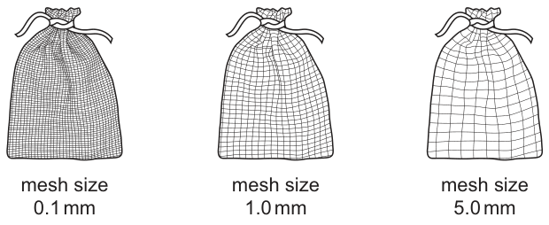
Each month for 5 months the bags were dug up and the total percentage loss in mass from the start of the experiment was calculated.
The results are shown.
| mesh size / | total percentage loss in mass | ||||
|---|---|---|---|---|---|
| month 1 | month 2 | month 3 | month 4 | month 5 | |
| 0.1 | 5 | 11 | 16 | 20 | 31 |
| 1.0 | 13 | 23 | 26 | 42 | 48 |
| 5.0 | 21 | 32 | 36 | 54 | 60 |
What can the students conclude from these results?
Options
A Decay is dependent on access to oxygen for the decomposers.
B The larger the mesh size the faster the rate of decay.
C Decay depends on how much water can get to the leaves.
D Nutrients diffuse away from the leaves more easily when mesh size increases.
Working
The table shows the total percentage loss in mass of leaves over 5 months for three different mesh sizes.
- At month 5, the mass loss for mesh size 0.1 mm is 31%.
- At month 5, the mass loss for mesh size 1.0 mm is 48%.
- At month 5, the mass loss for mesh size 5.0 mm is 60%.
The percentage loss in mass is an indication of the rate of decay (decomposition). The data shows a clear trend: as the mesh size increases, the percentage loss in mass increases. This means decay happens faster when the mesh is larger.
Let us evaluate the options:
- A: Oxygen can diffuse through all mesh sizes (even 0.1 mm is permeable to air/gases in soil). The mesh size is not primarily blocking oxygen. Incorrect.
- B: The data directly supports this. Larger mesh (5.0 mm) has the highest mass loss (60%), meaning the fastest rate of decay. Correct.
- C: Water can pass through all mesh sizes easily (water molecules are much smaller than 0.1 mm). Water access is not the variable being restricted significantly here. Incorrect.
- D: While nutrients might diffuse, the primary cause of mass loss in decay is the consumption of the leaf material by decomposers (respiration releasing CO2 and physical removal). The rate difference is due to access by organisms (like earthworms and insects in the larger mesh), not just diffusion of nutrients. Incorrect.
Answer
B
B
Walkthrough
- Analyze the experimental setup: Students buried bags of leaves with different mesh sizes (0.1 mm, 1.0 mm, 5.0 mm) in soil. The mesh size controls what can enter the bag. A 0.1 mm mesh is fine enough to stop most animals (like earthworms, woodlice, and insects) but allows water, air, and microorganisms (bacteria and fungi) to pass. A 5.0 mm mesh allows animals to enter as well.
- Interpret the data: Look at the "total percentage loss in mass" at the end of the experiment (month 5).
- Mesh 0.1 mm: 31% loss.
- Mesh 1.0 mm: 48% loss.
- Mesh 5.0 mm: 60% loss.
The percentage loss in mass represents how much of the leaf has been decomposed or removed. A higher percentage means decay is happening faster.
- Identify the trend: As the mesh size gets larger (0.1 → 1.0 → 5.0), the percentage loss in mass increases (31 → 48 → 60). Therefore, the rate of decay is faster with larger mesh sizes.
- Evaluate the options:
- Option A (Oxygen): Soil contains air, and oxygen can diffuse through even fine mesh (0.1 mm). The fine mesh does not create an anaerobic environment compared to the large mesh. So, oxygen access is not the main factor being varied here.
- Option B (Rate of decay): The data shows exactly this: larger mesh size corresponds to greater mass loss, which means a faster rate of decay. This is a direct conclusion from the results.
- Option C (Water): Water molecules are tiny and can pass through the 0.1 mm mesh easily. All bags are in the same soil environment, so water availability is roughly the same for all. This is not the conclusion supported by the variable change.
- Option D (Nutrient diffusion): While some soluble nutrients might leach out, the significant mass loss (30-60%) is primarily due to biological decay (decomposers breaking down organic matter and releasing CO2 via respiration) and physical removal by detritivores (animals that eat decaying matter). The difference in rate is due to the access of these larger organisms to the leaves in the larger mesh bags.
Key Takeaways
- Decomposition involves multiple organisms: Decay is not just done by bacteria and fungi (which can enter fine mesh); it is greatly accelerated by detritivores (earthworms, insects) which require larger holes to enter.
- Reading tables: Look for the trend across the independent variable (mesh size) and the dependent variable (mass loss). Here, they are positively correlated.
- Control variables: In this experiment, water and oxygen are available to all bags, so conclusions about them being the limiting factor are incorrect based on this data.
Common Mistakes
- Choosing A or C: Students might think fine mesh blocks air or water. In biology practicals, 0.1 mm mesh (like dialysis tubing or fine netting) is permeable to water and dissolved gases. It specifically blocks macro-organisms.
- Choosing D: Students might focus on "mass loss" and think of leaching. However, in the context of "decay of leaves", mass loss is a proxy for decomposition rate. Diffusion of nutrients alone wouldn't account for the massive 60% loss; biological consumption does.
- Misreading the table: Confusing the rows or columns. Ensure you are comparing the final values (month 5) or the overall trend across the months. In every month, the mass loss follows the order: 5.0 mm > 1.0 mm > 0.1 mm.
Things to Be Careful About
- Conclusion vs. Mechanism: The question asks what can be concluded from the results. Option B is a direct statement of the trend observed in the data. Options A, C, and D attempt to explain the mechanism but are either unsupported by the variable (mesh size doesn't block water/oxygen significantly) or incorrect (diffusion isn't the main cause of mass loss here). Always stick to what the data directly shows first.
- Mesh size purpose: Remember that in decay experiments, mesh size is used to exclude animals (detritivores) while allowing microbes and abiotic factors (water, air) to pass. Fine mesh = microbes only (slow decay). Large mesh = microbes + animals (fast decay).
Populations of animals are affected by disease, numbers of predators and the supply of food.
Which row would lead to the most growth in population size?
Options
| disease | number of predators | supply of food | |
|---|---|---|---|
| A | decrease | increase | increase |
| B | decrease | decrease | increase |
| C | increase | decrease | decrease |
| D | increase | increase | decrease |
Working
Population size grows most when the factors that reduce it are decreased and the factors that increase it are increased.
- Disease decreases population size, so fewer deaths happen when disease decreases.
- Predators decrease population size, so fewer deaths happen when the number of predators decreases.
- Food supply increases population size, so more births and survival happen when the food supply increases.
Option B has all three favourable changes: disease decrease, predator decrease, food supply increase.
Answer
B
B
Walkthrough
This question asks which combination of changes would cause the greatest growth in an animal population. Three factors are listed: disease, predators, and food supply.
Disease kills animals, so if disease decreases, fewer animals die and the population can grow. Predators kill animals too, so if the number of predators decreases, fewer animals are eaten and the population can grow. Food supply is needed for survival and reproduction, so if the food supply increases, more animals survive and can produce more offspring.
Look at each row:
- Row A: disease decreases (good), predators increase (bad), food increases (good). The increase in predators would reduce the population, so this is not the best.
- Row B: disease decreases (good), predators decrease (good), food increases (good). All three changes favour population growth.
- Row C: disease increases (bad), predators decrease (good), food decreases (bad). Two factors harm the population.
- Row D: disease increases (bad), predators increase (bad), food decreases (bad). All three changes harm the population.
So row B is the only one where every factor works in favour of population growth.
Key Takeaways
- Population size is controlled by factors that cause death, such as disease and predation, and factors that support survival and reproduction, such as food supply.
- To increase population size, death-causing factors should decrease and resources should increase.
- When comparing options, check each factor separately rather than guessing from the overall pattern.
Common Mistakes
- Choosing row A because disease decreases and food increases, while forgetting that an increase in predators would reduce the population.
- Confusing a decrease in predators with a decrease in food: predators reduce population size, so fewer predators help the population grow.
- Thinking that an increase in disease could help population growth; it cannot, because disease kills individuals.
Things to Be Careful About
- Read the question carefully: it asks which row leads to the MOST growth, so all three factors must be considered together.
- Match each factor to its effect: disease and predators reduce population; food supply increases population.
- The correct answer is B because it is the only row in which all three changes are favourable.
Which human activity has caused most damage to tropical rainforests?
Options
A burning fossil fuels
B flooding of land
C cutting down trees for industrial use
D searching for plants that can be used in medicine
Working
Cutting down trees for industrial use directly destroys the rainforest habitat on a massive scale. Burning fossil fuels causes pollution and climate change but does not directly cut down the forest; flooding land for dams destroys relatively small areas; searching for medicinal plants is non-destructive.
Answer
C
C
Walkthrough
This is a recall question about human impact on ecosystems. The phrase "most damage to tropical rainforests" points to direct destruction of the habitat. Option C, cutting down trees for industrial use, is the main direct cause of deforestation: timber is harvested and land is cleared on a huge scale. Burning fossil fuels (A) is a major cause of air pollution and global warming, but it does not directly remove rainforest. Flooding land (B), for example when dams are built, can destroy some forest but affects far smaller areas. Searching for plants that can be used in medicine (D) is generally non-destructive and can even encourage conservation. Therefore the correct answer is C.
Key Takeaways
- Deforestation is mainly caused by human activities such as logging and clearing land for agriculture and cattle ranching.
- It is important to distinguish direct causes of habitat destruction from indirect effects such as climate change.
- The syllabus asks about the causes and consequences of deforestation, and this question tests the main cause.
Common Mistakes
- Choosing A because burning fossil fuels is a well-known environmental problem; however, it causes climate change rather than direct rainforest destruction.
- Choosing B because dams flood land; this affects only local areas compared with industrial logging.
- Choosing D because searching for medicinal plants sounds harmless; it is not a major cause of damage.
- The mark scheme accepts only C.
Things to Be Careful About
- Read "most damage" carefully: direct destruction of the habitat is the key idea.
- Do not overthink; this is a one-mark recall question, so the simplest direct answer is correct.
Your score so far
Answer a question to start scoring
Your marks add up here as you work through the paper.