Biology 5090/22 — October/November 2013
Cambridge O-Level · Theory · worked solutions for every part, with the mark scheme
Topics Transport in Flowering Plants · Cell Structure and Organisation · Respiration · Classification · Cell Division and Reproduction · Coordination and Control · +12 more
Fig. 1.1 shows a demonstration related to blood circulation.
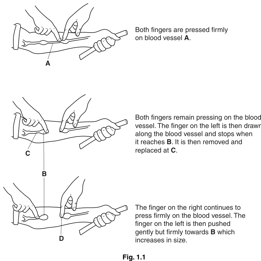
Name the type of blood vessel labelled A in Fig. 1.1.
______
Answer
vein
vein
Walkthrough
The vessel labelled A runs just beneath the skin of the forearm and shows visible swellings along its length. Valves are found only in veins — they are pockets of the inner wall that prevent backflow of blood, since blood in veins travels at low pressure. Arteries have thick muscular walls and no valves, and capillaries are microscopic, so neither could be pressed and emptied in this way. The swellings that appear at B are therefore the semilunar valves of a vein.
Key Takeaways
- Veins are superficial, low-pressure vessels containing valves.
- Valves appear as visible swellings under the skin, especially in the limbs.
Common Mistakes
- Writing "artery" — arteries carry blood at high pressure and have no valves, so they cannot be emptied and refilled by finger pressure.
- Writing "capillary" — capillaries are too small to see or press.
Things to Be Careful About
- The command word is "name", so one word — "vein" — earns the mark; no explanation is needed.
Name the structure leading to the effect shown in this blood vessel at position B.
______
Answer
valve
valve
Walkthrough
Position B is where the vein bulges when blood is pushed towards it. The bulge is caused by a valve: the pocket-like semilunar flaps of the valve fill with blood and balloon outwards, making the vein swell. This is exactly why veins show 'knotted' swellings under the skin.
Key Takeaways
- Valves in veins are semilunar pockets that fill with blood and prevent backflow.
- A swelling at a fixed point in a superficial vein indicates a valve.
Common Mistakes
- Naming the vessel again ("vein") instead of the structure at B.
- Writing "swelling" or "blood clot" — the swelling is the effect; the question asks for the structure causing it.
Things to Be Careful About
- "Name the structure" demands the biological name — "valve" — not a description of the effect.
In the space below, draw a longitudinal section (cut along its length) through the blood vessel as it appears at position B.
Answer
Longitudinal section drawing of the vein at B showing a valve with its two flaps touching and the vessel bulging on the correct side of the valve
Walkthrough
The question asks what the vein looks like in longitudinal section at B, where the blood has been pushed up against the valve. The valve consists of two semilunar pockets projecting from the walls into the lumen. When blood is pushed towards the valve, the pockets fill with blood and their free edges meet in the middle — the flaps touch — so blood cannot pass. Because the blood is trapped on the side the blood came from, the vein is widest (bulges) on that side of the valve, and the walls must be drawn continuing on both sides of the valve.
Marks: (1) a valve shown with its flaps touching; (1) the vessel widest at the correct side of the valve + walls shown both before and after the valve.
Key Takeaways
- A vein valve is two semilunar flaps whose free edges meet to block the lumen.
- In section, blood collects on the upstream side, so the vein bulges there.
Common Mistakes
- Drawing the flaps apart or as a solid plug — the mark requires the flaps touching.
- Drawing the bulge on the wrong side of the valve.
- Shading or sketchy lines — biological drawings must be clean continuous outlines.
- Omitting the vessel walls beyond the valve.
Things to Be Careful About
- Only two marks are available, so the drawing must show exactly the two credited features: touching flaps, and the bulge on the correct side with continuous walls.
Suggest why, at the end of the demonstration, the blood vessel is no longer visible between positions B and D.
Answer
- The vein is empty / contains no blood between B and D, because the blood has been pushed out of this section.
- Blood cannot flow back into it, because the valve at B prevents backflow.
- The finger pressing at D prevents blood from flowing in from that side.
- The walls of a vein are thin, so when it is empty it is not visible beneath the skin.
The vein is empty because the blood has been pushed out, the valve at B stops blood flowing back, the finger at D stops blood entering, and the empty thin-walled vein is not visible under the skin
Walkthrough
During the demonstration, the fingers milk the blood along the vein towards B. By the end, the section between B and D has been squeezed empty. Four separate ideas earn the four marks:
- The vein between B and D is empty — there is no blood in it, and it is the blood inside a superficial vein that makes it visible as a blue line under the skin.
- The blood has been physically pushed out by the fingers — this explains how it became empty.
- Blood cannot flow back in from B, because the valve there has its flaps pressed shut by any backward flow — valves only allow one-way flow towards the heart.
- The finger still pressing firmly at D blocks blood from re-entering from that side.
The final touch: veins have thin walls with little muscle, so an empty vein collapses flat and cannot be seen through the skin.
Key Takeaways
- A superficial vein is visible because of the blood inside it, not the wall itself.
- Vein valves allow flow in one direction only; backflow closes them.
- Veins have thin walls, so they collapse when empty.
Common Mistakes
- Saying the vein "disappeared" without explaining that it is the blood that makes it visible.
- Omitting the role of the valve in preventing backflow — this is the key link.
- Forgetting the finger at D, which blocks the other route for blood to return.
- Claiming the vein was cut or damaged — nothing in the demonstration injures it.
Things to Be Careful About
- This is a four-mark "suggest why": give four distinct linked points, each with its reason, not one idea repeated.
Suggest why a rod was repeatedly gripped tightly before carrying out this demonstration.
Answer
- Gripping the rod tightly contracts the forearm muscles.
- This increases blood pressure and blood flow / circulation, helping to fill the vein with blood so it is clearly visible.
Gripping the rod contracts the muscles, increasing blood pressure and blood flow so the vein fills with blood
Walkthrough
The demonstration needs the vein to be full of blood and easy to see. Squeezing a rod repeatedly is a form of exercise: the muscles of the forearm contract repeatedly. Muscle contraction squeezes the blood vessels and pushes more blood through the tissues, raising blood pressure and increasing the circulation in the arm. A fuller, higher-pressure vein stands out under the skin, making the demonstration easier to perform and see. (Muscle contraction also helps pump blood along the veins — the muscle pump — which suits the vein-filling purpose here.)
Key Takeaways
- Muscle contraction increases blood pressure and blood flow through the active tissue.
- Exercise makes superficial veins more prominent.
Common Mistakes
- Saying it "warms up the arm" without linking muscle contraction to blood flow.
- Saying it makes the vein "stronger" — no structural change occurs.
- Giving only one of the two credited halves: the mark needs muscle contraction AND the increase in blood pressure/flow.
Things to Be Careful About
- Both halves of each point are needed: the action (muscle contraction) and its effect (increased blood pressure/flow, filling the vein).
The rest of this paper
8 more questions- Q2Cell Structure and Organisation · Enzymes · Human Nutrition · Respiration · Biotechnology and Genetic Modification · Classification · Cell Division and Reproduction11M
- Q3Cell Structure and Organisation · Transport in Flowering Plants · Classification10M
- Q4Plant Nutrition · Respiration · Transport in Flowering Plants · Movement Into and Out of Cells11M
- Q5Organisms and Their Environment · Inheritance8M
- Q6Transport in Flowering Plants · Coordination and Control10M
- Q7Excretion · Coordination and Control10M
- Q8Sexual Reproduction in Plants · Sexual Reproduction in Humans · Cell Division and Reproduction10M
- Q9Disease and Immunity10M