Biology 5090/21 — October/November 2013
Cambridge O-Level · Theory · worked solutions for every part, with the mark scheme
Topics Organisms and Their Environment · Transport in Flowering Plants · Plant Nutrition · Coordination and Control · [Legacy] Support, Movement and Locomotion · Human Nutrition · +7 more
Fig. 1.1(a) and Fig. 1.1(b) show a section through the skin of a person at two different body temperatures.
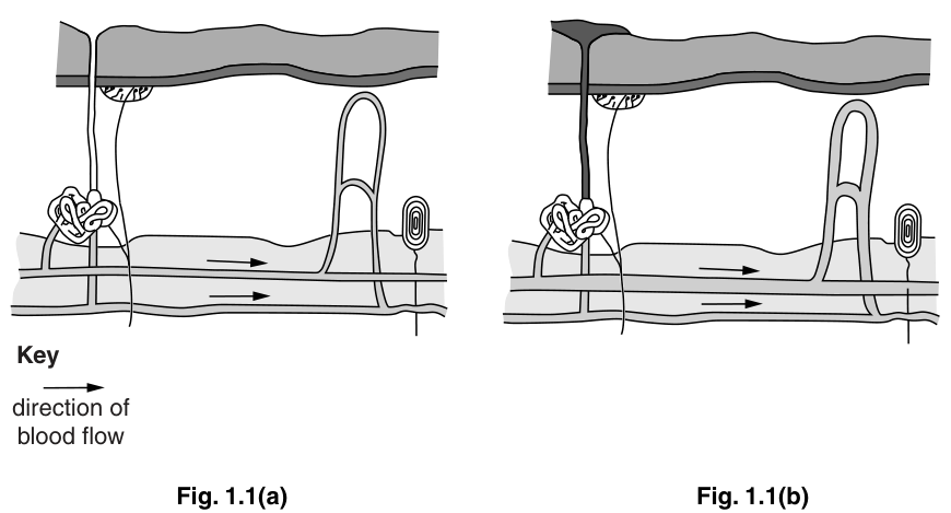
On Fig. 1.1(a), identify and label each of the following.
a sweat gland
Answer
Label the coiled glandular structure in the lower part of the dermis with a straight line touching the coil, and write 'sweat gland' next to the line.
Label the coiled glandular structure in the dermis as 'sweat gland'.
Walkthrough
The question asks to label a sweat gland on Fig. 1.1(a). A sweat gland consists of a coiled secretory portion located in the deeper dermis and a duct that leads to the surface. In the diagram, the coiled, lobulated structure at the bottom left is the secretory part of the sweat gland. The candidate must draw a straight label line that physically touches this coiled structure and write 'sweat gland'. The mark scheme explicitly states 'label line must touch the sweat gland'; a line pointing near it but not touching will not score.
Key Takeaways
Students must be able to recognise the anatomical layout of the skin: the epidermis (top layer), the dermis (middle layer containing hair follicles, sweat glands, and nerve endings), and the hypodermis (bottom layer). The sweat gland's coiled body is always in the lower dermis, while the duct passes through the epidermis to the surface.
Common Mistakes
- Label line not touching: The mark scheme requires the line to touch the structure. A floating label line scores zero.
- Labeling the duct: Students sometimes label the thin tube going up to the surface. The question asks for the 'sweat gland', which refers to the secretory coil at the base. Labeling the duct may not score depending on strictness, but the coil is the definitive part.
- Confusing with hair follicle: The hair follicle is the sheath around the hair root, distinct from the coiled sweat gland.
Things to Be Careful About
- Ensure the label line is straight and touches the specific structure.
- Write the label clearly outside the diagram if possible, or in a clear space.
- The term is 'sweat gland', not 'sweat duct' or 'perspiratory gland' (though the latter is sometimes accepted, stick to the syllabus term).
a temperature receptor.
Answer
Label the oval structure located in the upper part of the dermis (just below the epidermis/Malpighian layer) with a straight line touching it, and write 'temperature receptor' next to the line.
Label the oval structure in the upper dermis as 'temperature receptor'.
Walkthrough
Fig. 1.1(a) shows a temperature receptor on the right side. In the diagram, this is the oval/encapsulated structure located in the upper dermis, beneath the epidermis. The mark scheme specifies 'label line must touch some part of receptor under Malpighian layer'. The candidate must draw a straight line touching this oval structure and label it 'temperature receptor'.
Key Takeaways
Temperature receptors (thermoreceptors) are nerve endings located in the skin that detect changes in environmental temperature. They are typically found in the upper dermis. In diagrams, they are often shown as encapsulated structures or simple nerve endings.
Common Mistakes
- Labeling the wrong structure: The hair follicle or the blood vessel might be mistaken for the receptor. The receptor is the distinct oval structure on the right side of the diagram.
- Label line not touching: As with (a)(i), the line must touch the structure.
- Wrong name: 'Thermoreceptor' is acceptable, but 'temperature receptor' is the syllabus term used in the question.
Things to Be Careful About
- The Malpighian layer is the living layer of the epidermis. The receptor is just below this, in the dermis.
- Ensure the label is clear and legible.
State what has happened to the body temperature to cause the changes shown in Fig. 1.1(b) compared with Fig. 1.1(a).
______
Answer
The body temperature increases (or rises).
Temperature increases / rises.
Walkthrough
Compare Fig. 1.1(a) and Fig. 1.1(b). In (b), there is sweat being secreted onto the skin surface, and the superficial blood vessels (capillary loops) are dilated (wider). These are physiological responses to an increase in body temperature. The body is trying to lose heat. Therefore, the body temperature must have increased.
Key Takeaways
The skin responds to temperature changes. An increase in temperature triggers sweating and vasodilation to lose heat. A decrease triggers vasoconstriction and reduced sweating to conserve heat.
Common Mistakes
- Saying 'decreases': The changes shown (sweating, dilated vessels) are for heat loss, which happens when temperature rises. If temperature fell, the vessels would constrict and sweating would stop.
- Vague answers: 'It got hotter' is acceptable, but 'temperature increases' is more precise.
Things to Be Careful About
- The mark scheme accepts 'increases' or 'rises'. Rejects 'cooler' or 'decreases'.
- Base the answer on the evidence in the diagrams: sweat and dilated vessels.
State two reasons for the answer you have given in (b)(i).
- ______
- ______
Answer
- Sweat is produced / secreted onto the skin surface.
- Blood vessels (capillaries) in the dermis become wider (dilate), allowing more blood flow to the surface.
- Sweat is produced. 2. Blood vessels (capillaries) dilate / become wider.
Walkthrough
The question asks for two reasons for the answer in (b)(i), i.e., why the body temperature increased or what changes indicate the temperature increased. Actually, re-reading the mark scheme: 'State two reasons for the answer you have given in (b)(i)'. This is slightly ambiguously phrased in the question, but the mark scheme clarifies: it wants the evidence from the diagram that shows the temperature increased. The evidence is:
- Sweat is being produced/secreted (visible as the dark drop on the surface in Fig 1.1(b)).
- The blood vessels (capillaries) are wider/dilated (compare the U-shaped loop in (a) vs (b)).
These are the physiological responses that occur when body temperature rises.
Key Takeaways
When body temperature rises:
- Sweat glands produce sweat. Evaporation of sweat removes heat (latent heat of vaporisation).
- Arterioles dilate, increasing blood flow to superficial capillaries. Heat is lost from the blood to the environment by radiation.
Common Mistakes
- Saying 'veins': The mark scheme rejects 'veins'. The vessels that dilate are arterioles and the capillary loops in the dermis. Students often say 'veins dilate', which is incorrect in this context.
- Not mentioning sweat: One point must be about sweat.
- Not mentioning blood vessels: The other point must be about blood vessels becoming wider/more blood flow.
Things to Be Careful About
- The mark scheme accepts 'wider blood vessels', 'capillaries', or 'more blood'.
- Rejects 'veins'.
- Give exactly two points as requested.
On Fig. 1.1(a), label and name the type of blood vessel that constricts when the body temperature falls.
Answer
Label the upper horizontal blood vessel in the lower dermis (the vessel feeding the capillary loops) with a straight line touching it, and name it arteriole.
Arteriole.
Walkthrough
The question asks for the blood vessel that constricts when body temperature falls. This is the vessel that controls blood flow to the superficial capillaries. In the diagram, there is a horizontal vessel at the bottom with arrows showing blood flow. The upper horizontal vessel feeds into the capillary loops in the dermis. This is an arteriole. Constriction of the arteriole reduces blood flow to the capillaries, reducing heat loss.
The mark scheme specifies 'the upper horizontal blood vessel labelled'. In Fig 1.1(a), there are two horizontal vessels at the bottom. The upper one is the arteriole (carrying blood towards the capillaries), the lower is the venule (carrying blood away). The candidate must label the upper one and name it 'arteriole'.
Key Takeaways
- Arterioles are small branches of arteries that lead to capillaries. They have smooth muscle in their walls that can contract (vasoconstriction) or relax (vasodilation) to control blood flow.
- In the skin, arterioles control the amount of blood reaching the superficial capillaries, regulating heat loss.
Common Mistakes
- Saying 'artery': The mark scheme accepts 'Artery' as an alternative, but 'arteriole' is more precise. However, if a student says 'artery', it might score if the label is correct, but 'arteriole' is the correct term for the vessel controlling capillary flow.
- Labeling the wrong vessel: The lower horizontal vessel is a venule/vein. The upper one is the arteriole.
- Not labeling: The question says 'label and name', so both are required for the 2 marks.
Things to Be Careful About
- The mark scheme says 'the upper horizontal blood vessel labelled'. Ensure the label line touches the correct vessel.
- 'Arteriole' is the precise term. 'Artery' is accepted but less accurate.
- Do not label the capillary loops; label the feeding vessel.
Explain the effects caused by constriction of the blood vessel you have named in (c)(i).
Answer
- Constriction of the arteriole reduces blood flow to the superficial capillaries in the skin.
- Less blood reaches the skin surface, so less heat is carried to the surface and less heat is lost by radiation.
- Less blood flow to sweat glands means less sweat is produced.
- Less sweat means less heat is lost by evaporation (or latent heat of vaporisation).
See working.
Walkthrough
The question asks to explain the effects of constriction of the arteriole named in (c)(i). This is a chain of events:
- Arteriole constriction: The smooth muscle in the arteriole wall contracts, narrowing the lumen.
- Reduced blood flow to capillaries: Less blood is pushed into the superficial capillary loops in the dermis.
- Reduced heat loss by radiation: The capillaries are close to the skin surface. When blood flows through them, heat is lost to the environment by radiation. Less blood flow means less heat is brought to the surface, so less heat is lost. This helps conserve body heat.
- Reduced sweat production: Sweat glands also receive blood supply. Less blood flow to sweat glands means less water and salts are delivered, so less sweat is produced.
- Reduced heat loss by evaporation: Sweat evaporation removes a large amount of heat (latent heat of vaporisation). Less sweat means less heat loss by this mechanism.
The mark scheme credits up to 4 points:
- Less blood (flows);
- To capillaries (reject: capillaries constrict - the capillaries don't constrict, the arteriole does);
- To sweat glands;
- Blood carries heat / less heat carried;
- Less heat lost by skin / radiation;
- Less sweat produced;
- Ref to latent heat of vaporisation.
Key Takeaways
Vasoconstriction of arterioles in the skin is a heat-conserving mechanism:
- Reduces blood flow to superficial capillaries -> less heat lost by radiation.
- Reduces blood flow to sweat glands -> less sweat -> less heat lost by evaporation.
Common Mistakes
- Saying 'capillaries constrict': The mark scheme explicitly rejects this. The arteriole constricts, which reduces flow to the capillaries. The capillaries themselves don't actively constrict in this response.
- Not mentioning heat loss: The explanation must link the reduced blood flow to reduced heat loss (by radiation or evaporation).
- Not mentioning sweat: One of the effects is reduced sweat production, which reduces evaporative heat loss.
- Vague answers: 'It saves heat' is not enough. Must explain how (less blood flow, less radiation, less sweat).
Things to Be Careful About
- The mark scheme accepts 'less heat lost by blood' as an alternative to 'less heat lost by radiation', but 'radiation' is the specific mechanism.
- Must refer to 'latent heat of vaporisation' or 'evaporation' if discussing sweat.
- Give enough points to reach the 4-mark total. The explanation should be a clear chain: constriction -> less blood -> less heat carried to surface -> less radiation; AND less blood to sweat glands -> less sweat -> less evaporation.
The rest of this paper
8 more questions- Q2[Legacy] Support, Movement and Locomotion10M
- Q3Human Nutrition · Transport in Humans12M
- Q4Organisms and Their Environment · Transport in Flowering Plants · Excretion9M
- Q5Plant Nutrition · Enzymes8M
- Q6Transport in Flowering Plants · Plant Nutrition10M
- Q7Movement Into and Out of Cells · Respiration10M
- Q8Cell Division and Reproduction · Sexual Reproduction in Plants10M
- Q9Organisms and Their Environment10M