Biology 5090/62 — May/June 2013
Cambridge O-Level · Alternative to Practical · worked solutions for every part, with the mark scheme
Topics Experimental Contexts · Observations and Measurements · Use of Techniques, Apparatus and Materials · Microscopy and Biological Drawing · Planning Experiments and Investigations · Analysis, Conclusions and Evaluation
Some students investigated the effect of two different concentrations, and , of the same solution on strips of fresh Irish potato tissue.
Four strips of tissue, each measuring thick and long, were cut from a cleaned fresh potato. Two strips had the outer layer of skin left on them as shown in Fig. 1.1a; the other two strips had no skin, as shown in Fig. 1.1b.
The skin of a potato contains dead cells that have waterproof materials deposited in their walls.
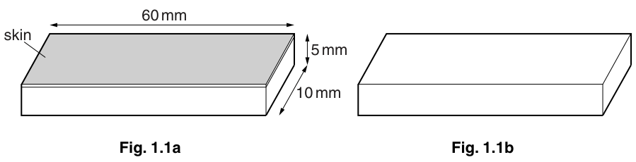
One strip with skin and one without skin were submerged in solution in a shallow dish. The remaining two strips were submerged in solution in a similar way.
The strips were measured at the start, after 10 minutes and after 20 minutes and these lengths were recorded in Table 1.1.
Calculate the change in length of the four potato strips after 20 minutes in the solutions and . Record your answers in Table 1.1.
Table 1.1
| time / mins | length of potato strip / mm in solution A with skin present | length of potato strip / mm in solution A without skin | length of potato strip / mm in solution B with skin present | length of potato strip / mm in solution B without skin |
|---|---|---|---|---|
| 0 [at start] | 60 | 60 | 60 | 60 |
| 10 | 60 | 58 | 60 | 62 |
| 20 | 58 | 56 | 62 | 65 |
| change in length / mm |
In the strips of potato without the skin, there was a change in texture as described in Table 1.2.
Table 1.2
| texture | time / mins | in solution A | in solution B |
|---|---|---|---|
| 20 | soft, flexible | hard, rigid |
Explain fully what had happened to cause the difference in texture between these potato strips.
In the strips of potato with the skin, there was a change in the shape as shown in Table 1.3.
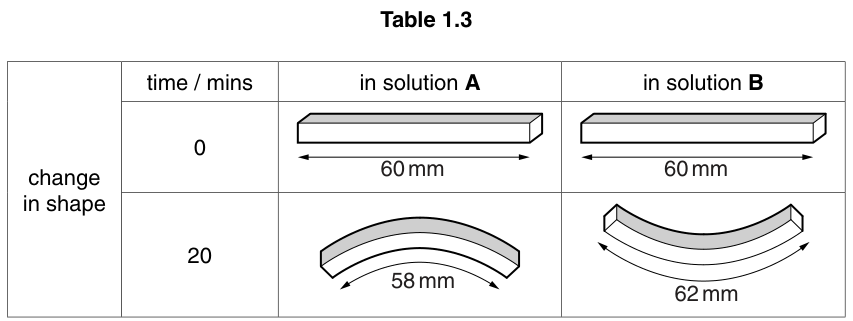
Using the information in Table 1.3, suggest how the presence of the skin has caused the differences in shape.
The rest of this paper
2 more questions- Q2Experimental Contexts · Observations and Measurements · Microscopy and Biological Drawing · Use of Techniques, Apparatus and Materials · Planning Experiments and Investigations · Analysis, Conclusions and Evaluation22M
- Q3Experimental Contexts · Use of Techniques, Apparatus and Materials · Observations and Measurements7M