Biology 5090/22 — May/June 2012
Cambridge O-Level · Theory · worked solutions for every part, with the mark scheme
Topics Human Nutrition · Sexual Reproduction in Plants · Human Gas Exchange · Coordination and Control · Cell Structure and Organisation · Transport in Flowering Plants · +6 more
Fig. 1.1 shows the human eye in horizontal section.
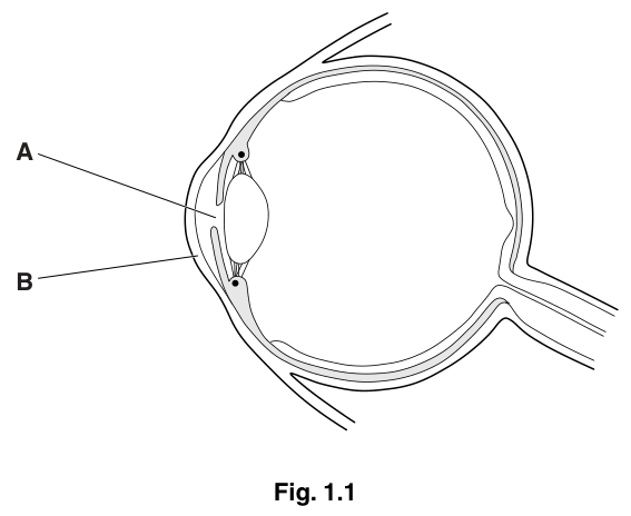
Identify A and B that are labelled on Fig. 1.1.
A ______
B ______
Answer
A pupil
B cornea
A = pupil; B = cornea
Walkthrough
The figure shows a horizontal section through the eye. Label A points to the opening in the centre of the iris, in front of the lens — this is the pupil, the hole through which light enters. Label B points to the transparent, curved layer at the very front of the eye — the cornea, which refracts most of the light entering the eye.
Key Takeaways
- The pupil is the aperture in the middle of the iris; the cornea is the transparent front window of the eye.
- The mark scheme also accepts 'aqueous humour' or 'anterior chamber' for A, because the label line passes through that region.
Common Mistakes
- Naming A as 'iris' — the iris is the coloured ring around the pupil, but the label line points to the central opening, so 'iris' is rejected.
- Confusing the cornea (B) with the sclera; the sclera is the tough white outer coat further back.
Things to Be Careful About
- Give the biological name, not just the letter. 'Pupil' is the safest single answer for A.
Describe what happens to A when light entering the eye becomes less intense.
______
Answer
The pupil dilates (becomes wider / enlarges) to let more light into the eye.
The pupil dilates / gets bigger
Walkthrough
When light intensity falls, the radial muscles of the iris contract and the circular muscles relax, so the pupil widens (dilates). This lets more light reach the retina so that vision is maintained in dim conditions. The mark scheme wants the word 'dilates' or an equivalent such as 'gets bigger', 'enlarges' or 'opens'.
Key Takeaways
- Dim light → pupil dilates (radial muscles contract); bright light → pupil constricts (circular muscles contract). This is the pupil reflex.
Common Mistakes
- Writing 'constricts' or 'gets smaller' — that is the response to bright light, the reverse of what is asked.
- Describing the muscle action when the question only asks what happens to A (the pupil) itself.
Things to Be Careful About
- One mark only — a single clear statement is enough; do not overwrite.
Place a letter Z on Fig. 1.1 where a response occurs as a result of a reflex action.
Answer
Place Z on the iris (or on the ciliary muscle) in Fig. 1.1 — the muscle that carries out the response.
Z placed on the iris or ciliary muscle
Walkthrough
A reflex action ends at an effector — the muscle or gland that carries out the response. In the pupil reflex, the response is the contraction of the muscles of the iris (changing pupil size); the ciliary muscles are also acceptable because they effect accommodation. So Z must be placed on the iris or ciliary muscle in the figure — the small circular structures shown attached to the lens by suspensory ligaments.
Key Takeaways
- In a reflex arc: receptor → sensory neurone → relay neurone → motor neurone → effector. The letter Z goes where the effector is, not where the receptor or the brain is.
Common Mistakes
- Placing Z on the retina (the receptor) or on the optic nerve/brain — the response does not occur there; the retina detects the stimulus, it does not respond.
- Placing Z on the lens — the lens is not a muscle and does not itself carry out the reflex response.
Things to Be Careful About
- The mark scheme accepts 'ciliary muscle / iris / external muscle' — any of these positions scores, but the lens, retina and optic nerve do not.
In some people's eyes, the retina becomes completely detached from the tissues beneath. Explain how this will affect their ability to see.
______
Answer
- The light receptors (rods and cones) of the retina become detached, so they are no longer in contact with the optic nerve.
- Therefore no nerve impulses are sent to the brain.
- No image / picture is formed, so the person cannot see (blindness in that eye).
Receptors detached from optic nerve, so no impulses reach the brain and no image is formed — blindness
Walkthrough
The retina contains the light-sensitive cells — the rods and cones. For sight to work, these receptors must pass their signals to the optic nerve, which carries impulses to the brain where the image is formed. If the retina peels away from the tissues beneath it, the receptors are physically separated from the optic nerve, so the chain is broken: no impulses leave the eye, the brain receives nothing, and no picture is formed — the person is blind in that eye. The mark scheme awards one mark for the receptors, one for the loss of contact with the optic nerve / no impulses, and one for the consequence (no picture / blindness). Note the scheme explicitly rejects 'blurred vision' — detachment causes total loss of vision, not blurring.
Key Takeaways
- Vision requires: light → receptors (rods/cones) in retina → impulses in optic nerve → brain forms image. Break any link and vision fails.
- The retina is the receptor layer; the optic nerve is the output pathway.
Common Mistakes
- Writing 'blurred vision' or 'reduced vision' — the mark scheme explicitly rejects (R) this; detachment causes blindness.
- Saying 'light cannot enter the eye' — light still enters; the problem is that signals cannot reach the brain.
- Naming the receptors vaguely as 'cells' without saying they are light receptors / rods and cones.
Things to Be Careful About
- 'Explain' means every point needs its reason — the detached receptors (cause) → no impulses to brain (mechanism) → no image (consequence). All three links are needed for the three marks.
As people get older, cloudy (opaque) patches sometimes form in the lens of the eye. These are called cataracts.
Suggest how cataracts might affect the ability of the lens to carry out its function.
______
Answer
- The cloudy patches stop light passing through the lens clearly, so the lens fails to focus the light rays properly.
- This produces a blurred image on the retina.
- The lens may also lose its elasticity, so it cannot change shape to accommodate (focus on near and distant objects).
Lens cannot focus light properly, giving a blurred image; loss of elasticity prevents accommodation
Walkthrough
The lens has two jobs: it is transparent, so light passes through it, and it can change shape (accommodate) to focus light from objects at different distances onto the retina. A cataract is an opaque patch, so it blocks, scatters and absorbs light rays — the light reaching the retina is reduced and spread out, so the rays cannot be brought to a sharp focus and the image is blurred. The scheme also credits the idea that the lens's elasticity may be affected, so accommodation is impaired, and that colours may appear faded. Any three of these points score.
Key Takeaways
- The lens must be transparent and elastic to function: transparency lets light through and allows refraction; elasticity allows accommodation for near and distant objects.
- 'Suggest' questions are answered by applying known structure–function biology to the new situation, not by recall.
Common Mistakes
- Saying the person becomes completely blind — a cloudy lens still lets some light through, so the credited consequence is a blurred image, not total blindness (unlike retinal detachment in part (b)).
- Confusing the lens with the cornea — both refract light, but the cataract is in the lens.
- Giving only 'vision is worse' without a mechanism — 'explain/suggest' marks sit in the because-clause.
Things to Be Careful About
- The mark scheme lists more points than the 3 marks available (max 3): failure to focus, blurred image, impaired passage of light, loss of elasticity/accommodation, faded colour vision. Give any three clear ones.
- Do not confuse this with part (b): detachment → blindness; cataract → blurred image.
The rest of this paper
8 more questions- Q2Human Nutrition9M
- Q3Cell Structure and Organisation · Transport in Flowering Plants11M
- Q4Sexual Reproduction in Plants · Cell Division and Reproduction · Inheritance11M
- Q5Enzymes9M
- Q6Sexual Reproduction in Plants10M
- Q7Organisms and Their Environment10M
- Q8Movement Into and Out of Cells · Human Gas Exchange10M
- Q9Excretion · Human Gas Exchange · Human Nutrition10M