Light microscopes, temporary preparations and biological drawing
“
make temporary preparations of cellular material suitable for viewing with a light microscope; draw cells from microscope slides and photomicrographs.
Why this comes first
Before you can calculate a magnification or name an organelle, you have to be able to get a specimen onto a slide and record what you see. Paper 2 tests this directly — "make a temporary preparation", "draw and label this cell" — and the drawing conventions it rewards are strict enough that careless candidates lose marks on drawings that look perfectly good. This section builds those two skills; the sections that follow turn what you see into numbers.
Making a temporary preparation
A temporary preparation is a fresh specimen mounted in a drop of liquid on a slide, ready to view straight away — as opposed to a permanent slide, which has been dehydrated and sealed to last years. The exam wants the method in the right order, with the reasons attached.
- 1
Take a very thin piece of the specimen — for onion, peel one single layer of epidermal cells with forceps.
Light must pass through the specimen to reach your eye, so anything too thick shows only a dark silhouette. Thinness is the step most often forgotten in written answers.
- 2
Place it flat in a drop of water on the centre of the slide.
The water keeps the cells alive and turgid, and fills the space under the coverslip so light passes uniformly.
- 3
Add a stain — for example iodine solution, which stains starch blue-black and makes nuclei more visible, or methylene blue for animal/nucleic material.
Most cell structures are colourless and translucent; without a stain many of them are invisible even when perfectly in focus.
- 4
Lower a coverslip slowly at an angle — rest one edge on the slide at about 45° and lower it gently with a mounted needle.
Lowering from an angle pushes air out sideways. Drop it flat and you trap air bubbles, which look like dark rings under the microscope and are the classic practical mistake — candidates routinely mistake them for cells.
- 5
Remove excess liquid with blotting paper before viewing at low power first, then higher power.
Floating wet mount slips under the objective; starting at low power finds the specimen and centres it before the tiny high-power field makes it unfindable.
Biological drawing: plan and detail
Paper 2 asks for two different kinds of drawing, and the conventions differ slightly:
- A low-power plan shows the distribution of tissues across a whole section — drawn as outlines only, no individual cells.
- A high-power detail drawing shows a few individual cells accurately — this is where labels earn their marks.
Both follow the same non-negotiable conventions, and mark schemes award (and withhold) marks for them specifically:
- a sharp pencil with single, clear, continuous lines — never sketchy or broken;
- no shading of any kind — if a structure looks dark, label its name instead of filling it in;
- drawing only what you actually see, in the correct proportions;
- labels written horizontally, with straight lines drawn with a ruler that touch the structure — never crossing each other, never with arrowheads;
- a title, and for drawings from micrographs a stated magnification.
Drawing a chromosome with correct conventions
The best time to obtain a clear image of chromosomes during a mitotic cell cycle is during the metaphase stage.
Fig. 6.1 is a scanning electron micrograph of a group of human chromosomes at metaphase.
Draw chromosome C in Fig. 6.1 in the space provided.
Label your drawing to show the structure of the chromosome.
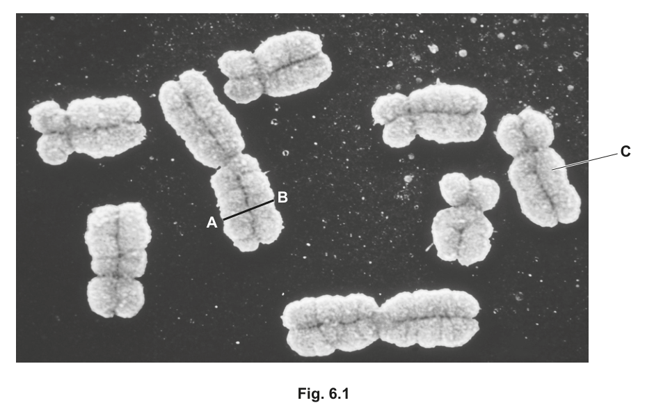
Fig. 6.1 — scanning electron micrograph of a group of human chromosomes at metaphase, with chromosome C and the line A–B labelled, as printed with the question.
Show full working
- 1
Decide what the drawing must show before starting. A chromosome at this stage of the cell cycle has already replicated, so it consists of two sister chromatids joined at one point — the centromere. That is the structure the labels must name.
Reading the biology first decides the whole drawing: two strands, one joint. Starting to draw before knowing this is how candidates produce a single sausage shape with nothing to label.
- 2
Draw the outline in sharp pencil: two chromatids lying side by side, joined at a centromere drawn as a visible constriction (a narrowing where the two touch), with the constriction placed off-centre so that the two arms are clearly unequal in length.
The mark scheme requires the centromere to be non-metacentric — off-centre, giving unequal arms. A centromere drawn in the exact middle loses that mark even though everything else is right.
- 3
Add labels with ruled lines touching the structures: one to either strand labelled (sister) chromatid, one to the constriction labelled centromere, and ones to the tips labelled telomere.
Telomeres must be labelled at or towards the ends of the chromatids — the same word labelled in the middle of an arm does not earn the mark. Label lines are drawn with a ruler, touch what they name, and carry no arrowheads.
A pencil outline of two unequal-armed chromatids joined at an off-centre constriction, labelled: sister chromatid, centromere (the constriction), telomere (at each end).
In every biological drawing, the labels are marks in their own right — budget time for them, and make each label line actually touch the structure it names.
Your turn
- 19700/22 F/M 2025 Q1(a)1 mark
Smilax china is a herbaceous plant.
Fig. 1.1 shows part of a transverse section of a root of S. china with root hair cells visible.
Name the type of microscope that has been used to obtain the image in Fig. 1.1.
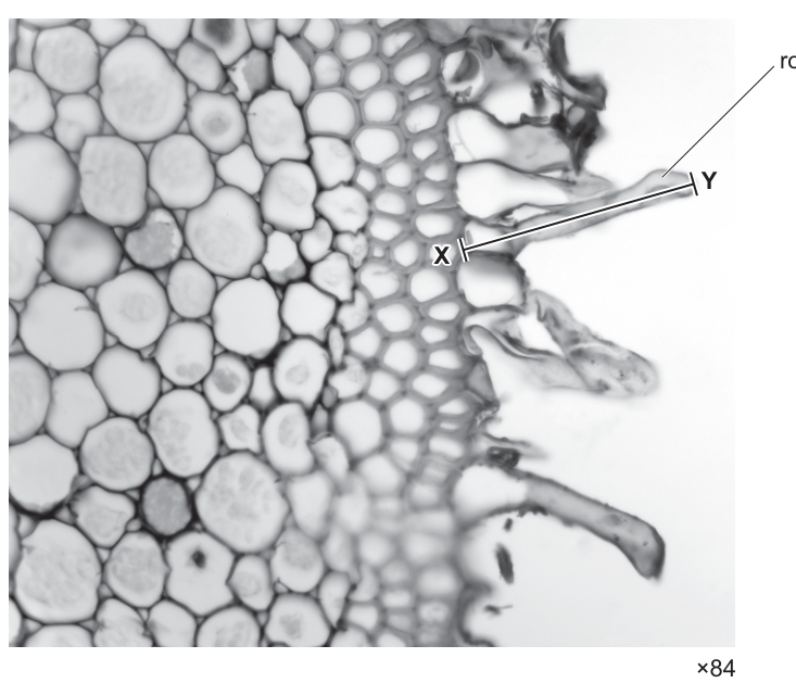
Fig. 1.1 — part of a transverse section of a root of S. china with root hair cells visible (×84), as printed with the question.
Show solution
- 1
The image shows a whole transverse section of tissue — many complete cells, each with visible walls and contents — but no internal ultrastructure: no individual membranes, no ribosomes, no organelle detail.
Judging microscope type from a micrograph is a resolution judgement, not a magnification one: ask what is the smallest thing visible? Here it is whole cells, not organelles.
- 2
Whole-cell detail without ultrastructure is exactly what the light (optical) microscope delivers — an electron micrograph of the same tissue would show membranes and organelles inside the cells.
The ×84 magnification alone cannot decide it — electron micrographs can be printed at low magnifications too. It is the level of detail, not the size, that identifies the instrument.
AnswerLight (optical) microscope — only whole-cell detail is visible, with no internal ultrastructure such as membranes or ribosomes.
- 1
- 2
Describe how to make a temporary preparation of onion epidermis suitable for viewing with the light microscope. Include the reason for each step.
Show solution
- 1
Peel a single, very thin layer of epidermis — light must pass through the specimen, so thick tissue shows only a silhouette.
- 2
Place it flat in a drop of water on a slide — water keeps the cells alive and supports them uniformly under the coverslip.
- 3
Stain with iodine solution — cell structures are otherwise colourless and translucent; iodine stains starch blue-black and darkens nuclei so they become visible.
- 4
Lower a coverslip at an angle with a mounted needle — lowering slowly from 45° pushes air out sideways and avoids trapping air bubbles, which otherwise look like dark rings and are easily mistaken for cells.
AnswerThin specimen → water mount → stain (e.g. iodine) → coverslip lowered at an angle to avoid air bubbles; reasons: light transmission, support, visibility, no bubbles.
- 1
- 3
A student hands in a drawing of a plant cell in which the vacuole is filled in with heavy pencil shading, three labels are joined to the cell by hand-drawn curved arrows, and one label line crosses another. State three things the student should do differently, and explain why each matters.
Stuck? Show hint
Each error breaks one of the drawing conventions — name the convention it breaks.
Show solution
- 1
Remove the shading. Conventions require single clear outlines only; shading hides line detail and cannot represent structure precisely — if a region looks dark, name the structure with a label instead.
- 2
Replace the curved arrows with ruled, straight label lines that touch the structure. Arrows are never used on biological drawings; each line must be drawn with a ruler and make contact with the exact part being named.
- 3
Rearrange the labels so no lines cross. Crossing lines are ambiguous — a reader cannot tell which structure the label names. Write labels horizontally and keep the lines separate.
AnswerNo shading (outline only); ruled straight label lines that touch the structure (no arrows); no crossing label lines, labels written horizontally.
- 1
Magnification and actual size
“
calculate magnifications of images and actual sizes of specimens from drawings, photomicrographs and electron micrographs (scanning and transmission).
What magnification actually is
Every drawing, photomicrograph and electron micrograph in this course shows a specimen larger than it really is. The magnification states exactly how many times larger, and calculating it — in both directions — is the most examined single skill in this topic (69 marks across Papers 1+2 in 2021–2025, more than any other calculation in AS Biology).
The definition is a division:
where image size is the length you measure on the drawing or micrograph (with a ruler), and actual size is the true length of the specimen in real life. Students often write the fraction upside down under pressure — anchor it with the common-sense check that a drawing times wider than the cell it shows is a magnification of , not .
A magnification is a ratio of two lengths measured in the same units — millimetres divided by millimetres, micrometres divided by micrometres. The units cancel, so the answer is a pure number written with a sign: , never . Mark schemes explicitly reject a magnification answer that carries units. The flip side: an actual size answer must always carry units (), because a bare number for a real length is meaningless.
The I–A–M triangle: cover the quantity you want and the triangle shows the operation — M = I ÷ A, A = I ÷ M, I = M × A. Magnification is a ratio of two lengths in the same units, so it never carries units.
The unit ladder: mm, µm, nm
The arithmetic of this topic is rarely hard — what loses marks is units that do not match. Biology measures lengths on a three-rung ladder, and every rung is a factor of :
- millimetre (mm) micrometres (µm) — about the width of a sharp pencil line; a eukaryotic cell is typically – µm across;
- micrometre (µm) nanometres (nm) — a bacterium is – µm, a virus – nm, a ribosome about nm.
Going down the ladder (mm → µm → nm) you multiply by 1000 at each step; going up, you divide. The rule that drives every worked example in this section: before you divide image size by actual size, convert both into the same units — and the safest habit is to convert the measured image length (in mm) into the units the actual size is given in.
The biological unit ladder with everyday anchors: each rung down multiplies by 1000. Cells live in µm; viruses and the finest cell structures live in nm.
- 1
Measure the image length with a millimetre ruler, as precisely as the ruler allows (to the nearest mm).
Measure the same feature the question names — the full length X–Y, not part of it. Mark the two ends on the paper with tiny pencil dots first if the feature is faint.
- 2
Convert the measured image length into the same units as the actual size, using the ladder — this is its own separate step, never folded into the division.
This is the step mark schemes watch hardest: dividing mm by µm without converting is the single most common way to be wrong by a factor of 1000.
- 3
Write the formula, then substitute: magnification = image size ÷ actual size, with each quantity now in matching units.
- 4
Evaluate and state the answer as ×N with no units.
Units on a magnification are actively penalised (R in the mark scheme). Give the answer to a sensible number of significant figures — usually 2 or 3.
A clean demonstration with invented numbers
A textbook drawing of a mitochondrion is measured to be mm long. The same organelle's actual length is µm. Find the magnification of the drawing.
Step 1 — measure the image. Already given: mm.
Step 2 — convert to matching units. The actual size is in µm, so convert the image measurement to µm — one rung down, multiply by 1000:
Step 3 — write the formula and substitute.
Step 4 — evaluate.
Notice the units cancelled — µm ÷ µm leaves a pure number, which is why the answer is written and nothing else.
Magnification from a measured drawing — formula and working required
Fig. 1.1 is a diagram drawn from a photomicrograph of a transverse section through part of a leaf.
The actual diameter of cell D in Fig. 1.1 along the length X–Y is .
Calculate the magnification of the image.
Write down the formula used to make your calculation. Show your working.
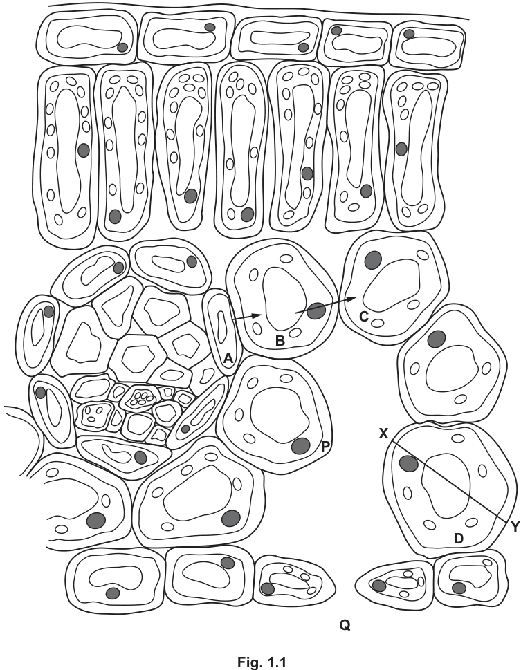
Fig. 1.1 — diagram drawn from a photomicrograph of a transverse section through part of a leaf, with cell D and the line X–Y marked, as printed with the question.
Show full working
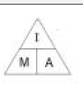
Mark-scheme figure: the magnification triangle (I over M × A), accepted in place of a written formula.
- 1
Write the formula first — the question awards a separate mark for it, so it must appear on its own line:
"Write down the formula" questions want the relationship in words or symbols before any numbers — jumping straight to the arithmetic drops this mark even if the answer is right.
- 2
Measure X–Y on the printed figure. With a millimetre ruler this measures mm (any measurement from to mm is acceptable to the examiners — each gives its own matching answer).
Human measurement varies slightly, which is why real mark schemes quote a range of acceptable answers rather than one value.
- 3
Convert the measurement into the units of the actual size — µm, one rung down the ladder:
- 4
Substitute into the formula:
- 5
Evaluate:
(The printed answers run from to for measurements of – mm — all acceptable.)
The mark scheme explicitly rejects the answer if units are given with the magnification. ×1480, full stop — no µm, no × symbol confusion.
magnification = image length ÷ actual length = 37 000 µm ÷ 25 µm = ×1480 (any value ×1440–×1560 accepted, matching your measurement) — no units.
When a magnification question says "write down the formula", treat the formula as a separate one-mark answer on its own line — it is marked independently of the number.
Actual size from a printed magnification
Fig. 3.1 is a scanning electron micrograph of a pair of human chromosomes in a stage of the mitotic cell cycle.
Calculate the actual length, to the nearest , of the chromosome in Fig. 3.1 indicated by the line X–Y.
Write the formula you used to make your calculation.
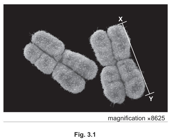
Fig. 3.1 — scanning electron micrograph of a pair of human chromosomes, with X–Y marked along the right-hand chromosome and magnification ×8625 printed below.
Show full working
- 1
Write the formula — rearranged this time, because the unknown is the actual size:
(A rearranged version of M = I ÷ A is accepted by the mark scheme.)
Cover A on the I–A–M triangle: what remains is I over M. Getting the division the right way up here is the classic trap — dividing by the magnification shrinks the image length down to the real length, which is what 'actual' means.
- 2
Read the magnification off the figure: it is printed on the micrograph as .
Before dividing, always hunt for a printed magnification or scale bar on the figure itself — the question is unanswerable without it.
- 3
Measure X–Y on the paper: mm.
The examiners' own measurement was 45 mm — yours may differ slightly, and the accepted answer range (5.0–5.4 µm) absorbs that.
- 4
Convert the measurement into µm so it matches the units the answer is asked for in:
- 5
Substitute and evaluate:
- 6
Round to the precision asked for — the nearest µm:
"To the nearest 0.1 µm" is an instruction about the final answer, and this answer — unlike a magnification — must carry its unit.
actual length = image length ÷ magnification = 45 000 µm ÷ 8625 = 5.2 µm (5.0–5.4 µm accepted)
Actual size = image ÷ magnification. If your 'actual size' ever comes out bigger than the image you measured, you have divided the wrong way round.
Magnification with nanometres in the mix
Fig. 6.1 is a scanning electron micrograph of a group of human chromosomes at metaphase.
The group of chromosomes shown in Fig. 6.1 is magnified many times.
The actual width of the human chromosome between A–B is .
Calculate the magnification of the scanning electron micrograph shown in Fig. 6.1.
Fig. 6.1 — scanning electron micrograph of a group of human chromosomes at metaphase, with the line A–B marked across one chromosome, as printed with the question.
Show full working
- 1
Measure A–B on the printed micrograph: mm (the examiners accepted – mm).
- 2
Convert the actual size into µm so both quantities can live on the same rung of the ladder:
Either quantity can be converted — the requirement is only that they match. Moving the small nm value up to µm keeps the numbers friendlier than pushing the mm value down to nm.
- 3
Convert the image measurement to µm as well:
- 4
Substitute and evaluate:
(Measurements of – mm give to — all accepted.)
Working in nm throughout gives the same result — 14 000 000 nm ÷ 1400 nm — but the extra zeros are exactly where slips happen. Matching units matter; which rung you meet them on does not.
magnification = 14 000 µm ÷ 1.4 µm = ×10 000 (×9286 to ×10 714 accepted for measurements of 13–15 mm) — no units.
It makes no difference which quantity you convert, only that both end up in the same units before you divide. Pick the direction that keeps the arithmetic kind.
The third direction: how big should the image be?
The triangle has one more corner — image size = magnification × actual size. It is the direction examiners use when they tell you a drawing must be drawn at a stated magnification, and it is the one students most often attack backwards. A clean demonstration with invented numbers:
A chloroplast is actually µm long. How long should its drawing be if it is drawn at ?
Step 1 — write the formula, covering I on the triangle:
Step 2 — match units by converting the actual size into the units a drawing is measured in — millimetres, because rulers measure in mm:
Step 3 — substitute and evaluate:
So the drawing's chloroplast should measure mm on the paper — about two ruler-widths, which passes the sanity check that of something microscopic fills a sensible chunk of page. Notice this answer must carry units: it is a real length on paper, not a ratio. The sanity check generalises — whatever direction you are working in, ask whether your answer is bigger than what you started from when magnification is large, and smaller when you divided by it.
Your turn
Every one of these is the same three moves — measure, convert, divide — but watch which quantity each question actually asks for.
- 19700/22 O/N 2016 Q4(c)3 marks
Fig. 4.1 is a cross-section of a human renal artery, a vessel that supplies blood to the kidney.
The actual diameter of the lumen of the renal artery at the point X–Y in Fig. 4.1 is .
Calculate the magnification of the image shown in Fig. 4.1. Write down the formula you will use to make your calculation and show your working.
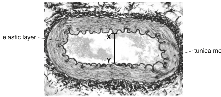
Fig. 4.1 — cross-section of a human renal artery, with X–Y marked across the lumen, as printed with the question.
Show solution
- 1
Formula:
- 2
X–Y measured on the figure: mm.
- 3
Both quantities are already in millimetres, so no conversion is needed — this question tests whether you convert only when the units actually differ.
Not every magnification question needs a conversion. Check the units first; converting unnecessarily wastes time and can introduce an error.
- 4
- 5
Rounded: (the mark scheme accepts to for measurements of – mm) — with no units.
Dividing a millimetre length by a millimetre length leaves nothing behind for a unit to attach to — the 'no units' rule applies here too, even though no conversion was needed.
Answermagnification = image ÷ actual = 18 mm ÷ 5.2 mm = ×3.5 (×3.3–×3.7 accepted) — no units.
- 1
- 29700/11 O/N 2025 Q11 mark
In a photomicrograph of magnification , a chloroplast measures mm in diameter. What is the actual diameter of the chloroplast?
A B C D
Stuck? Show hint
Convert the 25 mm into µm before dividing by 5000.
Show solution
- 1
The unknown is the actual size, so use .
- 2
Convert first: .
The options are all in µm — converting the measurement to µm before dividing lets you read the answer straight off the options.
- 3
AnswerD — 5 µm
- 1
- 39700/22 M/J 2024 Q1(b)(ii)1 mark
A student used a microscope fitted with a calibrated eyepiece graticule to estimate that the length of one smooth muscle cell was micrometres ().
The smallest object the student can see without the use of a microscope is in length.
Explain whether the student would be able to see a cell of length without the use of a microscope.
Stuck? Show hint
The comparison only works once both lengths are in the same units — and the mark scheme demands numbers and units in your answer.
Show solution
- 1
Convert the vision limit into µm: .
0.2 mm and 250 µm cannot be compared as written — one rung of the ladder separates them.
- 2
Compare: , so the cell is longer than the smallest object the student can see.
- 3
Yes — the student can see the cell unaided, because its length () exceeds the vision limit ().
The mark scheme requires the numerical values with units in the explanation — 'yes because it is bigger' alone does not earn the mark.
AnswerYes — 0.2 mm = 200 µm, and 250 µm is longer than 200 µm, so the cell is visible unaided.
- 1
- 49700/23 M/J 2020 Q4(a)1 mark
Saccharomyces cerevisiae is a unicellular fungus that is important in the brewing and baking industries.
Fig. 4.1 is a diagram of a transmission electron micrograph of S. cerevisiae.
A student was asked to calculate the magnification of the image shown in Fig. 4.1.
The student began by measuring the length of the scale bar in millimetres using a millimetre ruler.
State what the student should do next to obtain the correct answer.
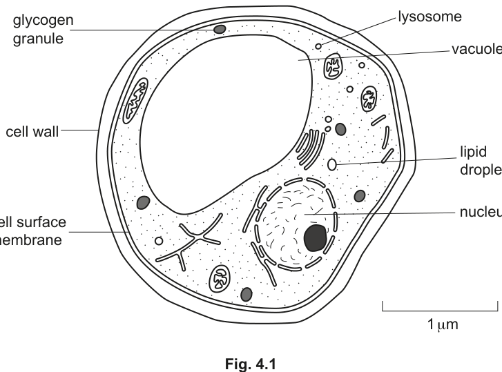
Fig. 4.1 — diagram of a transmission electron micrograph of S. cerevisiae, with labelled organelles and a 1 µm scale bar, as printed with the question.
Show solution
- 1
The next step is the conversion step: convert the measured scale-bar length from mm to µm (multiply by ) before dividing by the value the scale bar represents.
A scale bar's printed value is almost always in µm or nm, while your ruler reads mm — dividing before converting is being wrong by ×1000, which is precisely what this one-mark question is checking.
AnswerConvert the measured length from mm to µm (×1000) before using it in the magnification calculation.
- 1
- 59700/22 F/M 2025 Q1(b)1 mark
Smilax china is a herbaceous plant.
Fig. 1.1 shows part of a transverse section of a root of S. china with root hair cells visible.
Calculate the actual length, in micrometres (), of the root hair cell labelled in Fig. 1.1. Use the image length of the root hair cell along line X–Y in your calculation.
Fig. 1.1 — part of a transverse section of a root of S. china, with a root hair cell labelled, the line X–Y marked along it, and magnification ×84 printed below.
Show solution
- 1
Read the magnification printed below Fig. 1.1: . Measure X–Y with a ruler: about mm on the printed page.
The unknown is the actual size, so actual = image ÷ magnification — and the magnification has to come from the figure itself.
- 2
Convert: .
- 3
(The mark scheme accepts – µm, absorbing your measurement.)
Answeractual length = 39 000 µm ÷ 84 ≈ 460–464 µm (450–480 µm accepted)
- 1
The eyepiece graticule and stage micrometer
“
use an eyepiece graticule and stage micrometer scale to make measurements and use the appropriate units, millimetre (mm), micrometre (µm) and nanometre (nm).
Two scales, two different jobs
You cannot hold a ruler up to the image inside a microscope — the image exists only in your eye (or on a camera). To measure a specimen down the microscope you need two scales working together:
- The eyepiece graticule is a transparent plastic disc etched with a fine scale, sitting inside the eyepiece. It is fixed in the optics, so it stays put whatever you do to the slide — but its divisions are arbitrary: nobody knows what one division is worth until you calibrate it. We call one division an eyepiece unit (epu).
- The stage micrometer is a special slide carrying a scale of precisely known length — typically mm divided into parts (each division mm µm), or with divisions mm apart. It is the ruler; it just cannot be used to measure your specimen, because its markings are far too coarse for cells.
The measurement strategy is therefore: use the stage micrometer to find what one eyepiece unit is worth, then use the graticule to measure the specimen.
Calibrating means finding the real length (in µm) of one eyepiece unit at the magnification you are using. Line the two scales up, count how many eyepiece units span a known stage-micrometer length, and divide:
The calibration is only valid for the objective lens it was made with. Switch to a higher-power objective and the specimen image is stretched further, so each eyepiece unit now spans less real specimen — the µm-per-epu value falls. Measuring with a calibration made at a different objective is a silent factor-of-several error, and Paper 1 tests exactly this trap.
Calibration: the eyepiece graticule (arbitrary units) lined up against the stage micrometer (known µm). Here graticule marks 2 → 6 (4.0 epu) cover 50 → 130 µm of stage scale (80 µm), so 1 epu = 80 ÷ 4.0 = 20 µm — at that objective, and only that objective.
- 1
Place the stage micrometer on the stage and focus on its scale at the objective you intend to measure with.
Calibrating at a different objective invalidates everything downstream — same objective, always.
- 2
Line up the two scales so their zero marks coincide (or a convenient pair of marks coincides).
- 3
Count how many epu span a known stage-micrometer length — e.g. epu spanning mm µm.
- 4
Divide to find the calibration: µm.
One division of the sums: known length on top, epu count underneath. Writing the units (µm per epu) keeps the next step honest.
- 5
Replace the stage micrometer with the specimen slide and measure the specimen in epu, then multiply: actual size = (epu measured) × (µm per epu).
The graticule rides in the eyepiece, so it stays calibrated while slides change — only changing the objective changes the calibration.
A clean demonstration with invented numbers
At a certain objective, epu are found to span exactly µm of the stage micrometer.
Calibration. µm.
Measuring a specimen. A cell is seen to stretch across epu:
Two habits to carry into every graticule question: convert the stage-micrometer length into µm before dividing (a mm division is µm, not µm), and keep asking "which objective was this calibration made at?"
Reading a calibration off the printed diagram
The diagram shows a stage micrometer scale viewed with an eyepiece graticule, using a magnification of . Using the same magnification, a chloroplast is measured as 4 eyepiece graticule divisions long. How long is the chloroplast?
A B C D
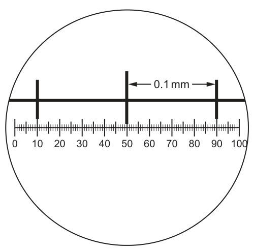
Fig. 1.1 — the graticule (0–100) with stage-micrometer marks falling on graticule units 10, 50 and 90; the arrowed stage division is labelled 0.1 mm.
Show full working
- 1
Read the alignment off the diagram. Stage-micrometer marks sit on graticule units , and — the arrowed stage division ( mm) spans from to : that is epu (and to is the same ).
The diagram is the data. The trap is assuming the marks line up with convenient round numbers without reading them — here they do, but only because you checked.
- 2
Convert the stage division to µm before dividing: µm.
Dividing 0.1 by 40 and trying to fix the units afterwards is where most candidates slip — convert first, then divide.
- 3
Calibrate:
- 4
Measure the chloroplast: epu µm per epu µm µm.
The ×200 magnification is context, not a second calculation — the calibration method bypasses it entirely, which is the whole point of a graticule.
A — 10 µm (40 epu = 0.1 mm = 100 µm, so 1 epu = 2.5 µm; 4 epu × 2.5 µm = 10 µm)
In every calibration diagram, find two marks that coincide and read both scales at those marks — the count between them is the only number you need.
Your turn
- 19700/13 O/N 2025 Q11 mark
The diagram shows a stage micrometer, with divisions mm apart, viewed through an eyepiece containing a graticule. The same eyepiece is now used to examine a blood smear. How many graticule divisions will cover the diameter of a lymphocyte of µm?
A 1 B 4 C 10 D 20
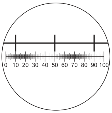
Fig. 1.1 — the graticule (0–100) with stage-micrometer marks falling on graticule units 10, 50 and 90.
Show solution
- 1
Read the alignment: stage marks fall on graticule and , so epu span one stage division of mm µm.
- 2
- 3
A lymphocyte of µm therefore covers graticule divisions.
This question runs the calibration backwards — instead of epu × calibration, it asks for epu = length ÷ calibration. Same two numbers, inverted operation.
AnswerB — 4 divisions
- 1
- 29700/13 O/N 2024 Q11 mark
Which steps are needed to find the actual width of a xylem vessel viewed in transverse section using a ×10 objective lens?
1 Convert from mm to µm by multiplying by .
2 Calibrate the eyepiece graticule using a stage micrometer on a ×4 objective lens.
3 Measure the width of the xylem vessel using an eyepiece graticule.
4 Multiply the number of eyepiece graticule units by the calibration of the eyepiece graticule.A 1, 2, 3 and 4 B 1 and 2 only C 2, 3 and 4 only D 3 and 4 only
Stuck? Show hint
Test each statement against the two rules: which objective must the calibration be made at, and which way does mm → µm go?
Show solution
- 1
Statement 1 is wrong. Converting mm to µm means multiplying by (), not : the µm value of a length is always its mm value, because mm µm. Multiplying by would shrink the number, which is backwards.
Statement 1 states the ladder direction wrongly — the classic conversion slip dressed up in standard form.
- 2
Statement 2 is wrong. The calibration must be made on the ×10 objective — the one the measurement will be made with. A calibration from the ×4 objective describes a different image stretch and gives a wrong answer on ×10.
This is the 'calibration is objective-specific' rule from earlier in the section — the most examined graticule fact on Paper 1.
- 3
Statements 3 and 4 are right. Measure the specimen in eyepiece units, then multiply by the µm-per-epu calibration: that is the measurement method.
- 4
So the needed steps are 3 and 4 only — option D.
AnswerD — 3 and 4 only (calibrate at the measuring objective; mm → µm is ×10³, not ×10⁻³)
- 1
Resolution versus magnification
“
define resolution and magnification and explain the differences between these terms, with reference to light microscopy and electron microscopy.
Two words that are not synonyms
Magnifying something and seeing it clearly are different achievements, and examiners probe the difference relentlessly — Resolution vs magnification; electron microscopy carried 53 marks across Papers 1+2 in 2021–2025. You met magnification in §02: how many times larger the image is than the specimen. Resolution (also called resolving power) is a different property entirely: it is the minimum distance two objects can be apart and still be seen as two separate objects rather than one blurred blob. A poster-size blow-up of a blurry photograph has enormous magnification and terrible resolution — everything is bigger, nothing is clearer.
Magnification = the number of times larger an image is than the actual specimen (, §02). It answers "how big does it look?" — and it carries no units. Resolution = the minimum distance apart that two points can be and still be distinguished as separate points. It answers "how close together can two things be before they merge?" — and it is a length, quoted like one ( for a school light microscope).
Why the light microscope hits a wall at about 0.25 µm
Resolution is limited by the wavelength of whatever is being used to form the image. Light travels as waves, and waves cannot turn sharply around objects much smaller than themselves — light passing beside a tiny object diffracts around it, smearing the image. As a working rule, two points closer together than roughly half the wavelength of the illuminating light blur into one.
Visible light has wavelengths of about – nm, so the finest detail a light microscope can resolve is around half of that: in practice about µm (= 250 nm). Quote this figure in the exam and attach the reason — because it is set by the wavelength of light — because that pairing is what mark schemes pay for.
This wall explains exactly which structures a light microscope can and cannot show:
- visible: anything larger than about 250 nm — nuclei, chloroplasts, mitochondria (as small ovals), whole bacteria, whole cells;
- invisible: anything smaller — ribosomes (~25 nm), cell surface membranes (~7 nm thick), ER membranes, the details inside a mitochondrion. These exist, but light cannot resolve them, so the cytoplasm looks featureless where they sit.
Why there is a maximum useful magnification of about ×1500
You could bolt lenses together until a light microscope reached — but you would only be magnifying the blur. If the finest resolvable detail is µm, enlarging beyond roughly adds size but no new information (empty magnification). This is the deepest consequence of the definition: past the resolution limit, extra magnification is wasted. Electron microscopes escape the wall precisely because their beam wavelength is far shorter, so their higher magnifications come with genuinely finer detail.
The same cell as a light microscope and an electron microscope show it. Below about 0.25 µm the light microscope merges neighbouring detail into one blob; the electron microscope's far shorter wavelength resolves membranes and ribosomes. Max useful magnification: about ×1500 (light) versus hundreds of thousands (electron).
Transmission and scanning electron microscopes
Electron microscopes fire a beam of electrons instead of light. Accelerated electrons have wavelengths thousands of times shorter than visible light, so the resolution wall moves from µm down to fractions of a nanometre — membranes and even ribosomes become visible. The two types point the beam at the specimen in different ways, and Paper 1 expects you to tell their products apart:
| TEM | SEM | |
|---|---|---|
| beam | passes through an ultrathin section | scans across the surface |
| what you see | internal ultrastructure, in a flat 2D slice | surface contours/topography, with a striking 3D appearance |
| resolving power / max useful magnification | finer (below 1 nm; hundreds of thousands of times) | coarser than TEM (but still far better than light) |
Both need elaborate preparation — specimens must be dried, fixed and placed in a vacuum, because air scatters electrons — so both work on dead material, unlike the light microscope.
- 1
Ask: what is the smallest structure visible? Ribosomes or membrane detail visible electron microscope — they are far below µm, so no light microscope could show them.
This is the single most reliable test, and the one the mark scheme rewards: it rests on the resolution argument, not on how big the picture looks.
- 2
If it is an EM, ask: through-the-inside or surface? Internal organelles shown as a flat sliced-through section TEM; surface contours with a 3D appearance SEM.
- 3
Never judge by magnification alone. Electron micrographs can be printed at low magnifications and light micrographs enlarged hugely — the level of detail, not the size on the page, identifies the instrument.
Options claiming 'a large magnification means…' are classic distractors; resolution decides, every time.
A clean demonstration with invented numbers
A light microscope resolves µm. Inside a bacterium µm long, two DNA loops lie µm apart. What can be seen?
Convert so both lengths share units (they already do): the bacterium is µm across, comfortably larger than µm, so it is detected. The two loops are only µm apart — less than the resolution — so however much you magnify, they show as one merged smear, not two loops. Detected but not resolved: magnifying harder never fixes resolution, because resolution belongs to the instrument, not to the enlargement.
Which viruses can a light microscope detect?
Norovirus has a diameter of .
Mimivirus has a diameter of .
Which viruses can be detected using a light microscope with a maximum resolution of ?
| Norovirus | Mimivirus | |
|---|---|---|
| A | ✓ | ✓ |
| B | ✗ | ✗ |
| C | ✗ | ✓ |
| D | ✓ | ✗ |
key: ✓ = can be detected, ✗ = cannot be detected
Show full working
- 1
Put all lengths on one rung of the ladder — convert each virus diameter from nm to µm:
Comparing 30 nm directly with 0.25 µm invites a factor-of-1000 slip; converting everything to µm first makes each comparison a glance.
- 2
Compare each with the resolution. Norovirus: — smaller than the smallest separable detail, so light from its edges diffracts and it cannot be picked out at all. Mimivirus: — larger than the resolution limit, so it can be detected (as a dot — none of its internal detail will show).
- 3
Answer: C — Norovirus ✗, Mimivirus ✓.
Notice the question says detected, not resolved. An object bigger than the resolution limit is detectable; nothing about its fine structure follows.
C — Mimivirus only (0.4 µm exceeds the 0.25 µm resolution; Norovirus at 0.03 µm is below it)
Any 'can it be seen with a light microscope?' question is one comparison: convert to matching units, compare with 0.25 µm. Bigger than the limit = detectable; smaller = invisible.
Identifying the microscope from what the micrograph shows
A cell is shown in the micrograph.
Which statement explains how it is possible to identify the type of microscope used to produce the micrograph?
A The nucleus is visible, so an electron microscope was used.
B The endoplasmic reticulum is not visible, so a light microscope was used.
C Chloroplasts are visible, so a light microscope was used.
D Ribosomes are visible, so an electron microscope was used.
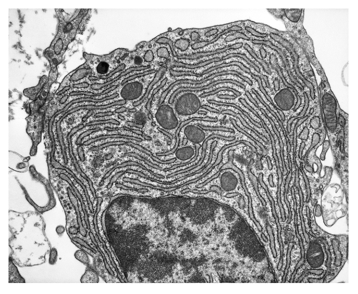
Fig. 2.1 — the micrograph of the cell, as printed with the question.
Show full working
- 1
Test each option against the resolution argument. Ribosomes are about nm across — far below the µm wall of any light microscope. They are clearly visible as dots along the ER membranes in Fig. 2.1, so the instrument must have been an electron microscope: option D is self-consistent and correct.
Visibility of the smallest organelle is the decisive evidence — it pins down the instrument's resolution, which is what 'identify the microscope' questions want.
- 2
See why the others fail. Option A: a nucleus is micrometres across — well within a light microscope's reach, so its visibility proves nothing. Option B is factually wrong about the image: ER is visible (the membrane sheets). Option C: no chloroplasts appear anywhere in this animal-type cell — and seeing one would identify the cell, not the microscope.
Each wrong option fails a different way — irrelevant evidence (A), misreading the image (B), and naming a structure that is not there (C). Reading options against the actual micrograph catches all three.
D — ribosomes (~25 nm) are below the light microscope's resolution, so their visibility shows an electron microscope was used
'How do you know which microscope?' always answers with the finest detail visible and the resolution needed to see it — never with the magnification printed on the figure.
Paper 1 recycles the resolution-versus-magnification distinction year after year, usually disguised as a "which statements are correct?" or "identify the microscope" stem. If you can convert units and compare with µm — plus name one structural giveaway for TEM and one for SEM — every variant of the question collapses into the same three moves.
Your turn
- 19700/24 M/J 2025 Q3(b)1 mark
Fig. 3.1 is a scanning electron micrograph of a pair of human chromosomes in a stage of the mitotic cell cycle.
Outline one feature of Fig. 3.1 that confirms the microscope used to obtain the image is a scanning electron microscope and not a transmission electron microscope.
Fig. 3.1 — scanning electron micrograph of a pair of human chromosomes (×8625), as printed with the question (the same figure as in §02).
Stuck? Show hint
What does an SEM scan, and what does that let it show that a TEM slice cannot?
Show solution
- 1
Look for evidence about surfaces: the chromosomes show their surface contours in a three-dimensional appearance, with no sign of a sliced-through interior — no internal detail of chromatids is visible, as it would be in a thin section under TEM.
The mark scheme accepts any one of: surface contours/topography visible; no thin section/internal detail; 3D appearance; good depth of field. Any single one earns the mark — outline it, don't just say 'it looks 3D' with no reference to surfaces.
- 2
One line earns the mark, e.g.: surface contours/topography are visible, giving a three-dimensional image — a TEM shows only a flat internal thin section.
AnswerSurface contours (topography) are visible / the image appears three-dimensional / no internal detail of a thin section is seen — all signatures of scanning, not transmission, EM.
- 1
- 29700/13 O/N 2025 Q41 mark
The image shown is produced using a microscope.
How many statements about this image are correct?
● It is an electron micrograph.
● It shows part of a eukaryotic cell.
● It shows at least one mitochondrion.
● It shows a specimen viewed at more than magnification.A 1 B 2 C 3 D 4
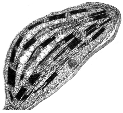
Fig. 4.1 — the microscope image, as printed with the question: an oval structure containing parallel internal membranes with dark stacked regions.
Show solution
- 1
Identify the organelle. Parallel stacks of internal membranes (grana) inside an oval boundary = a chloroplast.
- 2
Statement 1, true. Grana stacks are tens of nanometres across — far below µm — so only an electron microscope could resolve them.
- 3
Statement 2, true. Chloroplasts are organelles of eukaryotic cells (plant cells), so this is part of a eukaryotic cell.
- 4
Statement 3, false. No mitochondrion appears — the only organelle in view is the chloroplast. Recognising one organelle does not license others into the frame.
- 5
Statement 4, true. Resolving grana demands electron microscopy, and electron microscopes operate at magnifications far beyond .
Three true statements, one false — so the count is 3, option C. Notice how each statement leans on a different fact from this section: resolution limits, organelle identity, and what EM magnifications can reach.
AnswerC — three statements correct (electron micrograph: yes; eukaryotic cell: yes; mitochondrion: no; more than ×400: yes)
- 1
- 3
State whether each of the following would be visible (detected) using a light microscope of resolution µm, and give one reason in each case: (a) a chloroplast about µm long; (b) a ribosome about nm across; (c) a bacterium µm long; (d) the two membranes of a mitochondrion's envelope, nm apart.
Stuck? Show hint
Convert every size to µm first, then compare each with 0.25 µm.
Show solution
- 1
(a) µm µm: visible — a green oval, though internal grana detail will not resolve.
- 2
(b) nm µm µm: not visible — ten times smaller than the resolution limit.
- 3
(c) µm µm: visible as a rod-shaped object; nothing inside it will resolve.
- 4
(d) nm µm µm: not resolvable — the two membranes blur into a single line, so the double-membrane structure needs an electron micrograph.
Part (d) is the exam favourite: mitochondria are visible with a light microscope, but their double envelope is not. Size of the object and fineness of its internal detail are separate judgements.
Answer(a) yes (b) no (c) yes (d) no — compare each converted size with 0.25 µm; being detected and having resolvable internal detail are different questions.
- 1
Eukaryotic organelles — structure and function
“
recognise organelles and other cell structures found in eukaryotic cells and outline their structures and functions, limited to: cell surface membrane • nucleus, nuclear envelope and nucleolus • rough endoplasmic reticulum • smooth endoplasmic reticulum • Golgi body (Golgi apparatus or Golgi complex) • mitochondria (including the presence of small circular DNA) • ribosomes (80S in the cytoplasm and 70S in chloroplasts and mitochondria) • lysosomes • centrioles and microtubules • cilia • microvilli • chloroplasts (including the presence of small circular DNA) • cell wall • plasmodesmata • large permanent vacuole and tonoplast of plant cells.
The heart of the topic
This is the single heaviest sub-topic in AS Biology — 290 marks across Papers 1+2 in 2021–2025, more than magnification and resolution combined. The electron microscope (§04) is what made it examinable: once resolution dropped below µm, the "featureless cytoplasm" of light microscopy resolved into membranes, stacks, tubules and granules, each with a shape that betrays its job.
The skill being tested has two halves, and outline questions want both:
- recognise — name the structure from what an electron micrograph or drawing shows (a stack of flattened sacs with vesicles = Golgi body);
- link structure to function — say what it does in terms of what it looks like (its folded inner membrane gives a large surface area for respiration enzymes).
An organelle is a membrane-bound structure inside a cell with a specific function. Compartmentalisation is the point: membranes let incompatible reactions run side by side in the same cell, each in its own optimised space.
Animal cell ultrastructure as an electron microscope shows it. Use this as the map while reading the catalogue below — every labelled structure reappears as a mark-scheme answer somewhere in this topic.
Structure | Structure in brief | Function |
|---|---|---|
Nucleus + nuclear envelope | Largest organelle; chromatin (DNA + histones) inside a double-membrane envelope with nuclear pores; dark nucleolus within | Stores DNA genes and controls the cell's activities; pores let mRNA leave; the nucleolus assembles 80S ribosomes |
Rough ER | Flattened sheets of membrane, continuous with the nuclear envelope, studded with ribosomes | Folds and processes proteins made on its surface, then transports them in vesicles |
Smooth ER | Tubular membranes, no ribosomes attached | Synthesises lipids (e.g. triglycerides, cholesterol) |
Golgi body (apparatus / complex) | Stack of curved, flattened membrane sacs; vesicles pinch off its edges | Modifies and processes proteins and lipids, packages them into vesicles, forms lysosomes |
Mitochondrion | Double membrane; inner folded into cristae; matrix holds small circular DNA and 70S ribosomes | Site of aerobic respiration — makes ATP for the cell |
Ribosomes | Two subunits of RNA + protein; no membrane. 80S free or on rER in the cytoplasm; 70S inside mitochondria and chloroplasts | Protein synthesis — the site where mRNA is translated |
Lysosome | Small sac, single membrane, packed with hydrolytic digestive enzymes | Digests worn-out organelles and material engulfed by the cell |
Centrioles + microtubules | Pair of hollow cylinders, each of nine microtubule triplets, beside the nucleus (animal cells) | Grow the spindle fibres that move chromosomes in mitosis; microtubules also form the cell's internal scaffold |
Cilia | Hair-like projections beating outside the cell; microtubules in a 9+2 arrangement, anchored at a basal body | Rhythmic beating moves fluid or mucus over the cell surface |
Microvilli | Finger-like folds of the cell surface membrane, stiffened internally by microfilaments | Greatly increase surface area for absorption |
Cell wall (plant) | Rigid layer of cellulose fibres outside the membrane; fully permeable | Mechanical support; stops the cell bursting when water enters |
Plasmodesmata | Fine strands of cytoplasm running through pores in adjoining cell walls | Transport and communication between neighbouring plant cells |
Large permanent vacuole (plant) | Big sac bounded by a single membrane, the tonoplast, and filled with cell sap | Stores dissolved substances; keeps cells turgid |
Chloroplast | Double membrane; inner membranes stacked into grana within a fluid stroma; small circular DNA and 70S ribosomes | Photosynthesis — captures light energy to make sugars |
The full syllabus catalogue. Learn each row as a pair — what it looks like, what that shape is good for — because Paper 1 shows you one and asks for the other.
Three details the examiner always digs at
The catalogue above is the pass mark. The distinctions below are where the actual marks concentrate, because they separate candidates who memorised names from candidates who know the architecture.
1. Counting membranes. "Which organelles are enclosed by a double membrane?" is among the most repeated Paper 1 stems there is. Only three structures have envelopes of two membranes: the nucleus (the nuclear envelope — count its inner and outer membranes, pores notwithstanding), the mitochondrion and the chloroplast. Everything else membrane-bound is single: rER, sER, Golgi, lysosomes, the tonoplast, secretory vesicles. And some structures have no membrane at all: ribosomes, centrioles, microtubules, cilia and microvilli (they are built from membrane and protein, but they do not enclose anything).
Membrane census of the syllabus structures. Double envelope: nucleus, mitochondrion, chloroplast. Single membrane: rER, sER, Golgi, lysosome, tonoplast, secretory vesicles. No membrane of their own: ribosomes, centrioles, microtubules.
2. Ribosomes come in two sizes — and the small ones hide inside organelles. Cytoplasmic ribosomes of eukaryotes are 80S. But the ribosomes inside your own mitochondria and chloroplasts are 70S — the same size as bacterial ribosomes, and no coincidence (that story arrives in §07). A comparison-table question loves this row: bacteria 70S, eukaryotic cytoplasm 80S, mitochondria and chloroplasts 70S despite living inside a eukaryote.
3. Small circular DNA lives inside mitochondria and chloroplasts. Both organelles carry their own few genes as a small circle of naked DNA, quite separate from the linear chromosomes in the nucleus — another echo of bacteria, and the reason these organelles can synthesise some of their own enzymes using their own 70S ribosomes. Never call this DNA linear, and never say it is surrounded by a nuclear envelope.
The plant additions
Plants build four structures animal cells never carry: a cellulose cell wall, plasmodesmata threading through it, a large permanent vacuole with its tonoplast, and chloroplasts. Animal-only structures are just as short a list: centrioles (plants grow spindles without them) and, among the surface specialisations, cilia are typical of animal epithelia rather than plants. Everything else in the catalogue — nucleus, mitochondria, ER, Golgi, ribosomes, lysosomes, cell surface membrane — is shared.
Plant cell ultrastructure: cellulose wall with a plasmodesma passing through, tonoplast around the large permanent vacuole, chloroplasts with a double membrane and grana — plus the nucleus and a mitochondrion, shared with animal cells (ER, Golgi and ribosomes are present too, omitted here for clarity).
Cells use ATP from respiration for energy-requiring processes. That one-line syllabus statement earns marks across the whole course: active transport, endocytosis and exocytosis, mitosis, protein synthesis, organelle movement. The supply chain runs through the mitochondrion — glucose + oxygen are broken down in aerobic respiration there, and the ATP produced powers whatever the cell does next. So whenever a question asks why a cell has many mitochondria, the answer is a demand for ATP: muscle contraction, active uptake in gut epithelium, secretion. Match the organelle count to the energy bill.
Reading a cell's job from its organelle profile
Flip the catalogue around and it becomes a detective tool: a cell's function predicts which organelles it stocks heavily. Secretory cells (making and exporting protein, like the mucus-making goblet cells of the airways) need extensive rER to make the protein, an extensive Golgi body to process and package it, and crowds of secretory vesicles — which is why goblet cells dominate the answer to any "most Golgi / most single-membrane structures" question. Absorbing cells (small-intestine epithelium) pack microvilli for area and mitochondria to drive active transport. Muscle cells are stuffed with mitochondria for contraction. Paper 1 tests exactly this inference, usually as "which cell type…?" — reason from the job to the organelles, then match the options.
A clean demonstration before the past-paper questions
An electron micrograph of a secretory cell shows three unlabelled structures: (i) curved stacks of flattened membrane sacs with small round sacs budding off their edges; (ii) oval organelles with an inner membrane folded back on itself; (iii) sheets of membrane dotted with tiny dark granules.
Work each one through the catalogue: (i) stack + budding vesicles = Golgi body, packaging proteins for export; (ii) double membrane + folded cristae = mitochondrion, supplying the ATP that secretion costs; (iii) ribosome-studded sheets = rough ER, making the protein being exported. Membrane counts: single, double, single respectively — and the trio tells you the cell's trade before you read a word of stem: a busy protein-exporting cell. That two-move habit — recognise from structure, then link to function — is everything this section teaches.
Enzymes AND a double membrane
Which cell structures contain enzymes and are enclosed by a double membrane?
1 nucleus 2 mitochondrion 3 chloroplast
A 1, 2 and 3 B 1 and 2 only C 1 only D 2 and 3 only
Show full working
- 1
Run the double-membrane test on each structure. Nucleus — the nuclear envelope has inner and outer membranes: double. Mitochondrion — an envelope of outer and inner membranes (the inner one folded into cristae): double. Chloroplast — an envelope of outer and inner membranes (the thylakoids inside are extra, not part of the count): double.
This is the membrane census figure applied directly: exactly these three carry double envelopes. Students who count the nuclear envelope as single lose the question here.
- 2
Run the enzyme test. Nucleus — DNA polymerase and RNA polymerase work there during replication and transcription. Mitochondrion — aerobic respiration is enzyme-driven. Chloroplast — photosynthesis is enzyme-driven. All three contain enzymes.
- 3
Both tests pass for all three, so the answer is A — 1, 2 and 3.
The trap is assuming 'enzymes' means digestion or metabolism outside the nucleus — replication and transcription are enzyme-catalysed reactions, which puts enzymes firmly inside the nuclear envelope.
A — nucleus, mitochondrion and chloroplast each contain enzymes and are each bounded by a double membrane
Two-test questions like this are answered by intersecting two lists from memory: the double-membrane trio, and where named enzymes work. Build both lists once and reuse them forever.
Naming an organelle and earning the function mark
Fig. 1.2 is a drawing of a transmission electron micrograph (TEM) of a cell from the palisade mesophyll of a leaf.
The drawing does not show all of the organelles visible in a transmission electron micrograph.
Identify the organelle labelled X and state one function of this organelle.
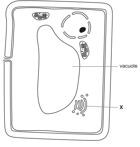
Fig. 1.2 — the palisade mesophyll cell drawing, with label X on a stack of curved, flattened membrane sacs with vesicles pinching off.
Show full working
- 1
Recognise X from its shape. A stack of curved, flattened membrane sacs, with small vesicles budding off the edges, sitting in the cytoplasm: this is the Golgi body (the mark scheme accepts Golgi apparatus or Golgi complex, and dictyosome).
Shape is the whole identification: no other organelle is drawn as a stack of flattened sacs shedding vesicles. Mitochondria are ovals with internal folds; nuclei are large and envelope-bound.
- 2
Give a function the mark scheme can pay for — any one of: modifies/processes proteins (or lipids), e.g. adding sugars; packages proteins into (Golgi) vesicles for transport or secretion; forms lysosomes.
'Transports' on its own is ignored by the mark scheme — say what the packaging is for. Naming the organelle earns one mark; the second is reserved for a processing or packaging function.
Golgi body — modifies and processes proteins (e.g. adds sugar chains) / packages proteins into vesicles / forms lysosomes
For every 'identify and state one function' pair, pick the function that names the process — modify, package, form — rather than a vague verb like 'transports' or 'helps'.
Your turn
Recognition first, then the structure–function link, then the membrane count — the same three moves in every item.
- 19700/21 O/N 2016 Q1(a)3 marks
Fig. 1.1 is a transmission electron micrograph of part of an animal cell. Name the structures A, B and C.
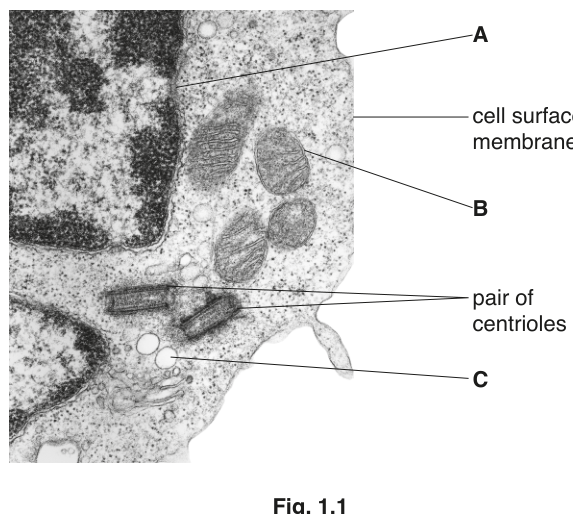
Fig. 1.1 — transmission electron micrograph of part of an animal cell, with structures A, B and C, the cell surface membrane and a pair of centrioles labelled, as printed with the question.
Stuck? Show hint
A wraps the chromatin; B has cristae; C is a small single-membrane sac near the Golgi body.
Show solution
- 1
A sits as a double membrane around the chromatin, with pores visible: nuclear envelope (also credited: nucleus, nuclear membrane).
The mark scheme ignores 'nuclear pore' as an answer for A — pore is a feature of the envelope, not its name.
- 2
B is oval with a double membrane whose inner one folds into cristae: mitochondrion.
- 3
C is a small, round, single-membraned sac near the Golgi stack: lysosome or (Golgi/secretory) vesicle — all credited.
'Vesicle' alone is accepted here; what loses marks is qualifying it wrongly ('phagocytic', 'temporary') — the plain structural name is what is being tested.
AnswerA nuclear envelope · B mitochondrion · C lysosome / Golgi vesicle / secretory vesicle
- 1
- 29700/13 M/J 2024 Q41 mark
Which type of cell will have the highest proportion of its volume taken up with cell structures bound by a single membrane?
A ciliated epithelial cell B goblet cell C red blood cell D companion cell
Stuck? Show hint
First list the single-membrane structures from the census. Then ask which cell's job needs the most of them.
Show solution
- 1
List the single-membrane structures: rER, sER, Golgi body, lysosomes and secretory vesicles. A cell that exports protein by exocytosis needs all of these, at scale.
The question is really about secretion: making and exporting mucus means rER to synthesise, Golgi to process, vesicles to carry, lysosomes to supply — a whole single-membrane production line.
- 2
The goblet cell is a professional secretory cell, so option B. The rivals fail cleanly: red blood cells have lost all organelles; ciliated epithelium and companion cells are ATP-hungry, which stocks them with double-membraned mitochondria instead.
Note the symmetry the examiner built in: high-energy cells fill up with double membranes (mitochondria), high-export cells fill up with single membranes.
AnswerB — goblet cell (secretion demands extensive rER, Golgi, vesicles and lysosomes — all single-membrane)
- 1
- 39700/11 O/N 2023 Q41 mark
Which animal cells would have the most extensive Golgi bodies?
A ciliated epithelial cells B goblet cells C red blood cells D smooth muscle cells
Stuck? Show hint
Extent of Golgi tracks the rate of protein processing and packaging.
Show solution
- 1
Match the organelle to the workload: the Golgi body modifies and packages proteins for export, so the cells processing and secreting the most protein have the most extensive Golgi.
This is the abundance-reasoning habit from this section applied to one organelle: no need to know any cell type's anatomy beyond its job.
- 2
Goblet cells secrete large quantities of mucus (a glycoprotein), so they run a huge Golgi throughput: B. Muscle cells mainly contract (mitochondria, not Golgi); ciliated epithelia mainly beat; red blood cells have no organelles at all.
AnswerB — goblet cells, because heavy secretion of mucus requires massive Golgi processing and packaging
- 1
- 49700/21 M/J 2025 Q2(a)2 marks
In mammals, the small intestine is the main site of absorption of the products of digestion.
Fig. 2.1 is a transmission electron micrograph of a longitudinal section (L.S.) of part of an epithelial cell from the small intestine of a mammal.
Fig. 2.2 is a transmission electron micrograph of a horizontal section made at the position indicated by the two arrows in Fig. 2.1.
Microvilli and cilia are cell structures.
Describe how the structure of cilia differs from the structure of the microvilli visible in Fig. 2.1 and Fig. 2.2.
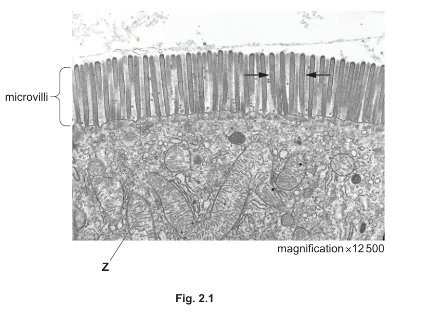
Fig. 2.1 — TEM of a longitudinal section of part of a small-intestine epithelial cell (×12 500), with the microvilli bracketed, two arrows marking the level of Fig. 2.2, and label Z.
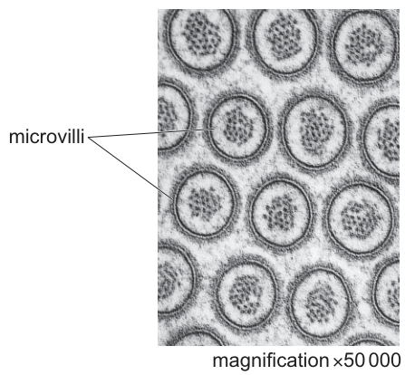
Fig. 2.2 — TEM of a horizontal section through the microvilli at the level of the arrows in Fig. 2.1 (×50 000).
Stuck? Show hint
Ask of each projection: what runs along inside it? One answer contains microtubules, the other does not.
Show solution
- 1
Say what is inside each. Cilia contain microtubules arranged in a characteristic 9+2 pattern, seen clearly in transverse section; microvilli are supported internally by microfilaments (actin), not microtubules — Fig. 2.2 shows only filament material inside each circular profile.
Giving microvilli microtubules is explicitly rejected by the mark scheme — the two answers must not swap skeletons. Any two of: microtubules vs microfilaments, the 9+2 arrangement, cilia extending from a basal body.
- 2
Add a second difference if unsure: each cilium grows from a basal body beneath the cell surface membrane, whereas microvilli are simple folds of the membrane itself with no such anchor.
AnswerAny two: cilia have microtubules (microvilli have microfilaments); microtubules in a 9+2 arrangement in cross-section; cilia extend from a basal body.
- 1
- 59700/21 M/J 2025 Q2(c)2 marks
In mammals, the small intestine is the main site of absorption of the products of digestion.
Fig. 2.1 is a transmission electron micrograph of a longitudinal section (L.S.) of part of an epithelial cell from the small intestine of a mammal.
Identify the organelle labelled Z in Fig. 2.1 and explain why there is a large number of these organelles in the epithelial cells of the small intestine.
Fig. 2.1 — TEM of a longitudinal section of part of a small-intestine epithelial cell (×12 500), with the microvilli bracketed and label Z, as printed with the question.
Stuck? Show hint
Z is oval with a folded inner membrane — and the cell absorbs against gradients.
Show solution
- 1
Identify Z: oval outline, double membrane with the inner one folded into cristae — a mitochondrion.
One mark for the name alone; keep the identification purely structural, since the explanation carries its own mark.
- 2
Explain the abundance through ATP demand: epithelial cells of the small intestine take up digested products by active transport, an energy-requiring process; mitochondria carry out aerobic respiration and so provide the ATP that active transport consumes — more transport, more mitochondria.
This is outcome 4 in action: cells use ATP from respiration for energy-requiring processes. Saying the mitochondria 'provide energy' without naming ATP risks losing the mark — name the currency.
AnswerZ = mitochondrion; numerous because active transport (absorption) in these cells requires ATP, which respiration in mitochondria provides
- 1
- 69700/22 O/N 2016 Q15 marks
Match the description for each of statements A to E to a correct cell structure.
A Double membrane-bound organelle, absent in animal cells, that produces ATP.
B Partially permeable membrane surrounding the large permanent vacuole of plant cells.
C Formed from microtubules during mitosis.
D Has peptidoglycan as one of its major components.
E Site of assembly of 80S ribosomes.Stuck? Show hint
Read every clause of each statement — one clause usually rules out the obvious-looking alternative.
Show solution
- 1
A = chloroplast. Double-membraned, makes ATP (from photosynthesis), and absent in animal cells — mitochondria also fit everything except that last clause.
'Absent in animal cells' is the discriminating phrase; read every clause before answering.
- 2
B = tonoplast (the vacuolar membrane). C = spindle (spindle fibres), grown from microtubules at mitosis. D = (prokaryotic) cell wall — peptidoglycan belongs to bacteria; crediting a cellulose plant wall here is explicitly rejected. E = nucleolus, where 80S ribosomes are assembled before export through nuclear pores.
D previews §07: peptidoglycan is the bacterial wall material, never cellulose. E explains why the nucleolus looks so dense in electron micrographs — it is a ribosome factory.
AnswerA chloroplast · B tonoplast · C spindle · D (prokaryotic/bacterial) cell wall · E nucleolus
- 1
Comparing plant and animal cells
“
describe and interpret photomicrographs, electron micrographs and drawings of typical plant and animal cells; compare the structure of typical plant and animal cells.
Sorting the catalogue into three lists
Everything in §05's catalogue belongs to exactly one of three groups, and compare questions are just those groups written down quickly:
- shared — nucleus with nuclear envelope and nucleolus, both ER types, Golgi body, mitochondria, ribosomes, lysosomes, cell surface membrane;
- plant only — cellulose cell wall, chloroplasts, large permanent vacuole with tonoplast, plasmodesmata;
- animal only — centrioles (and cilia as a surface specialisation of animal epithelia).
That is the whole comparison. The examiner's version adds two finer rows worth memorising: the carbohydrate store (starch grains in plants, glycogen granules in animals — visible in electron micrographs as dense particles), and the fixed, regular box-like shape a wall imposes on plant cells versus the irregular, flexible outline of animal cells.
Feature | Typical plant cell | Typical animal cell |
|---|---|---|
Cell wall | present — cellulose | absent |
Chloroplasts | present | absent |
Vacuole | large permanent vacuole, tonoplast-bound, at centre | none (small temporary vesicles only) |
Centrioles | absent | present, beside the nucleus |
Carbohydrate store | starch grains | glycogen granules |
Outer shape | regular, fixed by the wall | irregular, flexible — membrane only |
Everything else | nucleus, envelope, nucleolus, rER, sER, Golgi, ribosomes, lysosomes, mitochondria, cell surface membrane — identical | identical |
The complete plant–animal comparison. Three plant-only rows, one animal-only row, and one pair of store-and-shape details — everything else is shared.
When asked to outline the mature plant vacuole, mark schemes pay for exactly two statements: it is surrounded by the tonoplast, a single partially permeable membrane, and it contains cell sap — a solution of dissolved substances (salts, sugars) in water. Two answers are actively rejected (R): calling the tonoplast an envelope or double membrane, or describing it as a wall. The vacuole is a single-membrane sac, not an organelle with walls.
- 1
Look for the plant-only signatures first: a distinct wall outside the membrane boundary, chloroplasts (grana-stacked ovals), a dominant central vacuole pressing cytoplasm into a thin rim.
Any one of these settles 'plant'; their absence points to animal — but read the caution below before committing.
- 2
Confirm with shape and storage: a rigid, angular outline suggests wall (plant); irregular and rounded suggests membrane-only (animal).
- 3
Respect the thin-section caveat: an electron micrograph is one sliced plane (§04), so organelles outside that plane are simply not in view — few mitochondria visible does not mean few mitochondria present.
Examiners award marks specifically for this insight: visibility in a single thin section is evidence about the slice, not about the cell's inventory.
A clean demonstration before the past-paper question
An electron micrograph shows a cell with an angular outline, a thin layer of cytoplasm crowded with organelles around a huge empty-looking central sac, and several green-black oval bodies containing stacked internal membranes. Judge it. Wall → plant. Central sac bounded by a single membrane → large permanent vacuole (tonoplast). Ovals with stacks → chloroplasts (grana). Verdict: a photosynthesising plant cell, probably leaf mesophyll — you could also predict plenty of mitochondria for active transport even if none happen to lie in this particular slice. One pass through the three-lists table reads the entire cell.
Completing a three-column presence table
Animal cells, plant cells and prokaryotic cells have similarities and differences in their structure.
Table 1.1 lists five organelles found in cells.
Complete Table 1.1 by placing a tick (✓) to show whether the organelle is present in animal cells, plant cells and prokaryotic cells or a cross (✗) if the organelle is absent.
Put a tick (✓) or a cross (✗) in every box.
The first row has been completed for you.
Table 1.1
| organelle | animal cells | plant cells | prokaryotic cells |
|---|---|---|---|
| nucleus | ✓ | ✓ | ✗ |
| large permanent vacuole | |||
| rough endoplasmic reticulum | |||
| Golgi body | |||
| centrioles |
Show full working
- 1
Large permanent vacuole — read it off the 'plant only' list: absent from animal cells, present in plant cells, absent from prokaryotic cells → ✗ ✓ ✗.
This is the payoff of sorting the catalogue into three lists earlier in the section: each row is now a lookup, not a fresh decision.
- 2
Rough ER and Golgi body — shared by both eukaryotes: ✓ ✓ for the first two columns.
Both appear in the 'shared' list: every eukaryote runs the protein-processing pathway (rER → Golgi → vesicle) that §05 described.
- 3
…but give both a cross in the prokaryotic column: ✗. Every organelle named in this table except centrioles is a membrane-bounded compartment, and prokaryotic cells have no organelles surrounded by membranes.
This is the pattern that fills a whole prokaryotic column at once — and it is exactly the discrimination Paper 1 loves to test with Venn diagrams.
- 4
Centrioles — the animal-only row: ✓ ✗ ✗. Typical plant cells have no centrioles even though they still build a spindle at mitosis, and prokaryotes neither have them nor divide by mitosis.
Candidates often hand plants a tick here out of sympathy — resist it: centrioles are the single cleanest animal-only marker in the AS catalogue.
- 5
Sweep the table for blank boxes. Every one of the twelve remaining boxes now carries a tick or a cross.
The stem commands a mark in every box — a blank cannot score, and a swept-for-blanks table is how one mark per row (4 in total) is safely collected.
large permanent vacuole: ✗ ✓ ✗ · rough ER: ✓ ✓ ✗ · Golgi body: ✓ ✓ ✗ · centrioles: ✓ ✗ ✗ (nucleus row was given)
In presence tables, every membrane-bounded organelle gets a cross in the prokaryotic column — bacteria have none of those. Watch for the exceptions that are not membrane-bounded: ribosomes (70S) and the cell wall (peptidoglycan) do earn a tick for prokaryotes (§07 builds the full bacterium picture).
Your turn
- 19700/24 M/J 2025 Q1(b)(ii)2 marks
Outline the structure of a fully mature plant vacuole.
Stuck? Show hint
One mark lives in the membrane around it, one in what is inside it.
Show solution
- 1
Outside: the vacuole is bounded by the tonoplast — a single, partially permeable membrane.
Mark scheme: 'single (partially permeable) membrane / tonoplast'. Writing 'envelope' or 'double membrane' is explicitly rejected, and so is calling the tonoplast a wall.
- 2
Inside: it contains cell sap — a solution of dissolved substances (e.g. salts and sugars) in water.
'Contains cell sap' earns the second mark; adding what sap is (dissolved solutes in water) makes the outline genuinely structural rather than a label.
AnswerBounded by the tonoplast, a single partially permeable membrane; filled with cell sap — a solution of dissolved substances in water
- 1
- 2
Copy and complete the following table comparing a typical plant cell with a typical animal cell.
feature plant cell animal cell outer boundary layers carbohydrate storage product organelle that grows spindle fibres largest compartment Stuck? Show hint
Row 4 is the one students forget: which compartment occupies most of a mature plant cell's volume?
Show solution
- 1
Row 1: plant — cell surface membrane plus cellulose cell wall outside it; animal — cell surface membrane only.
- 2
Row 2: starch (as visible starch grains) in plants; glycogen (granules) in animals.
- 3
Row 3: centrioles grow the spindle in animal cells; plant cells form a spindle without centrioles, so the honest entry is 'centrioles' for animal and 'none (spindle still forms)' for plants.
- 4
Row 4: the large permanent vacuole dominates a mature plant cell; in an animal cell the nucleus or the cytoplasm generally takes the largest share.
Comparisons are marked feature-by-feature — an answer that fills every row with correct pairs collects every mark even if one cell type interests you more than the other.
Answer(1) membrane + cellulose wall vs membrane only · (2) starch vs glycogen · (3) none/spindle without centrioles vs centrioles · (4) large permanent vacuole vs cytoplasm/nucleus
- 1
- 3
An electron micrograph of a single cell shows: an irregular outline with no structure outside the cell surface membrane; dense glycogen granules in the cytoplasm; a Golgi body with many vesicles nearby; and no chloroplasts or vacuole anywhere in the section. State whether the cell is from a plant or an animal, and justify your answer with reference to two visible features and one limitation of the evidence.
Stuck? Show hint
Two of the visible features point one way; the final clause asks what a single thin section cannot show.
Show solution
- 1
Verdict: an animal cell. Glycogen granules are the animal carbohydrate store (plants store starch), and the absence of any wall, chloroplast or vacuole in the section fits the animal list.
Storage product is the quiet discriminator most candidates forget — it is visible in electron micrographs and appears in mark schemes regularly.
- 2
Limitation: this is one thin slice, so plant-only structures might exist elsewhere in the cell but simply not lie within the section plane; the verdict is a judgement from the available plane, not a certainty about the whole cell.
Stating the caveat is itself creditworthy — examiners use it to separate candidates who interpret micrographs from candidates who pattern-match.
AnswerAnimal — glycogen granules (not starch) and no wall/chloroplast/vacuole visible; but a single thin section cannot prove organelles outside the plane are absent.
- 1
Prokaryotic cells and the prokaryote–eukaryote comparison
“
outline key structural features of a prokaryotic cell as found in a typical bacterium, including: unicellular • generally 1–5 µm diameter • peptidoglycan cell walls • circular DNA • 70S ribosomes • absence of organelles surrounded by double membranes; compare the structure of a prokaryotic cell as found in a typical bacterium with the structures of typical eukaryotic cells in plants and animals.
A whole organism in one cell
Bacteria are single cells that must do everything — feeding, growing, responding, reproducing — with none of the compartmental equipment §05 gave eukaryotic cells. That austerity is the answer to every prokaryote question: unicellular, generally – µm across (a tenth of a typical eukaryotic cell or less), with no internal membranes worth the name. The syllabus names six features, and mark schemes quote them almost verbatim:
- unicellular — one cell carries out all the functions of life;
- generally 1–5 µm in diameter;
- a cell wall of peptidoglycan (never call it cellulose — cellulose is the plant wall material);
- circular DNA — a single loop of naked DNA, free in the cytoplasm with no nuclear envelope around it;
- 70S ribosomes (smaller than the 80S of eukaryotic cytoplasm);
- an absence of organelles surrounded by double membranes — no nuclear envelope, no mitochondria, no chloroplasts.
Beneath the wall sits a normal cell surface membrane, and everything happens directly in the cytoplasm: respiration enzymes are not housed in mitochondria because there are no mitochondria — the reactions simply run loose in the cell.
A typical bacterium at 1–5 µm. Note what is missing as much as what is present: peptidoglycan wall outside a cell surface membrane, a loop of naked circular DNA with no envelope around it, 70S ribosomes scattered in cytoplasm — and no membrane-bound organelles of any kind.
The full three-way comparison
With §05's catalogue and this section's six features, you can complete any plant–animal–bacterium grid. The rows that earn marks:
| Feature | Plant cell | Animal cell | Bacterium |
|---|---|---|---|
| Genetic material | linear chromosomes, inside a nuclear envelope | linear chromosomes, inside a nuclear envelope | circular DNA, naked, free in the cytoplasm |
| Ribosomes | 80S (plus 70S in chloroplasts and mitochondria) | 80S (plus 70S in mitochondria) | 70S only |
| Cell wall | cellulose | none | peptidoglycan |
| Double-membrane organelles | nucleus, mitochondria, chloroplasts | nucleus, mitochondria | none |
| Typical diameter | larger than animal cells | typically – µm | – µm |
The genetic-material row is the most examined line in the whole topic: it is not enough to know both groups have DNA — where it sits (envelope or not) and what shape it is (linear or circular) is exactly what the examiner asks you to distinguish.
Paper 1 loves three-circle Venn diagrams over structures like circular DNA, ribosome sizes and respiration. Two rules crack them. First, shared does not mean identical-size: prokaryotes and eukaryotes both have ribosomes, but 80S versus 70S makes them different structures — so 'has 80S ribosomes' belongs only in the eukaryote circle. Second, remember where §05 hid its surprises: mitochondria and chloroplasts carry small circular DNA and 70S ribosomes inside eukaryotic cells, so those two features genuinely straddle the circles — a typical eukaryotic cell contains circular DNA within its organelles even though its nuclear chromosomes are linear.
A clean demonstration before the past-paper questions
Sort these four claims into the right circle: (a) respires aerobically; (b) mRNA binds 80S ribosomes; (c) contains circular DNA; (d) surrounded by a peptidoglycan wall.
(a) both circles — bacteria respire, eukaryotic cells respire (even though eukaryotes do it in mitochondria). (b) eukaryote circle alone — bacterial ribosomes are 70S, so their mRNA binds 70S. (c) both circles — bacterial chromosome is circular, and eukaryotic mitochondria/chloroplasts carry small circular DNA too. (d) prokaryote circle alone (plants have walls, but of cellulose). Every claim lands by asking two questions: do both groups have it? and if so, in the same form?
A three-circle Venn over shared structures
Which features are found in typical eukaryotes and also in typical bacteria?
A region A B region B C region C D region D
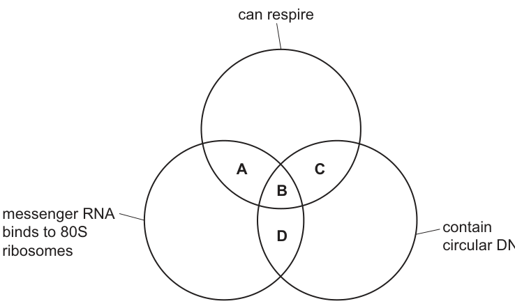
Fig. 3.1 — three overlapping circles: top, can respire; bottom left, messenger RNA binds to 80S ribosomes; bottom right, contain circular DNA. Region A = respire ∩ 80S; B = all three; C = respire ∩ circular DNA; D = 80S ∩ circular DNA.
Show full working
- 1
Eliminate every region containing 'mRNA binds to 80S ribosomes'. Regions A, B and D all require that feature — but bacterial ribosomes are 70S, so bacterial mRNA binds 70S, not 80S. No region with the 80S condition can include typical bacteria.
This single fact deletes three of the four options at a stroke — always look for the test that kills the most regions first.
- 2
Check region C ('can respire' ∩ 'contain circular DNA') against both groups. Typical eukaryotes: respire aerobically, and contain circular DNA inside their mitochondria (and chloroplasts) from §05. Typical bacteria: respire, and their chromosome is a circular loop. Both pass.
'Contain circular DNA' is deliberately worded to be true of whole eukaryotic cells via their organelles — the wording is what makes C work rather than trapping you into thinking eukaryotic DNA is only ever linear.
- 3
Answer: C.
C — both eukaryotes and bacteria respire, and both contain circular DNA (in mitochondria/chloroplasts, and as the bacterial chromosome respectively)
Venn questions fall to two checks per region: does group X have the feature, and does group Y have it in the named form? The form is where the traps live.
Your turn
- 19700/11 O/N 2023 Q61 mark
Which row about the genetic material in animal cells and prokaryotic cells is correct?
animal cells contain linear DNA prokaryotic genetic material is surrounded by a double membrane prokaryotic genetic material is double-stranded DNA A ✓ ✓ ✓ B ✓ ✓ ✗ C ✓ ✗ ✓ D ✗ ✗ ✓ (key: ✓ = correct, ✗ = not correct)
Stuck? Show hint
Judge each column separately before looking at the options.
Show solution
- 1
Column 1 — true. Animal genetic material is linear chromosomal DNA inside the nucleus.
- 2
Column 2 — false. Prokaryotic DNA lies free in the cytoplasm; there is no nuclear envelope of any kind, let alone a double membrane around it.
'Surrounded by a double membrane' describes the eukaryotic nuclear envelope — transplanting it onto bacteria is exactly the confusion this question harvests.
- 3
Column 3 — true. The circular bacterial chromosome is still double-stranded DNA — circular describes its shape, not its strands.
Columns 2 and 3 together give ✓ ✗ ✓ = option C. Note how option D tempts candidates who wrongly think prokaryotic DNA must be single-stranded because it is 'simple'.
AnswerC — animal cells have linear DNA; prokaryotic DNA is not membrane-surrounded; prokaryotic DNA is double-stranded
- 1
- 29700/11 O/N 2023 Q51 mark
The diagram shows three circles, 1, 3 and 5, and the shared structures, 2 and 4.
Which row correctly identifies the three circles and some of the structures that are shared between them?
circle 1 2 circle 3 4 circle 5 A chloroplasts circular DNA mitochondria 80S ribosomes prokaryotes B chloroplasts 80S ribosomes mitochondria circular DNA prokaryotes C prokaryotes circular DNA mitochondria circular DNA chloroplasts D prokaryotes 70S ribosomes chloroplasts 80S ribosomes mitochondria 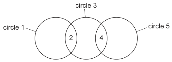
Fig. 5.1 — three circles in a row (1, then 3, then 5); 2 sits in the overlap of 1 and 3, 4 in the overlap of 3 and 5.
Stuck? Show hint
Each overlap must be true of BOTH neighbouring circles. Test overlaps 2 and 4 independently.
Show solution
- 1
Test option C first since the middle circle recurs: overlap 2 asks what prokaryotes and mitochondria share — both contain circular DNA ✓. Overlap 4 asks what mitochondria and chloroplasts share — both contain circular DNA ✓. Both overlaps hold, so C works.
Notice the examiner never claims the shared feature is exclusive — circular DNA appearing twice is fine, because each overlap is judged independently against its own two circles.
- 2
Kill the others on one failed overlap each. A: overlap 4 needs mitochondria ∩ prokaryotes to share 80S ribosomes — both are 70S, ✗. B: overlap 2 needs chloroplasts ∩ mitochondria to share 80S ribosomes — again both 70S, ✗. D: overlap 2 (prokaryotes ∩ chloroplasts share 70S) is true, but overlap 4 needs chloroplasts ∩ mitochondria to share 80S — false, ✗.
One wrong overlap disqualifies the whole row — which is why testing overlaps one at a time, rather than reading rows across, finds the answer faster and more safely.
AnswerC — prokaryotes and mitochondria share circular DNA; mitochondria and chloroplasts share circular DNA
- 1
- 3
List six key structural features of a typical bacterium (a prokaryotic cell).
Stuck? Show hint
Three concern what is present; two concern sizes or numbers; one concerns what is absent.
Show solution
- 1
Write the syllabus list out in full, one clause per line: unicellular; generally 1–5 µm in diameter; cell wall of peptidoglycan; circular DNA free in the cytoplasm (no nuclear envelope); 70S ribosomes; absence of organelles surrounded by double membranes.
Examiners award one mark per distinct feature — six short lines beat one long sentence, and nothing on the list may be swapped: 'linear DNA', '80S ribosomes' or a cellulose wall would each cancel a mark.
Answerunicellular · 1–5 µm diameter · peptidoglycan cell wall · naked circular DNA free in the cytoplasm · 70S ribosomes · no double-membrane-bound organelles
- 1
Viruses — non-cellular structures
“
state that all viruses are non-cellular structures with a nucleic acid core (either DNA or RNA) and a capsid made of protein, and that some viruses have an outer envelope made of phospholipids.
Not cells, and not quite alive
Seven sections in, every structure you have met has passed at least some of the tests that define a cell. The topic ends with the structures that fail all of them: all viruses are non-cellular. A virus has no cytoplasm, no ribosomes, no cell surface membrane of its own, and carries out no metabolism — it does not feed, respire, excrete or grow. It cannot even reproduce alone: it can only replicate by hijacking a host cell's machinery, which is precisely what makes it a parasite of cells and the bridge into this course's infectious-disease topics.
What a virus does have is a short parts list, and every mark-scheme answer on this sub-topic is assembled from exactly three items:
- a nucleic acid core — either DNA or RNA, never both (every true cell carries both; each virus commits to one);
- a capsid — a coat of protein enclosing the core;
- in some viruses, an outer envelope of phospholipids, stolen from the host cell's membranes as the virus exits.
A virus in cross-section. Core: nucleic acid — DNA or RNA, never both. Capsid: protein coat enclosing the core. Envelope: phospholipid bilayer, present in some viruses only.
Every virus: a nucleic acid core (DNA or RNA) and a protein capsid. Some viruses: a phospholipid envelope outside the capsid. No virus: cytoplasm, ribosomes, organelles, or both kinds of nucleic acid together. When a question says "state the key features of a virus", the safe three are the two all items plus non-cellular — anything from the no column loses the mark.
Why so few drugs have any effect on viruses
Designing a drug means finding a target the pathogen has and the patient lacks. Bacteria offer plenty — peptidoglycan walls, bacterial ribosomes, bacterial enzymes — which is why antibiotics exist. Viruses offer almost nothing:
- few targets: no cell wall, no cell surface membrane of their own (any envelope is host-derived phospholipid), no ribosomes;
- no or very few enzymes of their own to inhibit;
- no metabolism: antibiotics act against growing, living cells, and a virus outside its host does nothing at all;
- and even when it is replicating, the virus sits inside host cells, physically out of reach of most drugs — attacking it there would mean poisoning the patient's own tissue.
So the same austerity that defines a virus structurally explains why it is so hard to treat.
A clean demonstration before the past-paper questions
Suppose an electron micrograph shows an nm particle: a dense thread coiled inside a geometric shell, itself wrapped in a bubble-like outer layer. Read it through the census: the shell = capsid (protein); the thread inside = nucleic acid core — call it DNA or RNA, whichever evidence establishes, never both; the bubble layer = phospholipid envelope — present here, but remember it is a some-virus feature, not part of the definition. If asked for key features of this virus you could add its envelope; if asked for key features of viruses generally, stick to non-cellular + core + capsid.
Three key features of viruses
A virus named Pandoravirus salinus was discovered in 2013 by French scientists.
The virus was so large that the scientists initially thought that P. salinus was a bacterium.
P. salinus was confirmed to be a virus after further research.
List three key features of viruses.
Show full working
- 1
Feature 1 — they are non-cellular (acellular; not made of cells): no cytoplasm, no organelles, no metabolism of their own.
- 2
Feature 2 — a protein coat (capsid) surrounding the genetic material.
- 3
Feature 3 — a nucleic acid core: either DNA or RNA, one per virus.
These three are the mark scheme's named points. Alternatives credited include 'replicate only inside host cells', 'most are smaller than bacteria' — but the definition trio is the reliable route.
non-cellular (acellular) · protein coat/capsid · nucleic acid core (DNA or RNA)
'List' questions want distinct statements, not paraphrases of one idea — 'has no cells' twice will score once. Give the census trio and stop.
The plant–bacterium–virus comparison table
A student constructed a table to compare the structural features of a plant cell, a prokaryotic cell and a virus.
Complete Table 6.1.
Table 6.1
| feature | plant cell | prokaryotic cell | virus |
|---|---|---|---|
| external structure | cell wall composed of cellulose | cell wall composed of ______ | capsid composed of ______ |
| size of ribosomes | 80S and 70S | ______ | no ribosomes |
| nucleic acids | DNA and RNA | DNA and RNA | ______ |
Show full working
- 1
Blank 1 — prokaryotic wall material: peptidoglycan (also credited: murein).
Cellulose is the plant answer; writing it here is rejected outright. Peptidoglycan is one of the six syllabus features of a bacterium from §07.
- 2
Blank 2 — what the virus capsid is composed of: protein (also credited: polypeptides, or capsomeres).
Viruses are not cells, so they have no wall — their outer structure is the capsid from the census above, and a capsid is built of protein subunits.
- 3
Blank 3 — prokaryotic ribosome size: 70S.
The plant column already shows the pattern: 80S in cytoplasm plus 70S inside organelles. Bacteria run 70S only — the small type throughout.
- 4
Blank 4 — viral nucleic acids: DNA or RNA — one or the other, never both.
'DNA and RNA' is explicitly rejected (R) here: every cell carries both, but each virus particle carries exactly one kind. This asymmetry is the single most-tested virus fact on Paper 1.
peptidoglycan · (capsid composed of) protein · 70S · DNA or RNA (never both)
Comparison tables mark blank-by-blank — even if viruses feel unfamiliar, the four answers come straight from §05's ribosome split, §07's wall material, and the census above.
Your turn
- 19700/22 M/J 2020 Q1(a)2 marks
Picornaviruses are small viruses that are in diameter. Picornaviruses are able to enter the cells of mammals and birds and can replicate within these cells.
State the key features of a virus, such as picornavirus.
Stuck? Show hint
Any two distinct items from the definition trio.
Show solution
- 1
Any two of: protein coat / capsid; nucleic acid core — DNA or RNA; acellular / non-cellular.
Two marks, two different features — writing 'capsid made of protein' and then 'protein coat' is one feature stated twice and scores once.
Answercapsid (protein coat) · nucleic acid core (DNA or RNA) · non-cellular — any two
- 1
- 29700/23 O/N 2016 Q2(b)(iv)2 marks
Suggest why there are few drugs that have any effect on viruses.
Stuck? Show hint
Think like a drug designer: what could a drug actually bind to, and where is the virus when it is dangerous?
Show solution
- 1
Target argument: a virus offers almost nothing to bind to — no cell wall, no cell membrane of its own, no ribosomes, and no (or hardly any) enzymes.
This earns the first mark: 'few targets for drugs', with at least one concrete absence named. Vague answers like 'viruses are simple' do not score.
- 2
Reach/metabolism argument: antibiotics act on growing, living cells — a virus has no metabolism — and while replicating it is hidden inside host cells, out of reach of drugs without damaging those cells.
Either the no-metabolism point or the sheltered-inside-host-cells point completes the second mark. Saying antibodies fight viruses instead is explicitly rejected — antibodies are immune proteins, not drugs.
AnswerFew drug targets (no wall/membrane/ribosomes, few or no enzymes); no metabolism of their own; and inside host cells out of reach of drugs — any two points
- 1
- 3
Sort each of the following into all viruses, some viruses or no viruses: (a) a nucleic acid core; (b) a phospholipid envelope; (c) ribosomes; (d) a protein capsid; (e) DNA and RNA in the same particle; (f) both a capsid and an envelope in the same particle.
Stuck? Show hint
Only two of the six belong to every virus.
Show solution
- 1
(a) all viruses — every virus packages exactly one kind of nucleic acid. (d) all viruses — the protein capsid is universal. So the two universals are the core and the capsid.
- 2
(b) some viruses — envelopes are acquired from host membrane and many viruses (like picornaviruses) leave them out entirely. (f) some viruses too — capsid plus envelope together is simply what an enveloped virus looks like.
(b) and (f) are one fact asked two ways: the envelope is optional equipment — fitted to some viruses, missing from many.
- 3
(c) no viruses — protein synthesis happens only after the host's ribosomes are hijacked. (e) no viruses — the core is DNA or RNA, never both; carrying both would disqualify a structure from being a single virus at all.
Answerall: (a) core, (d) capsid · some: (b) phospholipid envelope, (f) capsid + envelope together · none: (c) ribosomes, (e) both nucleic acids together
- 1
Everything on one page
The M = I ÷ A triangle — magnification itself has NO units
Rearranged form when the printed magnification is given
The unit ladder — convert to matching units before dividing
Graticule calibration — valid only at the objective used to calibrate
The light microscope's wavelength-limited wall
The ribosome-size split that comparison tables always test
The six features every bacterium answer needs
The definition of every virus — never DNA and RNA together
Can you do all of these?
Make a temporary preparation (thin specimen, water, stain, angled coverslip) and justify each step
Draw cells or chromosomes to convention: sharp pencil, no shading, ruled touching labels, no arrowheads
Convert between mm, µm and nm in either direction without slipping
Calculate magnification, actual size or image size — writing the formula and showing every conversion step
State magnifications with no units, and actual sizes always with units
Calibrate an eyepiece graticule with a stage micrometer and use it to measure a specimen
Define resolution and magnification and explain the difference, including the 0.25 µm light-microscope limit
Decide whether a micrograph is light or electron, TEM or SEM, from what is visible in it
Name every syllabus organelle from its appearance, give its function, and count its membranes
Explain organelle abundance from cell function (secretory cells, muscle, small-intestine epithelium)
Compare plant with animal cells, and prokaryotes with eukaryotes, including DNA location and ribosome sizes
State the features of a virus and why DNA-or-RNA (never both), and explain why few drugs affect viruses