Testing for biological molecules
“
describe and carry out the Benedict's test for reducing sugars, the iodine test for starch, the emulsion test for lipids and the biuret test for proteins; describe and carry out a semi-quantitative Benedict's test on a reducing sugar solution by standardising the test and using the results (time to first colour change or comparison to colour standards) to estimate the concentration; describe and carry out a test to identify the presence of non-reducing sugars, using acid hydrolysis and Benedict's solution.
Why the tests come first
Before you can study a molecule you have to know it is there. Every investigation in this topic — following starch digestion in a gut, checking whether nectar contains glucose, finding out what phloem sap carries — begins with a food test, and the examiners know it: the tests are among the most examined practical skills on the course, asked about directly on Paper 2 ("outline the test… state the result…") and constantly behind the scenes on Paper 1 (a question tells you a solution turned lilac and expects you to know instantly what that means).
Each test follows the same three-part shape, and every mark-scheme answer is built from the same three parts:
- the reagent — what you add;
- the conditions — what you must do to it (heat? shake? nothing?);
- the positive result — the colour or appearance that says present, including the colour it started from.
Learn each test as that triple and you can answer "state the colour change" questions, "outline the procedure" questions and Paper 1 interpretation questions with the same memory.
- 1
Add Benedict's reagent (a blue solution of copper(II) compounds) to the sample in a test-tube.
Benedict's is supplied ready-made — never say 'add blue copper sulfate only'; the biuret reagents are the ones split into two solutions.
- 2
Heat the mixture — place in a boiling water-bath (or heat to at least 80 °C) for a stated time, typically a few minutes.
The heating step earns its own mark and is the one most often dropped. Without heat the reaction is far too slow to give a reliable result.
- 3
Watch the colour: a positive result moves from blue → green → yellow → orange → brick-red, ending as a coloured precipitate (copper(I) oxide) that settles out on standing.
Green/yellow/orange are all fully positive results — they simply mean less reducing sugar than a red one. The one wrong answer is 'no change': blue staying blue means no reducing sugar.
The Benedict's colour ladder. Blue means no reducing sugar; each step along green → yellow → orange → brick-red means more reducing sugar was present. The red end is a precipitate of copper(I) oxide, not just a colour change in solution.
- 1
Starch — iodine test. Add iodine solution (iodine in potassium iodide) to the sample at room temperature; orange/brown turning blue-black is positive. No heating, and the change is instant.
Iodine needs no heating — adding a boiling step here loses marks because it shows the tests have blurred together. The starting colour (orange/brown) is part of the answer: 'goes black' without it reads as incomplete.
- 2
Lipids — emulsion test. Dissolve the sample in ethanol (alcohol), then pour the ethanol solution into water in a clean tube. A cloudy white emulsion appearing is positive.
Order matters and the mark scheme polices it: ethanol first, then water. Do not heat and add nothing else — a lipid does not dissolve in water, which is precisely why it comes out of the ethanol as tiny droplets that scatter light.
- 3
Proteins — biuret test. Add biuret solution — or sodium/potassium hydroxide followed by dilute copper sulfate solution — at room temperature. Blue → lilac/purple/violet is positive.
Two acceptable reagent recipes exist; either scores. 'Pink' is not credited — the target words are lilac, purple or violet. Like iodine, this one runs cold.
Molecule tested | Reagent | Conditions | Positive result |
|---|---|---|---|
Reducing sugars | Benedict's solution | heat / boil | blue → green / yellow / orange → brick-red precipitate |
Starch | iodine solution | room temperature | orange/brown → blue-black |
Lipids | ethanol, then water | dissolve in ethanol first; no heating | cloudy white emulsion |
Protein | biuret solution (or NaOH/KOH then CuSO₄) | room temperature | blue → lilac / purple / violet |
The four qualitative tests as reagent–condition–result triples. Quote the start colour too — 'lilac' alone scores less reliably than 'blue to lilac'.
A colour change is only meaningful if you know what absence looks like. A negative result is the reagent's own colour staying put: Benedict's stays blue, iodine stays orange/brown, biuret stays blue, the water in an emulsion test stays clear. A control is a parallel tube run through exactly the same steps but known to lack the molecule (distilled water, for example). If the control stays negative while the sample goes positive, the colour really does report the molecule — not contaminated glassware or a warm day. When a question asks you to "justify a conclusion", it wants this pairing: sample positive, control negative, therefore the molecule is present in the sample alone.
Demo — three unlabelled bottles
A technician finds three unlabelled bottles. One holds a glucose solution, one a starch suspension and one a protein solution — the labels are gone, and only the tests from this section are available.
(a) Bottle P gives a brick-red precipitate with Benedict's reagent but stays orange/brown with iodine. Which solution is in P?
(b) Bottle Q tests negative with Benedict's reagent and negative with iodine, but turns lilac with biuret reagent. Which solution is in Q?
(c) Bottle R tests negative with Benedict's reagent — until it is boiled with dilute hydrochloric acid, cooled and neutralised, after which Benedict's reagent turns orange. What does this reveal about R?
Show full working
- 1
(a) Brick-red with Benedict's means a reducing sugar is present; iodine staying orange/brown rules out starch. P is the glucose solution.
One positive result names the chemical group; the second test closes the case. Pairing evidence with elimination is the habit part (c)'s logic chain builds on.
- 2
(b) Negative Benedict's rules out reducing sugars and negative iodine rules out starch — but lilac with biuret is the protein signature. Q is the protein solution.
Biuret needs no heating, so 'lilac arriving without a water bath' was already a clue — but the formal reasoning is two eliminations plus one positive.
- 3
(c) Negative first, positive after acid hydrolysis is exactly the non-reducing sugar signature: the acid broke glycosidic bonds, freeing reactive groups that now reduce Benedict's reagent.
This is the logic chain the section teaches: a non-reducing sugar hides its reactive group inside a glycosidic bond, so only hydrolysis exposes it. And note why the neutralising step matters — Benedict's responds properly only in alkaline conditions.
(a) glucose — reducing sugar present, starch absent. · (b) protein — biuret positive, both sugar tests negative. · (c) R contains a non-reducing sugar: acid hydrolysis released reducing groups, so Benedict's reagent now responds
Every food-test question is some arrangement of three moves — name the reagent, quote both colours, or reason from a negative-plus-positive pair.
Reagent and colour change for starch
Fig. 1.1 is a transmission electron micrograph of cells from the leaf of a plant.
Cell structure Y in Fig. 1.1 contains a large starch granule (grain).
Name the chemical reagent used to test for starch and state the colour change that will be seen if starch is present.
reagent ………
colour change ………
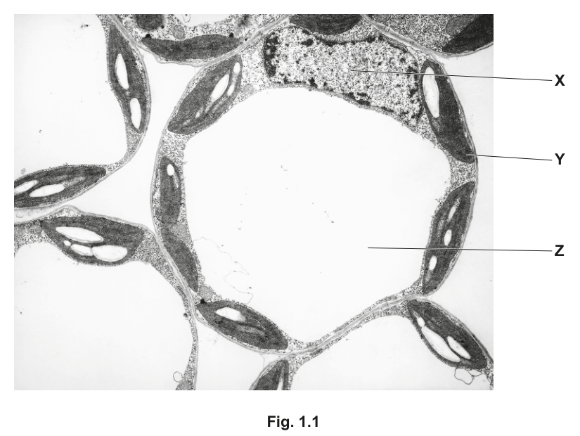
Fig. 1.1 — transmission electron micrograph of leaf cells, with structures X, Y and Z labelled.
Show full working
- 1
Reagent: iodine solution. (The mark scheme also credits iodine in potassium iodide solution — the reagent is iodine dissolved in potassium iodide, because iodine itself does not dissolve readily in water.)
One mark lives on the name alone. Writing 'iodine' is enough; writing 'Benedict's' here would score zero — the two sugar tests and the starch test are exactly the confusion this question harvests.
- 2
Colour change: from orange/brown to blue/black. The full creditable form quotes the starting colour and the end colour — orange/brown → blue-black.
The mark scheme accepts purple as an end colour too, but 'goes black' with no start colour risks reading as incomplete. Both colours are needed for the second mark.
reagent: iodine solution · colour change: orange/brown → blue-black
Every 'name the reagent and state the colour change' answer is two marks: one per half. Give both colours of the change, joined by an arrow.
Outline of the emulsion test
Lipase is an enzyme with many commercial uses. Some species of bacteria are of great interest as they produce large quantities of lipase.
Researchers carried out investigations into lipase extracted from a bacterium found in hot springs.
To measure the activity of the bacterial lipase during their investigations, the researchers used a method based on the biological test for triglycerides.
Outline a biological test that could be carried out to show the presence of triglyceride in a liquid mixture and describe the positive result for this test.
Show full working
- 1
Step 1 — dissolve the sample in ethanol (alcohol). Take a portion of the liquid mixture and shake it with ethanol so any triglyceride dissolves in the alcohol.
Lipids are insoluble in water but soluble in ethanol — dissolving them first is what allows a visible result at all. Adding the wrong substance to the alcohol loses the mark outright.
- 2
Step 2 — pour the ethanol solution into water in a second test-tube, in that order.
The sequence is marked: ethanol into water, two tubes involved. Tipping everything into one tube at once, or heating the mixture, caps the method marks.
- 3
Positive result — a cloudy white emulsion appears: the lipid leaves solution as a fine dispersion of droplets that scatter light, turning the water milky/opaque.
'Emulsion' is the credited word; milky, cloudy, opaque and white droplets are all accepted descriptions. 'Precipitate' is explicitly rejected — an emulsion is droplets suspended in the liquid, not a solid settling out.
Dissolve the sample in ethanol; add/pour the ethanol solution into water → cloudy white emulsion (milky/opaque) forms = positive
For every outline-the-test question, write the steps in order with the reagents named — the emulsion test in particular is marked on its sequence, not just its endpoint.
Your turn
Each item leans on one row of the tests table — reagent, condition or result.
- 19700/22 O/N 2021 Q2(b)2 marks
Name the chemical reagent or reagents used to test for proteins in a sample of blood plasma and state the colour change that will be seen if protein is present.
reagent or reagents ………
colour change ………
Show solution
- 1
Reagent: biuret solution. Equally credited: copper sulfate solution plus sodium hydroxide (or potassium hydroxide) — the two-solution recipe that biuret reagent bundles up.
Either recipe earns the mark; quoting only copper sulfate, or only the alkali, does not — the pair works together.
- 2
Colour change: from (light) blue to lilac (purple/violet also credited). The starting blue is the colour of the copper sulfate in the reagent itself.
Quoting the start colour makes the answer self-contained — 'turns lilac' implies something else was there first, and the examiner wants to read what it was.
Answerreagent: biuret solution (or NaOH/KOH + CuSO₄) · colour change: blue → lilac / purple / violet
- 1
- 29700/21 O/N 2025 Q3(b)(i)1 mark
A student carried out an experiment to study the synthesis of starch by phosphorylase found in potato tissue.
The student was provided with a solution, E, extracted from potato tissue. The extract was filtered to remove all the starch grains.
Extract E contained biological molecules from the potato tissue including phosphorylase but no starch.
Iodine solution was used to confirm that starch was not present in extract E.
State the colour observed when iodine solution was added to a sample of extract E.
Show solution
- 1
No starch is present, so no positive colour can develop: the iodine keeps its own colour — orange/brown (amber also credited).
This is the negative-result idea from the controls paragraph applied directly: absence is signalled by the reagent's own colour persisting. Yellow-brown and yellow-orange earn the mark; 'blue-black' would contradict the stem.
Answerorange / brown / amber — the iodine colour persists because no starch is present
- 1
- 39700/14 M/J 2025 Q8
Diastase is an enzyme that breaks down starch into maltose.
A sample of starch is treated with boiled diastase and left for 15 minutes.
Samples of the mixture are then tested with iodine solution and with Benedict's solution.
What is the correct result?
iodine solution Benedict's solution A blue-black blue B blue-black red C brown blue D brown red Show solution
- 1
Work out whether any digestion happened. Boiling denatures enzymes — the diastase's active site is destroyed, so it cannot catalyse anything. The mixture after 15 minutes is unchanged starch, no maltose.
The word 'boiled' is the whole question. Everything downstream follows from one fact you already know from practical work: heat kills enzymes.
- 2
Test each tube in imagination. Starch present → iodine gives its positive blue-black. No maltose means no reducing sugar added → Benedict's stays blue (negative).
Option D tempts candidates who assume enzyme + substrate must give product regardless. Option C fails because iodine without starch keeps its own orange/brown — which would mean the starch had gone, and it has not.
- 3
That pairing is option A.
AnswerA — boiled diastase cannot digest the starch, so iodine is positive (blue-black) and Benedict's is negative (blue)
- 1
Semi-quantitative Benedict's: turning a colour into a concentration
The qualitative test tells you a reducing sugar is present. A semi-quantitative test goes one step further and estimates how much — not to the fourth decimal place, but reliably enough to rank solutions or match them against known values. The trick is that the Benedict's colour ladder is not random: the more reducing sugar in the tube, the further along the ladder the colour travels (more copper reduced → deeper into orange/red), and the faster it gets there.
That only works if everything else is held constant. Standardising means fixing every variable except the one you are investigating:
- the same volume of Benedict's solution in every tube;
- the same volume of sample in every tube;
- the same heating time at the same temperature (or a defined end-point rule, below);
- the same tube size and mixing.
Change any of these and a colour difference could come from your procedure rather than from the samples. Two reading strategies then turn colours into numbers:
- Time to first colour change. Run each sample with Benedict's in the same water-bath and stop the clock when green first appears. A higher concentration gives a shorter time, because more reducing sugar reduces copper faster. Time can be read off against a calibration curve built from standard glucose solutions of known concentration.
- Comparison against colour standards. Heat every tube for an identical fixed time, then match each final colour against a set of standards made by running known concentrations side by side — or read the absorbance in a colorimeter. The closer to brick-red, the higher the concentration.
- 1
Decide the reading strategy first — timed end-point, or fixed-time colour comparison — because it dictates what must be standardised.
For timing you need equal temperatures and simultaneous starts; for colour comparison you need identical heating times so the only variable left is concentration.
- 2
Measure equal volumes: the same volume of Benedict's solution and the same volume of each sample, using the same measuring equipment.
'Same volume' is the phrase mark schemes look for — vague answers like 'add some Benedict's' cannot show the test was standardised.
- 3
Heat every tube identically — same water-bath, same temperature, same duration (or start all clocks together and record the time to the first colour change).
Heating is the engine of the reaction; unequal heating makes a dilute sample look concentrated. This is the step most often forgotten when students write up this practical.
- 4
Compare results with standards of known concentration (or plot time against known concentrations), and read off your unknown.
Semi-quantitative means anchored to something known — a colour ladder or a calibration curve built from standards, not eyeballed from memory.
A clean demonstration with invented numbers
Two unknown glucose solutions, X and Y, are tested with of Benedict's each, heated together in the same water-bath:
| solution | time to first (green) colour change | final colour after 5 min |
|---|---|---|
| X | 95 s | yellow |
| Y | 40 s | orange |
Both are positive, so both contain reducing sugar. Y turned green sooner and finished further along the ladder — two independent signals agreeing — so Y has the higher glucose concentration. If standards had been run alongside (say 0.5 % changing at 90 s, 1.0 % at 45 s, 2.0 % at 20 s), you could go further and estimate X near 0.5 % and Y near 1.0 %. Notice what made the comparison legal: equal volumes, equal heating, simultaneous timing. Without those, 55 seconds would mean nothing.
Which solution changes colour first?
A student was asked to carry out semi-quantitative Benedict's tests on two solutions.
- Solution A was extracted from the cytoplasm of cells in the mesophyll tissue of photosynthesising leaves.
- Solution B was extracted from the phloem sap in phloem sieve tubes.
The solutions were taken from the same plant, and other variables were standardised.
For each solution, the student measured the time taken for the first colour change to appear.
Suggest which of the two solutions, A or B, would change colour in the shortest time.
Explain your answer.
Show full working
- 1
Recall what each tissue actually carries. Mesophyll cytoplasm (solution A) holds glucose — a reducing sugar, either free or stored as starch. Phloem sap (solution B) transports sucrose — a non-reducing sugar.
This is the fact base the whole question rides on: photosynthesis makes glucose where it happens, but the loading form shipped around the plant is sucrose. §02 names sucrose non-reducing outright.
- 2
Apply the chemistry. Benedict's solution reacts only with reducing sugars — it does not react with sucrose. So solution B contributes little or nothing for Benedict's to work on, while solution A's glucose reacts readily.
The mark scheme's core line: 'Benedict's solution only reacts with reducing sugars'. If you have not yet met sucrose's non-reducing nature formally, hold the thought — §02 explains exactly why it fails the test.
- 3
Answer: solution A changes colour in the shortest time, because its (mainly) glucose/reducing sugars react immediately with Benedict's, whereas B's sucrose does not react.
(The examiners also credited the reverse argument if argued from different assumptions about sap contents — but the glucose-in-cytoplasm route is the expected one.)
Solution A — its sugar is mainly glucose (a reducing sugar), which reacts with Benedict's at once; B contains mainly sucrose, a non-reducing sugar that Benedict's does not react with
Semi-quantitative 'which is faster' questions are answered in two moves: identify the chemistry in each sample, then connect it to speed of colour change.
The non-reducing sugar test: unmasking sucrose
Some sugars — sucrose above all — sail through the Benedict's test with no colour change at all, yet are undeniably sugars. They are the non-reducing sugars: their reactive group is locked up inside the bond joining their two units (the full story of why arrives in §02). The test for them is therefore a transformation: break that bond artificially, and the reducing halves appear.
The logic chain is what Paper 2 actually examines:
Benedict's negative first → hydrolyse with dilute acid → neutralise → Benedict's positive second ⇒ a non-reducing sugar was present.
Each clause earns its own mark, and each exists for a reason:
- the first negative rules out ordinary reducing sugars — without it you cannot tell whether the second positive came from them;
- dilute hydrochloric acid, heated, catalyses the hydrolysis of the sugar into its reducing monosaccharides (for sucrose: glucose and fructose);
- cooling comes before neutralising so you are not adding alkali to near-boiling acid;
- neutralising (with sodium hydrogencarbonate or sodium hydroxide) matters because Benedict's only reacts properly around neutral pH — acid left behind would spoil the re-test;
- the re-test then goes green→yellow→orange→red, because the hydrolysis products are reducing sugars.
- 1
Run an ordinary Benedict's test on the sample. Result: stays blue — negative.
This first result is part of the answer, not a warm-up: it establishes that any reducing sugars present are below detection, so the second test can only be responding to something new.
- 2
Take a fresh sample and boil it with dilute hydrochloric acid (a few minutes in a water-bath) to hydrolyse any non-reducing sugar into its monosaccharides.
'Hydrolysis' is the credited word — the acid plus heat splits bonds using water. Fresh sample, because the first tube already contains Benedict's reagent.
- 3
Cool the tube, then neutralise by adding sodium hydrogencarbonate (or sodium hydroxide) a little at a time.
Cooling before neutralising avoids spitting and protects the reagents; neutralising restores the pH Benedict's needs. Checking with universal indicator paper that pH ≈ 7 was specifically credited in mark schemes.
- 4
Re-test with Benedict's, heating as usual. A positive result now (green → yellow → orange → brick-red precipitate) shows the hydrolysis released reducing sugars — so a non-reducing sugar was present in the original solution.
Positive-after-hydrolysis-only is the signature: if the first test had also been positive you could not conclude this, because glucose would explain both results on its own.
Three ways this question eats marks
Forgetting to neutralise. The re-test fails in acidic conditions, so an unneutralised tube can look falsely negative — and the method loses its mark even when the idea of hydrolysis was right. Skipping the first Benedict's test. Writing 'boil with acid, then test' answers a different question — without the initial negative there is no logic chain. Heating the wrong step. In the sequence, acid hydrolysis needs heat and each Benedict's stage needs heat; but nothing else does. Mark schemes cap credit when steps are out of order — learn the order: first test → acid + boil → cool → neutralise → re-test.
Outlining the procedure and results for trehalose
Saccharomyces cerevisiae is a unicellular fungus that is important in the brewing and baking industries.
A disaccharide, trehalose, is a reserve store of energy for S. cerevisiae when glycogen stores decrease. The monomer of glycogen and trehalose is α-glucose.
A student carried out tests on a solution of trehalose and correctly concluded that trehalose is a non-reducing sugar.
Outline the procedure carried out by the student and state the results that were obtained.
Show full working
- 1
Procedure, in order: heat the trehalose solution with Benedict's solution → no colour change / remains blue (negative); then boil a fresh sample with (hydrochloric) acid to hydrolyse it; cool and neutralise with alkali; finally heat again with Benedict's.
The mark scheme's credited points are exactly these: Benedict's first with a stated negative result, then boiling with acid (or hydrolysing with acid/enzyme). Credit was also given for cooling before neutralising and checking neutrality with indicator paper — the finishing touches of a careful write-up.
- 2
Results: negative first (stays blue), then a positive result after hydrolysis — a coloured precipitate (green/yellow/orange/brown/red all accepted).
The final positive must be stated to complete the conclusion — the coloured precipitate is what lets the student claim a non-reducing sugar. Negative-then-positive together is the whole argument.
Heat with Benedict's → remains blue (negative); boil fresh sample with dilute HCl, cool, neutralise with alkali; re-test with Benedict's → coloured precipitate forms (positive) ⇒ non-reducing sugar present
'Outline the procedure AND state the results' means your answer alternates: do this → saw this → did this → saw this. A procedure with no observations leaves the conclusion hanging.
Paper 1 recycles the non-reducing sugar test almost every sitting as a "put the steps in order" question — and the distractors are always the same three swaps: neutralise before the acid, omit the first Benedict's test, or heat in the wrong place. If you can write the five-step chain from memory, every variant collapses. Across the bank, 115 questions (99 Paper 1 MCQs, 16 Paper 2 parts) touch the food tests, 2010–2025.
Your turn
These run the semi-quantitative and non-reducing material back at you — watch the order of operations.
- 19700/11 O/N 2025 Q6
Solution X was tested for the presence of non-reducing sugars. It did not contain reducing sugars.
Some steps that can be used to test for the presence of biological molecules are listed.
1 Add Benedict's solution to the test-tube.
2 Add dilute hydrochloric acid to the test-tube.
3 Add sodium hydrogencarbonate to the test-tube.
4 Heat the test-tube in a water-bath.Which order of steps to identify the presence of non-reducing sugars in solution X is correct?
A 1 → 4
B 2 → 3 → 1 → 4
C 2 → 4 → 3 → 1 → 4
D 3 → 2 → 4 → 1Stuck? Show hint
Acid must act hot before anything neutralises it; Benedict's goes in last, and needs its own heating.
Show solution
- 1
Start with the acid. Hydrolysis comes first, so step 2 opens the correct sequences — options B, C survive, A and D fall away (A never hydrolyses at all; D adds the alkali before the acid).
Neutralising before hydrolysing (D) defeats the point: there is no acid yet to neutralise, and the alkali would then have to be overwhelmed by the acid added next.
- 2
The acid needs heat to work: step 4 follows step 2 directly — option C's opening (2 → 4). Option B puts the sodium hydrogencarbonate in before any heating, so the acid would be neutralised while cold, having done nothing.
Hydrolysis is being catalysed by the acid but needs the energy of heating to happen at a useful rate — the pair travels together.
- 3
Then neutralise (3), then Benedict's + heat (1 → 4): the full chain is 2 → 4 → 3 → 1 → 4, which is option C. Note step 4 appears twice — once for the hydrolysis, once for the final Benedict's test, which itself requires heating.
Two separate heatings is exactly the trap: candidates who forget the Benedict's stage needs its own boil pick B-style orders where the only heat is on the acid.
AnswerC — 2 → 4 → 3 → 1 → 4 (acid, heat, neutralise, add Benedict's, heat again)
- 1
- 29700/23 M/J 2019 Q4(b)2 marks
Sucrose is transported in phloem sieve tubes.
A student carried out a test to identify the presence of sucrose in a sample of sap taken from inside a phloem sieve tube. One of the steps in the procedure instructed the student to heat the phloem sap with hydrochloric acid.
Explain why it is necessary to carry out this step in the procedure.
Stuck? Show hint
What does the acid treatment do to sucrose, and what can Benedict's react with afterwards?
Show solution
- 1
First mark — what the acid does: the hot acid hydrolyses sucrose, breaking its glycosidic bond to release the smaller sugars glucose and fructose.
'Hydrolyses / breaks glycosidic bonds' and 'produces reducing sugars (glucose and fructose)' are two separate marking points on the scheme — stating both already secures the two marks.
- 2
Second mark — why that matters: the products are reducing sugars, so they give a positive Benedict's test, whereas sucrose itself is non-reducing — Benedict's cannot react with it as it stands.
The examiner wants the causal link made explicit: hydrolysis is necessary because the original sugar cannot be detected directly. Saying just 'to test for sucrose' restates the aim instead of explaining the mechanism.
AnswerTo hydrolyse sucrose — breaking glycosidic bonds and producing the reducing sugars glucose and fructose — which Benedict's can then detect; sucrose itself gives no reaction
- 1
- 39700/21 M/J 2018 Q2(b)2 marks
When Benedict's solution is added to a sucrose solution and put into a boiling water-bath, no change in colour is observed.
State why no colour change is observed.
Show solution
- 1
Point 1: sucrose is a non-reducing sugar, so it does not react with Benedict's solution.
Naming the class is one mark. The examiner accepts 'no reducing sugars present' here — the stem has already ruled out other sugars.
- 2
Point 2 — the deeper reason: sucrose's reactive group is not available — it is tied up in the bond joining its two units, so it cannot reduce the blue copper(II) ions to the coloured copper(I) state. No acid has been added to break that bond and release the reactive groups.
Any of these phrasings earned credit: reactive groups unavailable, cannot donate electrons, will not reduce Cu²⁺ to Cu⁺, no free aldehyde/ketone group, sucrose not hydrolysed. Pick whichever wording you can reproduce under pressure — §02 develops the full story.
AnswerSucrose is a non-reducing sugar whose reactive groups are tied up in its glycosidic bond, so it cannot reduce copper(II) ions; without hydrolysis Benedict's shows no colour change
- 1
The rest of this note
Can you do all of these?
State reagent, conditions and positive result for Benedict's, iodine, emulsion and biuret tests — and give a negative result meaning
Describe a controlled, standardised semi-quantitative Benedict's test and read concentration from time-to-colour-change or colour standards
Classify glucose, fructose and maltose as reducing and sucrose as non-reducing, and say why — then sequence the non-reducing sugar test and interpret a negative-first-test, positive-after-hydrolysis pair
Use the terms monomer, polymer, macromolecule, mono-/di-/polysaccharide precisely — including why stearin is not a polymer
Draw α- and β-glucose rings with every OH placed correctly and consistently (all H kept or all H dropped)
Explain condensation and hydrolysis of the glycosidic bond, including where the water comes from and goes
Describe starch as amylose plus amylopectin (α-1,4 and α-1,6 bonds) and match branching, compactness and insolubility to storage
Describe cellulose's straight β-1,4 chains, hydrogen-bonded microfibrils, tensile strength and permeability in cell walls
Build a triglyceride from glycerol and three fatty acids — three ester bonds, three waters — and distinguish saturated from unsaturated chains
Describe a phospholipid's hydrophilic phosphate head and hydrophobic fatty-acid tails, and contrast it with a triglyceride
Draw the general amino acid and show the peptide bond forming by condensation (water out) and breaking by hydrolysis
Define primary, secondary, tertiary and quaternary structure with the interactions holding each, and explain how primary structure determines tertiary shape and therefore function
Sort proteins into globular (soluble, physiological roles) and fibrous (insoluble, structural roles); describe haemoglobin — two α-globins, two β-globins, four haem groups whose Fe²⁺ binds one O₂ molecule each — and collagen's triple helix with glycine every third position, its molecules parallel, staggered and covalently cross-linked into fibrils and fibres, relating both structures to function
Explain hydrogen bonding between water molecules using δ⁻ oxygen / δ⁺ hydrogen partial charges, and relate water's properties to its three examined roles: solvent action on polar/ionic substances, high specific heat capacity keeping blood temperature stable, and high latent heat of vaporisation cooling by evaporation