The chloroplast — where the two stages of photosynthesis happen
“
describe the relationship between the structure of chloroplasts, as shown in diagrams and electron micrographs, and their function; state that within a chloroplast, the thylakoids (thylakoid membranes and thylakoid spaces), which occur in stacks called grana, are the site of the light-dependent stage and the stroma is the site of the light-independent stage.
Why the chloroplast is shaped the way it is
A chloroplast is the only organelle in a plant cell that can convert light energy into chemical energy, and almost every feature of its structure can be read as an adaptation to that one job. A typical mesophyll chloroplast is about 3–10 µm long, lens-shaped, and bounded by a double membrane envelope (an outer and an inner membrane, with a thin intermembrane space between them). Inside the envelope is the stroma — a protein-rich soluble phase not unlike the cytoplasm of a bacterial cell, and the place where the light-independent stage runs. Suspended in the stroma is a third, internal membrane system, the thylakoid membrane, which is folded into flattened disc-like sacs called thylakoids, and the thylakoids are stacked into piles called grana (singular: granum). Grana are connected to each other by intergranal lamellae — single thylakoids that run between the stacks. The space inside each thylakoid disc is the thylakoid space (or lumen).
The first rule of the topic is that the light-dependent stage happens on the thylakoid membrane, and the light-independent stage (Calvin cycle) happens in the stroma. The reason for the split is functional: the thylakoid membrane is the only place in the cell that can build a proton gradient, and the stroma is the only place where the Calvin cycle's enzymes are free to diffuse. The two stages are linked by ATP and reduced NADP, which are made on the thylakoid membrane and used in the stroma.
Fig 13.1 A cut-away diagram of a chloroplast. The double-membrane envelope encloses the stroma. Inside the stroma are stacks of thylakoids (grana) connected by intergranal lamellae. A starch grain is shown in the middle as the storage product of photosynthesis.
The five structural features and what each one is for
A chloroplast is small, but every part has a function, and the mark schemes expect you to know which part does what. The five features that come up most often are:
- Double membrane envelope. A protein-rich outer membrane and a selectively permeable inner membrane, with an intermembrane space between them. The envelope keeps the chloroplast's contents — the stroma enzymes, the thylakoid membrane, the DNA and ribosomes — separate from the rest of the cell. The inner membrane is the only barrier that small molecules like glucose and CO₂ have to cross to get in or out.
- Grana (stacks of thylakoids). Thylakoid membranes are folded into disc-shaped vesicles and stacked into grana. Stacking gives a very large surface area for the photosystems, electron carriers and ATP synthase to be embedded in, which is essential because the light-dependent stage is a membrane-bound process. Grana also bring photosystem II (PSII) and photosystem I (PSI) close enough to share mobile electron carriers (plastoquinone, plastocyanin).
- Intergranal lamellae (also called stroma thylakoids or stroma lamellae). Single thylakoids that connect adjacent grana, and that bear ATP synthase and PSI. They are the highway between grana for the mobile carriers.
- Stroma. The protein-rich soluble phase surrounding the thylakoids. The stroma contains the enzymes of the Calvin cycle (notably rubisco, the most abundant protein on Earth), the chloroplast's own DNA and ribosomes (chloroplasts make some of their own proteins), and the starch grains that store the product of photosynthesis.
- Starch grains. Large, pale, dense deposits of starch that build up in the stroma in the light. They are not part of the photosynthetic machinery, but they are an obvious diagnostic feature on an electron micrograph, and they are the storage form of the triose phosphate made by the Calvin cycle.
Feature | What it is | Function in photosynthesis |
|---|---|---|
Double membrane envelope | Outer + inner membrane, intermembrane space between | Holds the stroma and thylakoid system; controls what enters and leaves the chloroplast |
Granum (plural: grana) | A stack of thylakoid discs | Holds the photosystems, electron carriers and ATP synthase; large surface area for the light-dependent stage |
Intergranal lamella | A single thylakoid connecting two grana | Carries ATP synthase and PSI between grana; route for mobile electron carriers |
Thylakoid space (lumen) | Space inside a thylakoid disc | Accumulates H⁺ during the light-dependent stage; site of the proton gradient that drives chemiosmosis |
Stroma | Soluble phase inside the envelope but outside the thylakoids | Site of the Calvin cycle; contains rubisco, other Calvin-cycle enzymes, chloroplast DNA, ribosomes, starch grains |
Starch grain | Deposit of stored starch in the stroma | Stores the triose-phosphate product of the Calvin cycle as starch; not a functional part of the photosynthetic machinery |
The five structural features of a chloroplast. Each one is a feature, not just a name — the mark scheme will not credit 'granum' without a function.
What a chloroplast looks like under the electron microscope
Under a transmission electron microscope, a chloroplast has a distinctive appearance that the mark schemes describe in four pieces:
- The envelope is two parallel dark lines, the outer and inner membranes, separated by a thin lighter space.
- The grana are the densest dark regions, because the thylakoid membranes are stacked tightly and they stain heavily with osmium. Each granum looks like a stack of dark stripes.
- The stroma is the lighter, granular material between the grana. The granularity is from rubisco (which is very abundant) and the chloroplast's own ribosomes.
- Starch grains are pale, dense, often lens-shaped bodies in the stroma. They are not membrane-bound, so they look different from the grana — uniformly electron-dense rather than striped.
The MS phrasing for a "describe the appearance of a chloroplast under the EM" answer is "envelope (double membrane), grana (stacks of thylakoid membranes), stroma (lighter, granular), starch grains (pale, dense, lens-shaped)" — a structured list, not a paragraph.
Fig 13.2 A chloroplast under the transmission electron microscope. The double-membrane envelope, dark grana stacks, lighter granular stroma and pale starch grains are all visible.
Linking structure to function
The single most common mark lost in this section is the failure to link structure to function. The mark scheme wants answers like "the granum is a stack of thylakoids, which gives a large surface area for the light-dependent stage", not "chloroplasts have grana". Every structural feature is an answer to a functional question:
- Envelope? Holds the chloroplast together; controls entry and exit.
- Stacked grana? Large surface area for the photosystems and ATP synthase; close packing for the mobile carriers.
- Lamellae? Connect grana; route for mobile carriers; bear PSI and ATP synthase.
- Stroma? Soluble enzymes of the Calvin cycle; large volume; no membrane to slow substrate diffusion.
- Starch grains? Storage; can be made and broken down without affecting the rest of the chloroplast.
If a question asks "describe and explain the structure of a chloroplast" the answer is describe (a feature) and explain (a function). A list of structures without functions loses half the marks.
Identifying the parts of a chloroplast on a micrograph
Fig. 1.1 shows a transmission electron micrograph of part of a chloroplast.
Table 1.1 describes some functions that occur in different parts of a chloroplast.
Complete Table 1.1 by identifying the letter on Fig. 1.1 that is a location matching the description. Each letter may be used once, more than once, or not at all.
| description | letter |
|---|---|
| accumulates (builds up) a high concentration of protons | |
| makes triose phosphate | |
| makes some chloroplast proteins | |
| pumps protons |
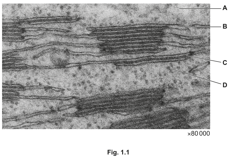
Fig. 1.1 — A transmission electron micrograph of part of a chloroplast. Letters A, B, C and D label four different parts of the chloroplast.
Show full working
- 1
Accumulates a high concentration of protons → B (the thylakoid space). Protons (H⁺) are pumped from the stroma into the thylakoid space during the light-dependent stage, so the thylakoid space is the place where the proton concentration builds up. The letter B is on a thylakoid space.
Mark 1. The MS credits the link between proton gradient and thylakoid space. The candidate must say thylakoid space (or lumen), not just 'inside'.
- 2
Makes triose phosphate → A (the stroma). The Calvin cycle runs in the stroma, and the reduction of GP to triose phosphate (TP) is the second of its three stages. The stroma is where the Calvin-cycle enzymes (including rubisco) are dissolved.
Mark 2. The MS rewards 'stroma' as the site of the Calvin cycle.
- 3
Makes some chloroplast proteins → D (a chloroplast ribosome). Chloroplasts have their own DNA and ribosomes; they make some of their own proteins, particularly the very hydrophobic ones embedded in the thylakoid membrane. The letter D labels a small dense body in the stroma — a ribosome.
Mark 3. The MS credits 'ribosome' or 'chloroplast ribosome'.
- 4
Pumps protons → C (the thylakoid membrane). Protons are pumped across the thylakoid membrane by the electron-transport chain carriers (between PSII and PSI). The letter C is on the thylakoid membrane.
Mark 4. The MS credits 'thylakoid membrane' (or a specific protein in it, e.g. cytochrome b₆f).
Accumulates a high concentration of protons: B (thylakoid space). Makes triose phosphate: A (stroma). Makes some chloroplast proteins: D (chloroplast ribosome). Pumps protons: C (thylakoid membrane).
On a 'match a description to a letter' question, the four answers are independent: do not let an obvious one influence the others, and check that the letter you have written down actually appears on the diagram. The MS often allows 'once, more than once, or not at all' — so a letter that fits no row is a deliberate red herring.
The rest of this note
Can you do all of these?
Describe the relationship between the structure of the chloroplast and its function (envelope, grana, lamellae, stroma, starch grains, thylakoid space)
State that the light-dependent stage occurs on the thylakoid membrane and the light-independent stage (Calvin cycle) occurs in the stroma
Describe the four chloroplast pigments (chlorophyll a, chlorophyll b, carotene, xanthophyll) and their role in light absorption in thylakoids (chlorophyll a is the primary pigment, the others are accessory pigments in the antenna complex)
Interpret absorption spectra of chloroplast pigments and action spectra for photosynthesis (absorption peaks: chlorophyll a 430/660 nm, chlorophyll b 455/640 nm, carotenoids blue only)
Describe and use chromatography to separate and identify chloroplast pigments, including calculating Rf and identifying each pigment from its Rf in a known solvent
State that cyclic and non-cyclic photophosphorylation both occur during the light-dependent stage
Explain cyclic photophosphorylation: only PSI is involved; photoactivation of chlorophyll occurs; ATP is synthesised (no reduced NADP, no photolysis, no O₂)
Explain non-cyclic photophosphorylation: both PSI and PSII are involved; photoactivation at both photosystems; photolysis of water at PSII (oxygen-evolving complex); ATP and reduced NADP are synthesised
Explain that during photophosphorylation: energetic electrons release energy along the ETC; the energy pumps H⁺ into the thylakoid space; H⁺ flows back to the stroma through ATP synthase; the energy of flow drives ATP synthesis (chemiosmosis)
Outline the three stages of the Calvin cycle: carbon fixation (CO₂ + RuBP → 2 GP, catalysed by rubisco); reduction (GP → TP using reduced NADP and ATP); regeneration (RuBP regenerated from TP using ATP)
State that Calvin-cycle intermediates are used to produce other molecules, limited to GP → some amino acids and TP → carbohydrates, lipids and amino acids
State that light intensity, CO₂ concentration and temperature are limiting factors of photosynthesis
Explain the effects of changes in light intensity, CO₂ concentration and temperature on the rate of photosynthesis (each gives a rise-and-plateau curve; temperature gives an additional fall above the optimum due to denaturation)
Describe and carry out investigations using DCPIP / methylene blue and isolated chloroplasts to determine the effect of light intensity and light wavelength on the rate of the light-dependent stage
Describe and carry out investigations using whole plants (especially aquatic plants such as Hydrilla) to determine the effect of light intensity, CO₂ concentration and temperature on the rate of photosynthesis